|
Electron Energy Loss Spectroscopy
In electron energy loss spectroscopy (EELS) a material is exposed to a beam of electrons with a known, narrow range of kinetic energies. Some of the electrons will undergo inelastic scattering, which means that they lose energy and have their paths slightly and randomly deflected. The amount of energy loss can be measured via an electron spectrometer and interpreted in terms of what caused the energy loss. Inelastic interactions include phonon excitations, inter- and intra-band transitions, plasmon excitations, inner shell ionizations, and Cherenkov radiation. The inner-shell ionizations are particularly useful for detecting the elemental components of a material. For example, one might find that a larger-than-expected number of electrons comes through the material with 285 eV less energy than they had when they entered the material. This is approximately the amount of energy needed to remove an inner-shell electron from a carbon atom, which can be taken as evidence t ... [...More Info...] [...Related Items...] OR: [Wikipedia] [Google] [Baidu] |
Electron Energy Loss Spectrum Feature Overview
The electron (, or in nuclear reactions) is a subatomic particle with a negative one elementary electric charge The elementary charge, usually denoted by is the electric charge carried by a single proton or, equivalently, the magnitude of the negative electric charge carried by a single electron, which has charge −1 . This elementary charge is a funda .... Electrons belong to the first generation (particle physics), generation of the lepton particle family, and are generally thought to be elementary particles because they have no known components or substructure. The electron's Invariant mass, mass is approximately Proton-to-electron mass ratio, 1/1836 that of the proton. Quantum mechanics, Quantum mechanical properties of the electron include an intrinsic angular momentum (spin (physics), spin) of a half-integer value, expressed in units of the Planck constant#Reduced Planck constant, reduced Planck constant, . Being fermions, no two electrons can occupy the same q ... [...More Info...] [...Related Items...] OR: [Wikipedia] [Google] [Baidu] |
Electron Energy Loss Spectroscopy Coreloss Lsmo
The electron (, or in nuclear reactions) is a subatomic particle with a negative one elementary electric charge. Electrons belong to the first generation of the lepton particle family, and are generally thought to be elementary particles because they have no known components or substructure. The electron's mass is approximately 1/1836 that of the proton. Quantum mechanical properties of the electron include an intrinsic angular momentum (spin) of a half-integer value, expressed in units of the reduced Planck constant, . Being fermions, no two electrons can occupy the same quantum state, per the Pauli exclusion principle. Like all elementary particles, electrons exhibit properties of both particles and waves: They can collide with other particles and can be diffracted like light. The wave properties of electrons are easier to observe with experiments than those of other particles like neutrons and protons because electrons have a lower mass and hence a longer de Broglie ... [...More Info...] [...Related Items...] OR: [Wikipedia] [Google] [Baidu] |
BIOS Scientific Publishers
BIOS Scientific Publishers was an English publisher.Publisher Information: BIOS Scientific Publishers(Imprint of Taylor & Francis Group) |
Scanning Transmission Electron Microscopy
A scanning transmission electron microscope (STEM) is a type of transmission electron microscope (TEM). Pronunciation is tɛmor �sti:i:ɛm As with a conventional transmission electron microscope (CTEM), images are formed by electrons passing through a sufficiently thin specimen. However, unlike CTEM, in STEM the electron beam is focused to a fine spot (with the typical spot size 0.05 – 0.2 nm) which is then scanned over the sample in a raster illumination system constructed so that the sample is illuminated at each point with the beam parallel to the optical axis. The rastering of the beam across the sample makes STEM suitable for analytical techniques such as Z-contrast annular dark-field imaging, and spectroscopic mapping by energy dispersive X-ray (EDX) spectroscopy, or electron energy loss spectroscopy (EELS). These signals can be obtained simultaneously, allowing direct correlation of images and spectroscopic data. A typical STEM is a conventional transmission ... [...More Info...] [...Related Items...] OR: [Wikipedia] [Google] [Baidu] |
Transmission Electron Microscopy
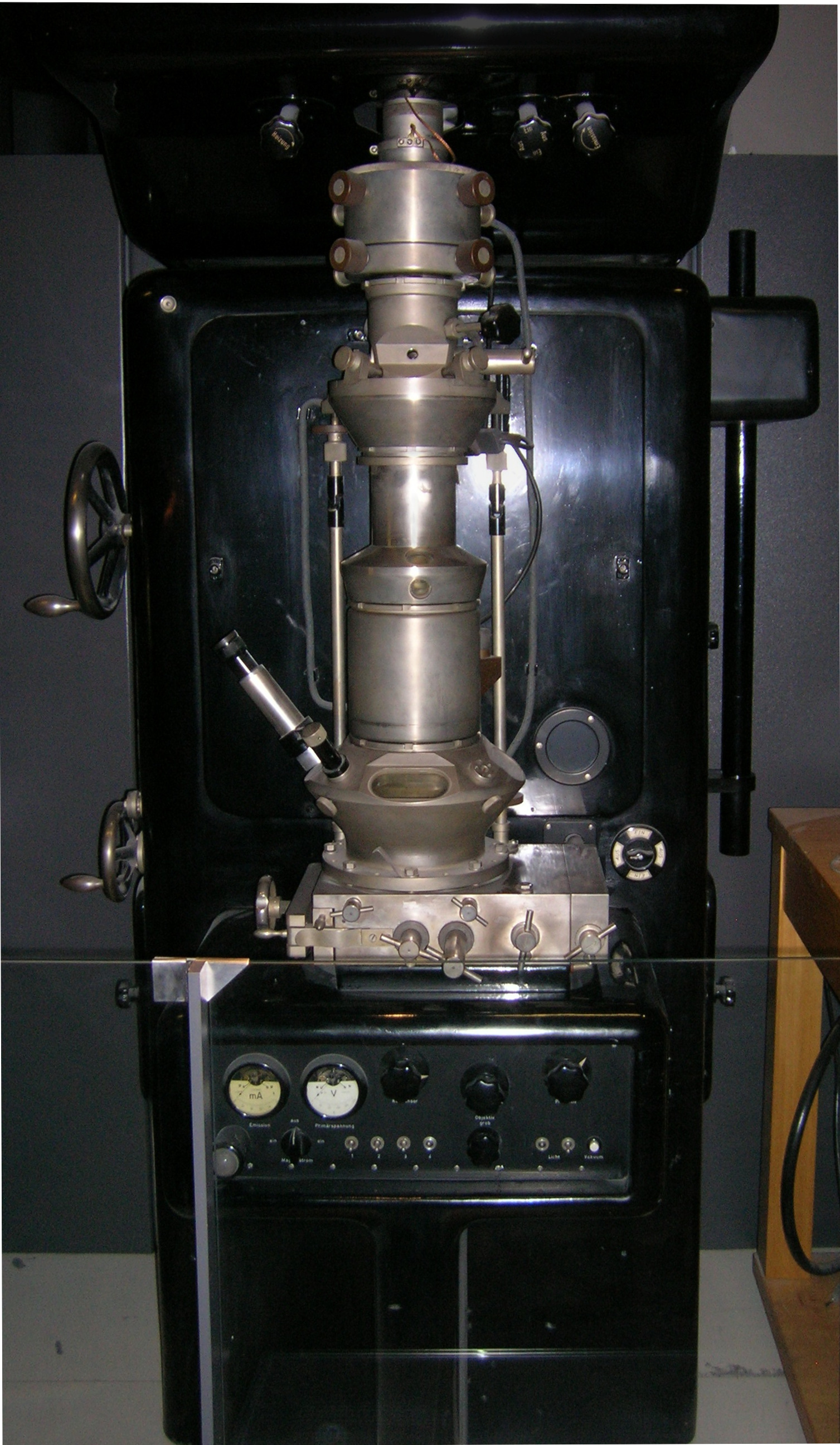
Transmission electron microscopy (TEM) is a microscopy technique in which a beam of electrons is transmitted through a specimen to form an image. The specimen is most often an ultrathin section less than 100 nm thick or a suspension on a grid. An image is formed from the interaction of the electrons with the sample as the beam is transmitted through the specimen. The image is then magnified and focused onto an imaging device, such as a fluorescent screen, a layer of photographic film, or a sensor such as a scintillator attached to a charge-coupled device. Transmission electron microscopes are capable of imaging at a significantly higher resolution than light microscopes, owing to the smaller de Broglie wavelength of electrons. This enables the instrument to capture fine detail—even as small as a single column of atoms, which is thousands of times smaller than a resolvable object seen in a light microscope. Transmission electron microscopy is a major analytical method ... [...More Info...] [...Related Items...] OR: [Wikipedia] [Google] [Baidu] |
Magic Angle (EELS)
The magic angle is a particular value of the collection angle of an electron microscope at which the measured energy-loss spectrum "magically" becomes independent of the tilt angle of the sample with respect to the beam direction. The magic angle is not uniquely defined for isotropic Isotropy is uniformity in all orientations; it is derived . Precise definitions depend on the subject area. Exceptions, or inequalities, are frequently indicated by the prefix ' or ', hence ''anisotropy''. ''Anisotropy'' is also used to describe ... samples, but the definition is unique in the (typical) case of small angle scattering on materials with a "c-axis", such as graphite. The "magic" angle depends on both the incoming electron energy (which is typically fixed) and the energy loss suffered by the electron. The ratio of the magic angle \theta_M to the characteristic angle \theta_E is roughly independent of the energy loss and roughly independent of the particular type of sample conside ... [...More Info...] [...Related Items...] OR: [Wikipedia] [Google] [Baidu] |
Energy Filtered Transmission Electron Microscopy
Energy-filtered transmission electron microscopy (EFTEM) is a technique used in transmission electron microscopy, in which only electrons of particular kinetic energies are used to form the image or diffraction pattern. The technique can be used to aid chemical analysis of the sample in conjunction with complementary techniques such as electron crystallography. Principle If a very thin sample is illuminated with a beam of high-energy electrons, then a majority of the electrons will pass unhindered through the sample but some will interact with the sample, being scattered elastically or inelastically (phonon scattering, plasmon scattering or inner shell ionisation). Inelastic scattering results in both a loss of energy and a change in momentum, which in the case of inner shell ionisation is characteristic of the element in the sample. If the electron beam emerging from the sample is passed through a magnetic prism, then the flight path of the electrons will vary depending on their ... [...More Info...] [...Related Items...] OR: [Wikipedia] [Google] [Baidu] |
Scanning Confocal Electron Energy Loss Microscopy
Scan may refer to: Acronyms * Schedules for Clinical Assessment in Neuropsychiatry (SCAN), a psychiatric diagnostic tool developed by WHO * Shared Check Authorization Network (SCAN), a database of bad check writers and collection agency for bad checks * Space Communications and Navigation Program (SCaN) * Social Cognitive and Affective Neuroscience (journal) * Scientific content analysis (SCAN), also known as statement analysis Businesses * Scan Furniture, Washington, D.C., US chain * SCAN Health Plan, not-for-profit health care company based in Long Beach, California * Scan AB or Scan Foods UK Ltd, the Swedish and UK subsidiaries of the Finnish HKScan Oyj * Seattle Community Access Network, Seattle, Washington, US TV channel * Scan (company), a software company based in Provo, Utah, US Electronics or computer related * 3D scanning * Counter-scanning, in physical micro and nanotopography measuring instruments like scanning probe microscope * Elevator algorithm (also SCAN ... [...More Info...] [...Related Items...] OR: [Wikipedia] [Google] [Baidu] |
Diamond Anvil Cell
A diamond anvil cell (DAC) is a high-pressure device used in geology, engineering, and materials science experiments. It enables the compression of a small (sub-millimeter-sized) piece of material to extreme pressures, typically up to around 100–200 gigapascals, although it is possible to achieve pressures up to 770 gigapascals (7,700,000 bars or 7.7 million atmospheres). The device has been used to recreate the pressure existing deep inside planets to synthesize materials and phases not observed under normal ambient conditions. Notable examples include the non-molecular ice X, polymeric nitrogen and metallic phases of xenon, lonsdaleite, and potentially metallic hydrogen. A DAC consists of two opposing diamonds with a sample compressed between the polished culets (tips). Pressure may be monitored using a reference material whose behavior under pressure is known. Common pressure standards include ruby fluorescence, and various structurally simp ... [...More Info...] [...Related Items...] OR: [Wikipedia] [Google] [Baidu] |
Scanning Transmission Electron Microscopy
A scanning transmission electron microscope (STEM) is a type of transmission electron microscope (TEM). Pronunciation is tɛmor �sti:i:ɛm As with a conventional transmission electron microscope (CTEM), images are formed by electrons passing through a sufficiently thin specimen. However, unlike CTEM, in STEM the electron beam is focused to a fine spot (with the typical spot size 0.05 – 0.2 nm) which is then scanned over the sample in a raster illumination system constructed so that the sample is illuminated at each point with the beam parallel to the optical axis. The rastering of the beam across the sample makes STEM suitable for analytical techniques such as Z-contrast annular dark-field imaging, and spectroscopic mapping by energy dispersive X-ray (EDX) spectroscopy, or electron energy loss spectroscopy (EELS). These signals can be obtained simultaneously, allowing direct correlation of images and spectroscopic data. A typical STEM is a conventional transmission ... [...More Info...] [...Related Items...] OR: [Wikipedia] [Google] [Baidu] |
Transmission Electron Microscopy
Transmission electron microscopy (TEM) is a microscopy technique in which a beam of electrons is transmitted through a specimen to form an image. The specimen is most often an ultrathin section less than 100 nm thick or a suspension on a grid. An image is formed from the interaction of the electrons with the sample as the beam is transmitted through the specimen. The image is then magnified and focused onto an imaging device, such as a fluorescent screen, a layer of photographic film, or a sensor such as a scintillator attached to a charge-coupled device. Transmission electron microscopes are capable of imaging at a significantly higher resolution than light microscopes, owing to the smaller de Broglie wavelength of electrons. This enables the instrument to capture fine detail—even as small as a single column of atoms, which is thousands of times smaller than a resolvable object seen in a light microscope. Transmission electron microscopy is a major analytical method ... [...More Info...] [...Related Items...] OR: [Wikipedia] [Google] [Baidu] |
Vibrational Spectroscopy
Infrared spectroscopy (IR spectroscopy or vibrational spectroscopy) is the measurement of the interaction of infrared radiation with matter by absorption, emission, or reflection. It is used to study and identify chemical substances or functional groups in solid, liquid, or gaseous forms. It can be used to characterize new materials or identify and verify known and unknown samples. The method or technique of infrared spectroscopy is conducted with an instrument called an infrared spectrometer (or spectrophotometer) which produces an infrared spectrum. An IR spectrum can be visualized in a graph of infrared light absorbance (or transmittance) on the vertical axis vs. frequency, wavenumber or wavelength on the horizontal axis. Typical units of wavenumber used in IR spectra are reciprocal centimeters, with the symbol cm−1. Units of IR wavelength are commonly given in micrometers (formerly called "microns"), symbol μm, which are related to the wavenumber in a reciprocal wa ... [...More Info...] [...Related Items...] OR: [Wikipedia] [Google] [Baidu] |


