|
Eimeria Tenella
''Eimeria tenella'' is a species of ''Eimeria'' that causes hemorrhagic cecum, cecal coccidiosis in young poultry. It is found worldwide. Description This species has a Monoxenous development, monoxenous life cycle with the only definitive host as chickens; it is extremely host-specific. Acquired via fecal contamination of food and water (oral-fecal route), it undergoes endogenous merogony in the crypts of Lieberkuhn (intestinal ceca of chicken) and gametogony in epithelial cells of the small intestines. Fusion of microgamete and macrogamete forms results in unsporulated zygotes, which are released with feces of chicken. The zygote sporulates after one to five days, and becomes infective. Diagnosis is based on finding oocysts in feces. While no effective treatment exists, the rate of infection can be reduced via prophylactics, anticoccidial drugs and vaccination of baby chicks. Life cycle ''Eimeria tenella'' has a monogenetic life cycle, that is, the life cycle involves a s ... [...More Info...] [...Related Items...] OR: [Wikipedia] [Google] [Baidu] |
Eimeria
''Eimeria'' is a genus of apicomplexan parasites that includes various species capable of causing the disease coccidiosis in animals such as cattle, poultry and smaller ruminants including sheep and goats. ''Eimeria'' species are considered to be monoxenous because the life cycle is completed within a single host, and stenoxenous because they tend to be host specific, although a number of exceptions have been identified. Species of this genus infect a wide variety of hosts. Thirty-one species are known to occur in bats (Chiroptera), two in turtles, and 130 named species infect fish. Two species (''E. phocae'' and ''E. weddelli'') infect seals. Five species infect llamas and alpacas: ''E. alpacae'', ''E. ivitaensis'', ''E. lamae'', ''E. macusaniensis'', and ''E. punonensis''. A number of species infect rodents, including ''E. couesii'', ''E. kinsellai'', ''E. palustris'', ''E. ojastii'' and ''E. oryzomysi''. Others infect poultry (''E. necatrix'' and ''E. tenella''), rabbits (''E. ... [...More Info...] [...Related Items...] OR: [Wikipedia] [Google] [Baidu] |
Golgi Bodies
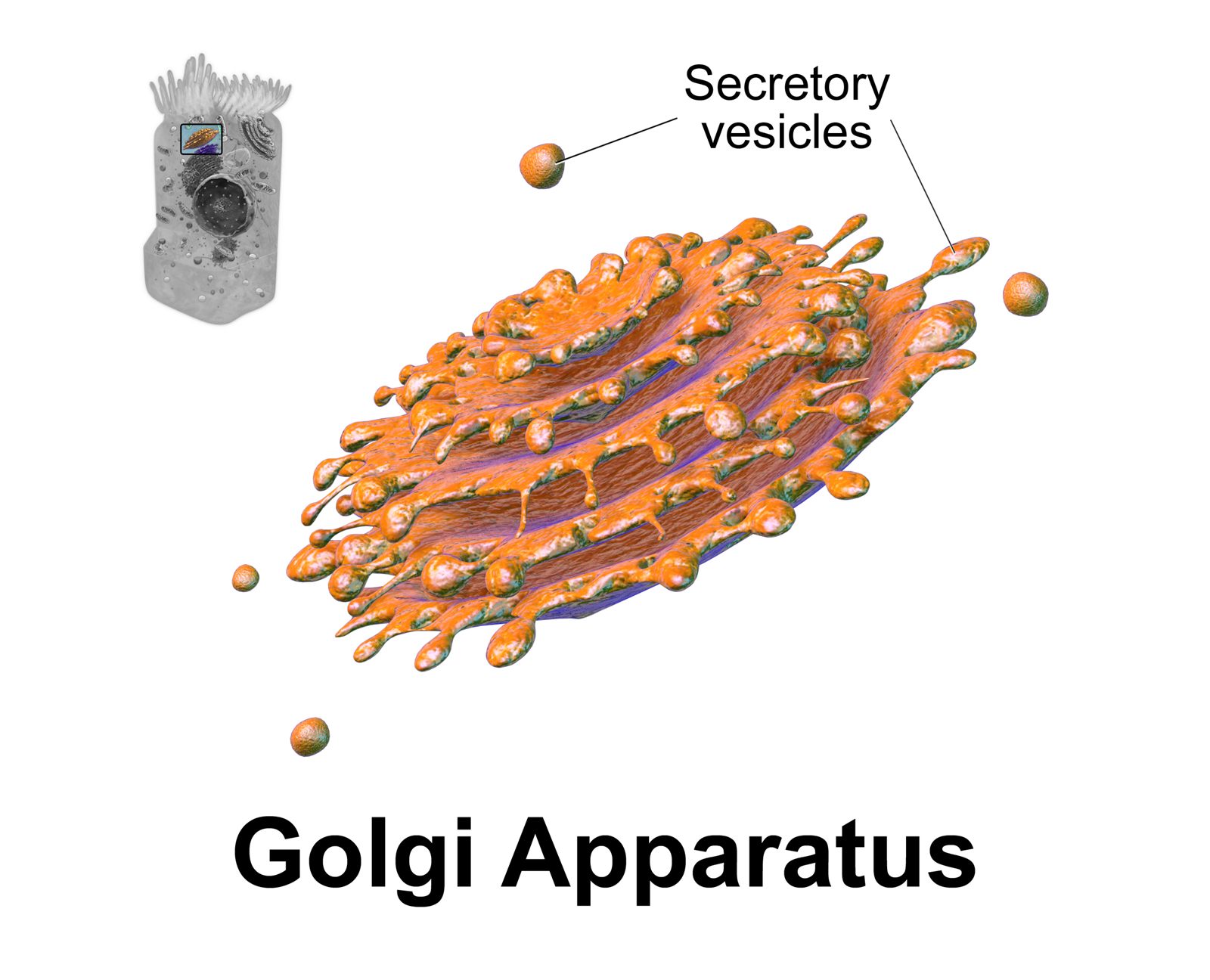
The Golgi apparatus (), also known as the Golgi complex, Golgi body, or simply the Golgi, is an organelle found in most eukaryotic cells. Part of the endomembrane system in the cytoplasm, it packages proteins into membrane-bound vesicles inside the cell before the vesicles are sent to their destination. It resides at the intersection of the secretory, lysosomal, and endocytic pathways. It is of particular importance in processing proteins for secretion, containing a set of glycosylation enzymes that attach various sugar monomers to proteins as the proteins move through the apparatus. It was identified in 1897 by the Italian scientist Camillo Golgi and was named after him in 1898. Discovery Owing to its large size and distinctive structure, the Golgi apparatus was one of the first organelles to be discovered and observed in detail. It was discovered in 1898 by Italian physician Camillo Golgi during an investigation of the nervous system. After first observing it under ... [...More Info...] [...Related Items...] OR: [Wikipedia] [Google] [Baidu] |
Mitochondrion
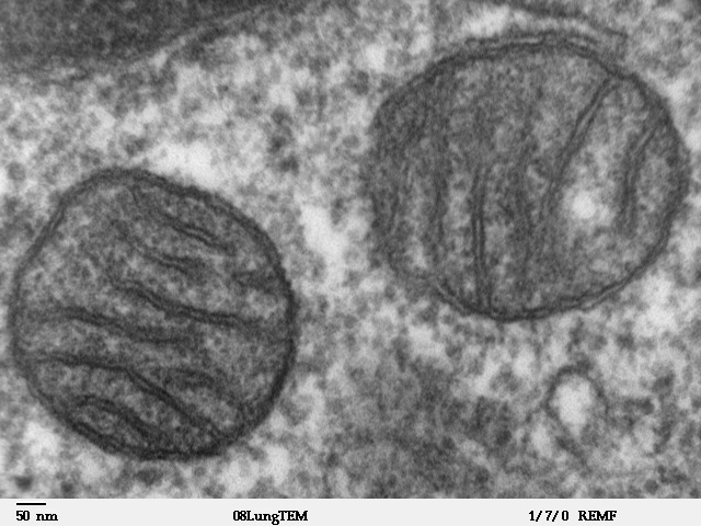
A mitochondrion (; ) is an organelle found in the cells of most Eukaryotes, such as animals, plants and fungi. Mitochondria have a double membrane structure and use aerobic respiration to generate adenosine triphosphate (ATP), which is used throughout the cell as a source of chemical energy. They were discovered by Albert von Kölliker in 1857 in the voluntary muscles of insects. The term ''mitochondrion'' was coined by Carl Benda in 1898. The mitochondrion is popularly nicknamed the "powerhouse of the cell", a phrase coined by Philip Siekevitz in a 1957 article of the same name. Some cells in some multicellular organisms lack mitochondria (for example, mature mammalian red blood cells). A large number of unicellular organisms, such as microsporidia, parabasalids and diplomonads, have reduced or transformed their mitochondria into other structures. One eukaryote, ''Monocercomonoides'', is known to have completely lost its mitochondria, and one multicellular organism, ... [...More Info...] [...Related Items...] OR: [Wikipedia] [Google] [Baidu] |
Cytoplasm
In cell biology, the cytoplasm is all of the material within a eukaryotic cell, enclosed by the cell membrane, except for the cell nucleus. The material inside the nucleus and contained within the nuclear membrane is termed the nucleoplasm. The main components of the cytoplasm are cytosol (a gel-like substance), the organelles (the cell's internal sub-structures), and various cytoplasmic inclusions. The cytoplasm is about 80% water and is usually colorless. The submicroscopic ground cell substance or cytoplasmic matrix which remains after exclusion of the cell organelles and particles is groundplasm. It is the hyaloplasm of light microscopy, a highly complex, polyphasic system in which all resolvable cytoplasmic elements are suspended, including the larger organelles such as the ribosomes, mitochondria, the plant plastids, lipid droplets, and vacuoles. Most cellular activities take place within the cytoplasm, such as many metabolic pathways including glycolysis, ... [...More Info...] [...Related Items...] OR: [Wikipedia] [Google] [Baidu] |
Microtubule
Microtubules are polymers of tubulin that form part of the cytoskeleton and provide structure and shape to eukaryotic cells. Microtubules can be as long as 50 micrometres, as wide as 23 to 27 nm and have an inner diameter between 11 and 15 nm. They are formed by the polymerization of a dimer of two globular proteins, alpha and beta tubulin into protofilaments that can then associate laterally to form a hollow tube, the microtubule. The most common form of a microtubule consists of 13 protofilaments in the tubular arrangement. Microtubules play an important role in a number of cellular processes. They are involved in maintaining the structure of the cell and, together with microfilaments and intermediate filaments, they form the cytoskeleton. They also make up the internal structure of cilia and flagella. They provide platforms for intracellular transport and are involved in a variety of cellular processes, including the movement of secretory vesicles, ... [...More Info...] [...Related Items...] OR: [Wikipedia] [Google] [Baidu] |
Pellicle (biology)
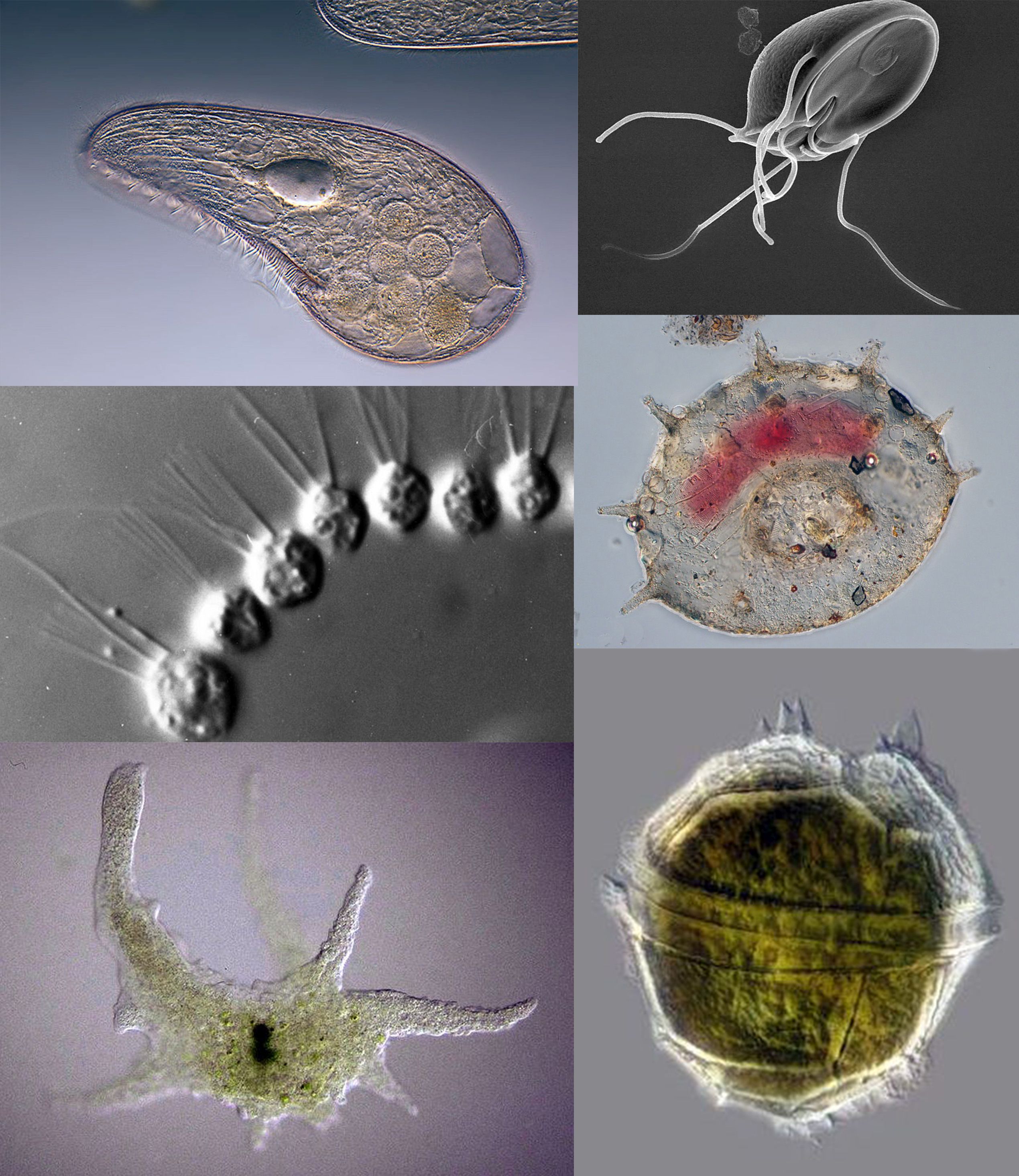
Protozoa (singular: protozoan or protozoon; alternative plural: protozoans) are a group of single-celled eukaryotes, either free-living or parasitic, that feed on organic matter such as other microorganisms or organic tissues and debris. Historically, protozoans were regarded as "one-celled animals", because they often possess animal-like behaviours, such as motility and predation, and lack a cell wall, as found in plants and many algae. When first introduced by Georg Goldfuss (originally spelled Goldfuß) in 1818, the taxon Protozoa was erected as a class within the Animalia, with the word 'protozoa' meaning "first animals". In later classification schemes it was elevated to a variety of higher ranks, including phylum, subkingdom and kingdom, and sometimes included within Protoctista or Protista. The approach of classifying Protozoa within the context of Animalia was widespread in the 19th and early 20th century, but not universal. By the 1970s, it became usual to requi ... [...More Info...] [...Related Items...] OR: [Wikipedia] [Google] [Baidu] |
Merozoite
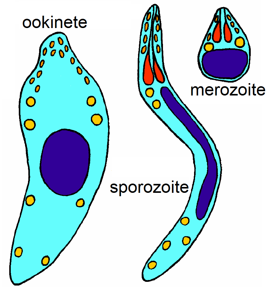
Apicomplexans, a group of intracellular parasites, have life cycle stages that allow them to survive the wide variety of environments they are exposed to during their complex life cycle. Each stage in the life cycle of an apicomplexan organism is typified by a ''cellular variety'' with a distinct morphology and biochemistry. Not all apicomplexa develop all the following cellular varieties and division methods. This presentation is intended as an outline of a hypothetical generalised apicomplexan organism. Methods of asexual replication Apicomplexans (sporozoans) replicate via ways of multiple fission (also known as schizogony). These ways include , and , although the latter is sometimes referred to as schizogony, despite its general meaning. Merogony is an asexually reproductive process of apicomplexa. After infecting a host cell, a trophozoite ( see glossary below) increases in size while repeatedly replicating its nucleus and other organelles. During this process, the org ... [...More Info...] [...Related Items...] OR: [Wikipedia] [Google] [Baidu] |
Enterocyte
Enterocytes, or intestinal absorptive cells, are simple columnar epithelial cells which line the inner surface of the small and large intestines. A glycocalyx surface coat contains digestive enzymes. Microvilli on the apical surface increase its surface area. This facilitates transport of numerous small molecules into the enterocyte from the intestinal lumen. These include broken down proteins, fats, and sugars, as well as water, electrolytes, vitamins, and bile salts. Enterocytes also have an endocrine role, secreting hormones such as leptin. Function The major functions of enterocytes include: *Ion uptake, including sodium, calcium, magnesium, iron, zinc, and copper. This typically occurs through active transport. *Water uptake. This follows the osmotic gradient established by Na+/K+ ATPase on the basolateral surface. This can occur transcellularly or paracellularly. *Sugar uptake. Polysaccharides and disaccharidases in the glycocalyx break down large sugar molecul ... [...More Info...] [...Related Items...] OR: [Wikipedia] [Google] [Baidu] |
Intestinal Villus
Intestinal villi (singular: villus) are small, finger-like projections that extend into the lumen of the small intestine. Each villus is approximately 0.5–1.6 mm in length (in humans), and has many microvilli projecting from the enterocytes of its epithelium which collectively form the striated or brush border. Each of these microvilli are about 1 µm in length, around 1000 times shorter than a single villus. The intestinal villi are much smaller than any of the circular folds in the intestine. Villi increase the internal surface area of the intestinal walls making available a greater surface area for absorption. An increased absorptive area is useful because digested nutrients (including monosaccharide and amino acids) pass into the semipermeable villi through diffusion, which is effective only at short distances. In other words, increased surface area (in contact with the fluid in the lumen) decreases the average distance travelled by nutrient molecules, so effect ... [...More Info...] [...Related Items...] OR: [Wikipedia] [Google] [Baidu] |
Sporozoite
Apicomplexans, a group of intracellular parasites, have life cycle stages that allow them to survive the wide variety of environments they are exposed to during their complex life cycle. Each stage in the life cycle of an apicomplexan organism is typified by a ''cellular variety'' with a distinct morphology and biochemistry. Not all apicomplexa develop all the following cellular varieties and division methods. This presentation is intended as an outline of a hypothetical generalised apicomplexan organism. Methods of asexual replication Apicomplexans (sporozoans) replicate via ways of multiple fission (also known as schizogony). These ways include , and , although the latter is sometimes referred to as schizogony, despite its general meaning. Merogony is an asexually reproductive process of apicomplexa. After infecting a host cell, a trophozoite ( see glossary below) increases in size while repeatedly replicating its nucleus and other organelles. During this process, the org ... [...More Info...] [...Related Items...] OR: [Wikipedia] [Google] [Baidu] |
Haploid
Ploidy () is the number of complete sets of chromosomes in a cell, and hence the number of possible alleles for autosomal and pseudoautosomal genes. Sets of chromosomes refer to the number of maternal and paternal chromosome copies, respectively, in each homologous chromosome pair, which chromosomes naturally exist as. Somatic cells, tissues, and individual organisms can be described according to the number of sets of chromosomes present (the "ploidy level"): monoploid (1 set), diploid (2 sets), triploid (3 sets), tetraploid (4 sets), pentaploid (5 sets), hexaploid (6 sets), heptaploid or septaploid (7 sets), etc. The generic term polyploid is often used to describe cells with three or more chromosome sets. Virtually all sexually reproducing organisms are made up of somatic cells that are diploid or greater, but ploidy level may vary widely between different organisms, between different tissues within the same organism, and at different stages in an organism's life cycle. Hal ... [...More Info...] [...Related Items...] OR: [Wikipedia] [Google] [Baidu] |


