|
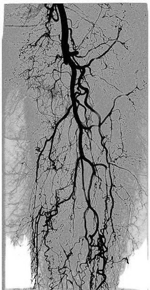
Digital Variance Angiography
Digital variance angiography (DVA) is a novel image processing method based on kinetic imaging, which allows the visualization of motion on image sequences generated by penetrating radiations. DVA is a specific form of kinetic imaging: it requires angiographic image series, which are created by X-ray or fluoroscopic imaging and by the administration of contrast media during various medical procedures. The resulting single DVA image visualizes the path of contrast agent with relatively low background noise. Between 2017 and 2019, two clinical studies have been performed to investigate the clinical usability of DVA and these studies have found that it has the potential to be used for low-dose radiographic imaging and carbon-dioxide angiography in the future. DVA is currently under development by Kinetic Health Ltd. and Semmelweis University (Budapest, Hungary). Lower extremity DSA comparison study In 2018 Gyánó M. et al. compared the quality of DVA and DSA (digital subtra ... [...More Info...] [...Related Items...] OR: [Wikipedia] [Google] [Baidu] |
Fig Dva Wiki
The fig is the edible fruit of ''Ficus carica'', a species of small tree in the flowering plant family Moraceae. Native to the Mediterranean and western Asia, it has been cultivated since ancient times and is now widely grown throughout the world, both for its fruit and as an ornamental plant.''The Fig: its History, Culture, and Curing'', Gustavus A. Eisen, Washington, Govt. print. off., 1901 ''Ficus carica'' is the type species of the genus '' Ficus'', containing over 800 tropical and subtropical plant species. A fig plant is a small deciduous tree or large shrub growing up to tall, with smooth white bark. Its large leaves have three to five deep lobes. Its fruit (referred to as syconium, a type of multiple fruit) is tear-shaped, long, with a green skin that may ripen toward purple or brown, and sweet soft reddish flesh containing numerous crunchy seeds. The milky sap of the green parts is an irritant to human skin. In the Northern Hemisphere, fresh figs are in season ... [...More Info...] [...Related Items...] OR: [Wikipedia] [Google] [Baidu] |
Kinetic Imaging
Kinetic imaging is an imaging technology developed by Szabolcs Osváth and Krisztián Szigeti in the Department of Biophysics and Radiation Biology at Semmelweis University (Budapest, Hungary). The technology allows the visualization of motion based on an altered data acquisition and image processing algorithm combined with imaging techniques that use penetrating radiation (e.g., X-rays). Kinetic imaging has the potential for use in a wide variety of areas including medicine, engineering, and surveillance. For example, physiological movements, such as the circulation of blood or motion of organs(e.g., palpitations, arrhythmia) can be visualized using kinetic imaging. Because of the reduced noise and the motion-based image contrast, kinetic imaging can be used to reduce X-ray dose and/or amount of required contrast agent in medical imaging (e.g., X-ray angiography). In fact, clinical trials are underway in the fields of vascular surgery and interventional radiology. Non-medic ... [...More Info...] [...Related Items...] OR: [Wikipedia] [Google] [Baidu] |
Semmelweis University
Ignaz Philipp Semmelweis (; hu, Semmelweis Ignác Fülöp ; 1 July 1818 – 13 August 1865) was a Hungarian physician and scientist, who was an early pioneer of antiseptic procedures. Described as the "saviour of mothers", he discovered that the incidence of puerperal fever (also known as "childbed fever") could be drastically reduced by requiring hand disinfection in obstetrical clinics. Puerperal fever was common in mid-19th-century hospitals and often fatal. He proposed the practice of washing hands with chlorinated lime solutions in 1847 while working in Vienna General Hospital's First Obstetrical Clinic, where doctors' wards had three times the mortality of midwives' wards. He published a book of his findings in ''Etiology, Concept and Prophylaxis of Childbed Fever''. Despite various publications of results where hand-washing reduced mortality to below 1%, Semmelweis's observations conflicted with the established scientific and medical opinions of the time and his idea ... [...More Info...] [...Related Items...] OR: [Wikipedia] [Google] [Baidu] |
Fig201
The fig is the edible fruit of ''Ficus carica'', a species of small tree in the flowering plant family Moraceae. Native to the Mediterranean and western Asia, it has been cultivated since ancient times and is now widely grown throughout the world, both for its fruit and as an ornamental plant.''The Fig: its History, Culture, and Curing'', Gustavus A. Eisen, Washington, Govt. print. off., 1901 ''Ficus carica'' is the type species of the genus '' Ficus'', containing over 800 tropical and subtropical plant species. A fig plant is a small deciduous tree or large shrub growing up to tall, with smooth white bark. Its large leaves have three to five deep lobes. Its fruit (referred to as syconium, a type of multiple fruit) is tear-shaped, long, with a green skin that may ripen toward purple or brown, and sweet soft reddish flesh containing numerous crunchy seeds. The milky sap of the green parts is an irritant to human skin. In the Northern Hemisphere, fresh figs are in season ... [...More Info...] [...Related Items...] OR: [Wikipedia] [Google] [Baidu] |
Digital Subtraction Angiography
Digital subtraction angiography (DSA) is a fluoroscopy technique used in interventional radiology to clearly visualize blood vessels in a bony or dense soft tissue environment. Images are produced using contrast medium by subtracting a "pre-contrast image" or ''mask'' from subsequent images, once the contrast medium has been introduced into a structure. Hence the term "digital ''subtraction'' angiography. Subtraction angiography was first described in 1935 and in English sources in 1962 as a manual technique. Digital technology made DSA practical starting in the 1970s. Procedure DSA and fluoroscopy In traditional angiography, images are acquired by exposing an area of interest with time-controlled x-rays while injecting a contrast medium into the blood vessels. The image obtained includes the blood vessels, together with all overlying and underlying structures. The images are useful for determining anatomical position and variations, but unhelpful for visualizing blood vessels acc ... [...More Info...] [...Related Items...] OR: [Wikipedia] [Google] [Baidu] |
Signal-to-noise Ratio (imaging)
Signal-to-noise ratio (SNR) is used in imaging to characterize image quality. The sensitivity of a (digital or film) imaging system is typically described in the terms of the signal level that yields a threshold level of SNR. Industry standards define sensitivity in terms of the ISO film speed equivalent, using SNR thresholds (at average scene luminance) of 40:1 for "excellent" image quality and 10:1 for "acceptable" image quality. SNR is sometimes quantified in decibels (dB) of signal power relative to noise power, though in the imaging field the concept of "power" is sometimes taken to be the power of a voltage signal proportional to optical power; so a 20 dB SNR may mean either 10:1 or 100:1 optical power, depending on which definition is in use. Definition of SNR Traditionally, SNR is defined to be the ratio of the average signal value \mu_\mathrm to the standard deviation of the signal \sigma_\mathrm: : \mathrm = \frac when the signal is an optical intensity, or ... [...More Info...] [...Related Items...] OR: [Wikipedia] [Google] [Baidu] |
CIRSE
The Cardiovascular and Interventional Radiological Society of Europe is a learned society of interventional radiologists from Europe and overseas. The society has its headquarters in Vienna (Austria) and was founded in 1985 by the merging of the European College of Angiography and European Society of Cardio-Vascular Radiology and Interventional Radiology. It currently has approximately 4,200 members from around the world, including 24 national societies. CIRSE's objective is to provide continuing education to physicians and scientists with an active interest in interventional radiology and to promote research as well as registries. CIRSE organises an annual congress with more than 5,000 participants, scientific meetings such as the European Conference on Embolotherapy and the European Conference on Interventional Oncology, as well as educational activities such as courses focusing on specific procedures. It publishes a bimonthly medical journal A medical journal is a peer-rev ... [...More Info...] [...Related Items...] OR: [Wikipedia] [Google] [Baidu] |
Radiography
Radiography is an imaging technique using X-rays, gamma rays, or similar ionizing radiation and non-ionizing radiation to view the internal form of an object. Applications of radiography include medical radiography ("diagnostic" and "therapeutic") and industrial radiography. Similar techniques are used in airport security (where "body scanners" generally use backscatter X-ray). To create an image in conventional radiography, a beam of X-rays is produced by an X-ray generator and is projected toward the object. A certain amount of the X-rays or other radiation is absorbed by the object, dependent on the object's density and structural composition. The X-rays that pass through the object are captured behind the object by a detector (either photographic film or a digital detector). The generation of flat two dimensional images by this technique is called projectional radiography. In computed tomography (CT scanning) an X-ray source and its associated detectors rotate around the su ... [...More Info...] [...Related Items...] OR: [Wikipedia] [Google] [Baidu] |
Fluoroscopy
Fluoroscopy () is an imaging technique that uses X-rays to obtain real-time moving images of the interior of an object. In its primary application of medical imaging, a fluoroscope () allows a physician to see the internal structure and function of a patient, so that the pumping action of the heart or the motion of swallowing, for example, can be watched. This is useful for both diagnosis and therapy and occurs in general radiology, interventional radiology, and image-guided surgery. In its simplest form, a fluoroscope consists of an X-ray source and a fluorescent screen, between which a patient is placed. However, since the 1950s most fluoroscopes have included X-ray image intensifiers and cameras as well, to improve the image's visibility and make it available on a remote display screen. For many decades, fluoroscopy tended to produce live pictures that were not recorded, but since the 1960s, as technology improved, recording and playback became the norm. Fluoroscopy is s ... [...More Info...] [...Related Items...] OR: [Wikipedia] [Google] [Baidu] |
Angiography
Angiography or arteriography is a medical imaging technique used to visualize the inside, or lumen, of blood vessels and organs of the body, with particular interest in the arteries, veins, and the heart chambers. Modern angiography is performed by injecting a radio-opaque contrast agent into the blood vessel and imaging using X-ray based techniques such as fluoroscopy. The word itself comes from the Greek words ἀγγεῖον ''angeion'' 'vessel' and γράφειν ''graphein'' 'to write, record'. The film or image of the blood vessels is called an ''angiograph'', or more commonly an ''angiogram''. Though the word can describe both an arteriogram and a venogram, in everyday usage the terms angiogram and arteriogram are often used synonymously, whereas the term venogram is used more precisely. The term angiography has been applied to radionuclide angiography and newer vascular imaging techniques such as CO2 angiography, CT angiography and MR angiography. The term ''isotope a ... [...More Info...] [...Related Items...] OR: [Wikipedia] [Google] [Baidu] |


