|
Deep Cervical Fascia
The deep cervical fascia (or fascia colli in older texts) lies under cover of the platysma, and invests the muscles of the neck; it also forms sheaths for the carotid vessels, and for the structures situated in front of the vertebral column. Its attachment to the hyoid bone prevents the formation of a dewlap. The investing portion of the fascia is attached behind to the ligamentum nuchæ and to the spinous process of the seventh cervical vertebra. The ''alar fascia'' is a portion of the ''deep cervical fascia''. Divisions The deep cervical fascia is often divided into a superficial, middle, and deep layer. The superficial layer is also known as the investing layer of deep cervical fascia. It envelops the trapezius, sternocleidomastoid, and muscles of facial expression. It also contains the submandibular and parotid salivary gland as well as the muscles of mastication (the masseter, pterygoid, and temporalis muscles). The middle layer is also known as the pretracheal fascia. I ... [...More Info...] [...Related Items...] OR: [Wikipedia] [Google] [Baidu] |
Platysma
The platysma muscle is a superficial muscle of the human neck that overlaps the sternocleidomastoid. It covers the anterior surface of the neck superficially. When it contracts, it produces a slight wrinkling of the neck, and a "bowstring" effect on either side of the neck. Structure The platysma muscle is a broad sheet of muscle arising from the fascia covering the upper parts of the pectoralis major muscle and deltoid muscle. Its fibers cross the clavicle, and proceed obliquely upward and medially along the side of the neck. This leaves the inferior part of the neck in the midline deficient of significant muscle cover. Fibres at the front of the muscle from the left and right sides intermingle together below and behind the mandibular symphysis, the junction where the two lateral halves of the mandible are fused at an early period of life (although not a true symphysis). Fibres at the back of the muscle cross the mandible, some being inserted into the bone below the oblique li ... [...More Info...] [...Related Items...] OR: [Wikipedia] [Google] [Baidu] |
Angle Of The Mandible
__NOTOC__ The angle of the mandible (gonial angle) is located at the posterior border at the junction of the lower border of the ramus of the mandible. The angle of the mandible, which may be either inverted or everted, is marked by rough, oblique ridges on each side, for the attachment of the masseter laterally, and the pterygoideus internus (medial pterygoid muscle) medially; the stylomandibular ligament is attached to the angle between these muscles. The forensic term for the midpoint of the mandibular angle is the gonion. The gonion is a cephalometric landmark located at the lowest, posterior, and lateral point on the angle. This site is at the apex of the maximum curvature of the mandible, where the ascending ramus becomes the body of the mandible. The mandibular angle has been named as a forensic tool for gender determination, but some studies have called into question whether there is any significant sex difference in humans in the angle. See also *Ohngren's line Add ... [...More Info...] [...Related Items...] OR: [Wikipedia] [Google] [Baidu] |
Lymph Gland
A lymph node, or lymph gland, is a kidney-shaped organ of the lymphatic system and the adaptive immune system. A large number of lymph nodes are linked throughout the body by the lymphatic vessels. They are major sites of lymphocytes that include B and T cells. Lymph nodes are important for the proper functioning of the immune system, acting as filters for foreign particles including cancer cells, but have no detoxification function. In the lymphatic system a lymph node is a secondary lymphoid organ. A lymph node is enclosed in a fibrous capsule and is made up of an outer cortex and an inner medulla. Lymph nodes become inflamed or enlarged in various diseases, which may range from trivial throat infections to life-threatening cancers. The condition of lymph nodes is very important in cancer staging, which decides the treatment to be used and determines the prognosis. Lymphadenopathy refers to glands that are enlarged or swollen. When inflamed or enlarged, lymph nodes can be fi ... [...More Info...] [...Related Items...] OR: [Wikipedia] [Google] [Baidu] |
Anterior Jugular Veins
The anterior jugular vein is a vein in the neck. Structure The anterior jugular vein lies lateral to the cricothyroid ligament. It begins near the hyoid bone by the confluence of several superficial veins from the submandibular region. Its tributaries are some laryngeal veins, and occasionally a small thyroid vein. It descends between the median line and the anterior border of the sternocleidomastoid muscle, and, at the lower part of the neck, passes beneath that muscle to open into the termination of the external jugular vein, or, in some instances, into the subclavian vein. Just above the sternum the two anterior jugular veins communicate by a transverse trunk, the venous jugular arch, which receive tributaries from the inferior thyroid veins; each also communicates with the internal jugular. There are no valves in this vein. The pretracheal lymph nodes follow the anterior jugular vein on each side of the midline. Variation The anterior jugular vein varies considerably ... [...More Info...] [...Related Items...] OR: [Wikipedia] [Google] [Baidu] |
Manubrium Sterni
The sternum or breastbone is a long flat bone located in the central part of the chest. It connects to the ribs via cartilage and forms the front of the rib cage, thus helping to protect the heart, lungs, and major blood vessels from injury. Shaped roughly like a necktie, it is one of the largest and longest flat bones of the body. Its three regions are the manubrium, the body, and the xiphoid process. The word "sternum" originates from the Ancient Greek στέρνον (stérnon), meaning "chest". Structure The sternum is a narrow, flat bone, forming the middle portion of the front of the chest. The top of the sternum supports the clavicles (collarbones) and its edges join with the costal cartilages of the first two pairs of ribs. The inner surface of the sternum is also the attachment of the sternopericardial ligaments. Its top is also connected to the sternocleidomastoid muscle. The sternum consists of three main parts, listed from the top: * Manubrium * Body (gladiolus) * X ... [...More Info...] [...Related Items...] OR: [Wikipedia] [Google] [Baidu] |
Clavicle
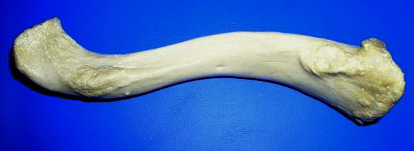
The clavicle, or collarbone, is a slender, S-shaped long bone approximately 6 inches (15 cm) long that serves as a strut between the shoulder blade and the sternum (breastbone). There are two clavicles, one on the left and one on the right. The clavicle is the only long bone in the body that lies horizontally. Together with the shoulder blade, it makes up the shoulder girdle. It is a palpable bone and, in people who have less fat in this region, the location of the bone is clearly visible. It receives its name from the Latin ''clavicula'' ("little key"), because the bone rotates along its axis like a key when the shoulder is abducted. The clavicle is the most commonly fractured bone. It can easily be fractured by impacts to the shoulder from the force of falling on outstretched arms or by a direct hit. Structure The collarbone is a thin doubly curved long bone that connects the arm to the trunk of the body. Located directly above the first rib, it acts as a strut to k ... [...More Info...] [...Related Items...] OR: [Wikipedia] [Google] [Baidu] |
Acromion
In human anatomy, the acromion (from Greek: ''akros'', "highest", ''ōmos'', "shoulder", plural: acromia) is a bony process on the scapula (shoulder blade). Together with the coracoid process it extends laterally over the shoulder joint. The acromion is a continuation of the scapular spine, and hooks over anteriorly. It articulates with the clavicle (collar bone) to form the acromioclavicular joint. Structure The acromion forms the summit of the shoulder, and is a large, somewhat triangular or oblong process, flattened from behind forward, projecting at first lateralward, and then curving forward and upward, so as to overhang the glenoid fossa.''Gray's Anatomy'' 1918, see infobox It starts from the base of acromion which marks its projecting point emerging from the spine of scapula. Surfaces Its superior surface, directed upward, backward, and lateralward, is convex, rough, and gives attachment to some fibers of the deltoideus, and in the rest of its extent is subcutaneous. ... [...More Info...] [...Related Items...] OR: [Wikipedia] [Google] [Baidu] |
Mastication
Chewing or mastication is the process by which food is crushed and ground by teeth. It is the first step of digestion, and it increases the surface area of foods to allow a more efficient break down by enzymes. During the mastication process, the food is positioned by the cheek and tongue between the teeth for grinding. The muscles of mastication move the jaws to bring the teeth into intermittent contact, repeatedly occluding and opening. As chewing continues, the food is made softer and warmer, and the enzymes in saliva begin to break down carbohydrates in the food. After chewing, the food (now called a bolus) is swallowed. It enters the esophagus and via peristalsis continues on to the stomach, where the next step of digestion occurs. Increasing the number of chews per bite increases relevant gut hormones. Studies suggest that chewing may decrease self-reported hunger and food intake. Chewing gum has been around for many centuries; there is evidence that northern Europeans c ... [...More Info...] [...Related Items...] OR: [Wikipedia] [Google] [Baidu] |
Lateral Pterygoid Plate
The pterygoid processes of the sphenoid (from Greek ''pteryx'', ''pterygos'', "wing"), one on either side, descend perpendicularly from the regions where the body and the greater wings of the sphenoid bone unite. Each process consists of a medial pterygoid plate and a lateral pterygoid plate, the latter of which serve as the origins of the medial and lateral pterygoid muscles. The medial pterygoid, along with the masseter allows the jaw to move in a vertical direction as it contracts and relaxes. The lateral pterygoid allows the jaw to move in a horizontal direction during mastication (chewing). Fracture of either plate are used in clinical medicine to distinguish the Le Fort fracture classification for high impact injuries to the sphenoid and maxillary bones. The superior portion of the pterygoid processes are fused anteriorly; a vertical groove, the pterygopalatine fossa, descends on the front of the line of fusion. The plates are separated below by an angular cleft, the pt ... [...More Info...] [...Related Items...] OR: [Wikipedia] [Google] [Baidu] |
Pterygospinous Ligament
The pterygospinous ligament stretches from the upper part of the posterior border of the lateral pterygoid plate to the spinous process of the sphenoid. Structure Variation It occasionally ossifies, and in such cases, between its upper border and the base of the skull, a foramen is formed - pterygospinous foramen (Civinini) which transmits the branches of the mandibular nerve to the muscles of mastication. See also * Pterygoalar ligament References {{Authority control Joints ... [...More Info...] [...Related Items...] OR: [Wikipedia] [Google] [Baidu] |
Stylomandibular Ligament
The stylomandibular ligament is the thickened posterior portion of the investing cervical fascia around the neck. It extends from near the apex of the styloid process of the temporal bone to the angle and posterior border of the angle of the mandible, between the masseter muscle and medial pterygoid muscle. The stylomandibular ligament limits mandibular movements, such as preventing excessive opening. Structure The stylomandibular ligament extends from near the apex of the styloid process of the temporal bone to the angle and posterior border of the angle of the mandible, between the masseter muscle and medial pterygoid muscle. From its deep surface, some fibers of the styloglossus muscle originate. Although classed among the ligaments of the temporomandibular joint, it can only be considered as accessory to it. Function The stylomandibular ligament, along with the sphenomandibular ligament, limits mandibular movements, such as preventing excessive opening. Clinical sign ... [...More Info...] [...Related Items...] OR: [Wikipedia] [Google] [Baidu] |
.jpg)


