|
Dysphagia Lusoria
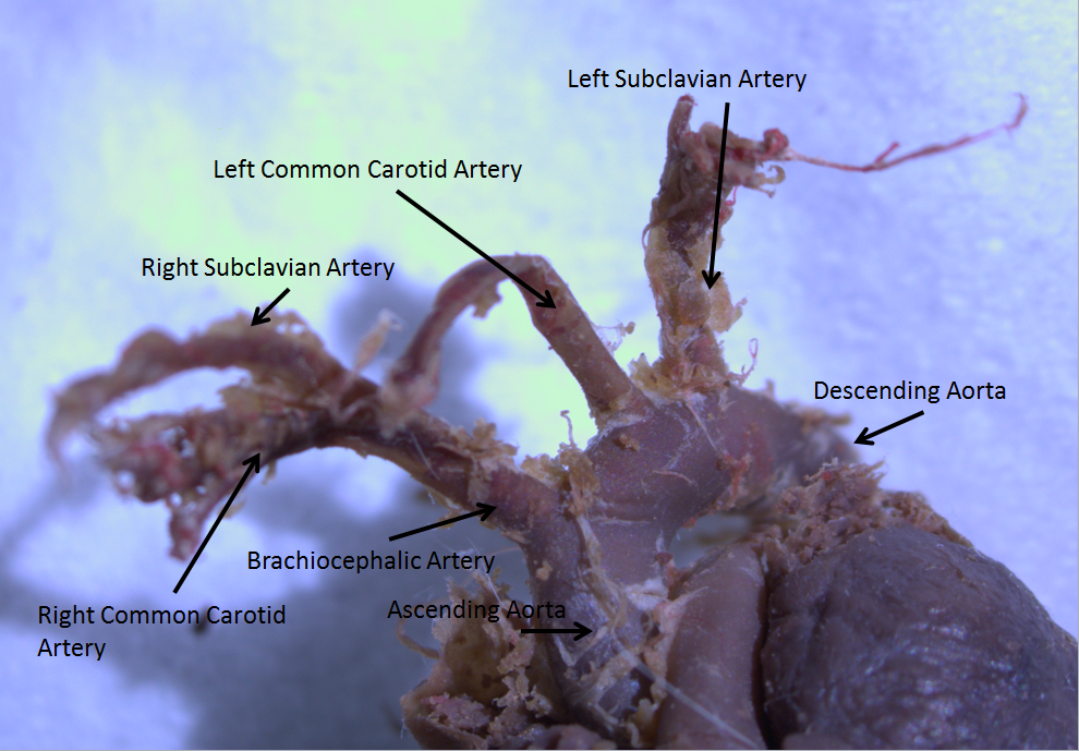
Dysphagia lusoria (or Bayford-Autenrieth dysphagia) is an abnormal condition characterized by difficulty in swallowing caused by an aberrant right subclavian artery. It was discovered by David Bayford in 1761 and first reported in a paper by the same in 1787. Pathophysiology During development of aortic arch, if the proximal portion of the right fourth arch disappears instead of distal portion, the right subclavian artery will arise as the last branch of aortic arch. It then courses behind the esophagus (or rarely in front of esophagus, or even in front of trachea) to supply blood to right arm. This causes pressure on esophagus and results in dysphagia. It can sometimes result in upper gastrointestinal tract bleeding. Investigation of choice - CT angiography Treatment Surgical repair is performed. Reconstruction or ligation of aberrant right subclavian artery by sternotomy/by neck approach. Eponym David Bayford called it dysphagia lusoria because in Latin, ''lusus naturæ'' mean ... [...More Info...] [...Related Items...] OR: [Wikipedia] [Google] [Baidu] |
Subclavian Artery
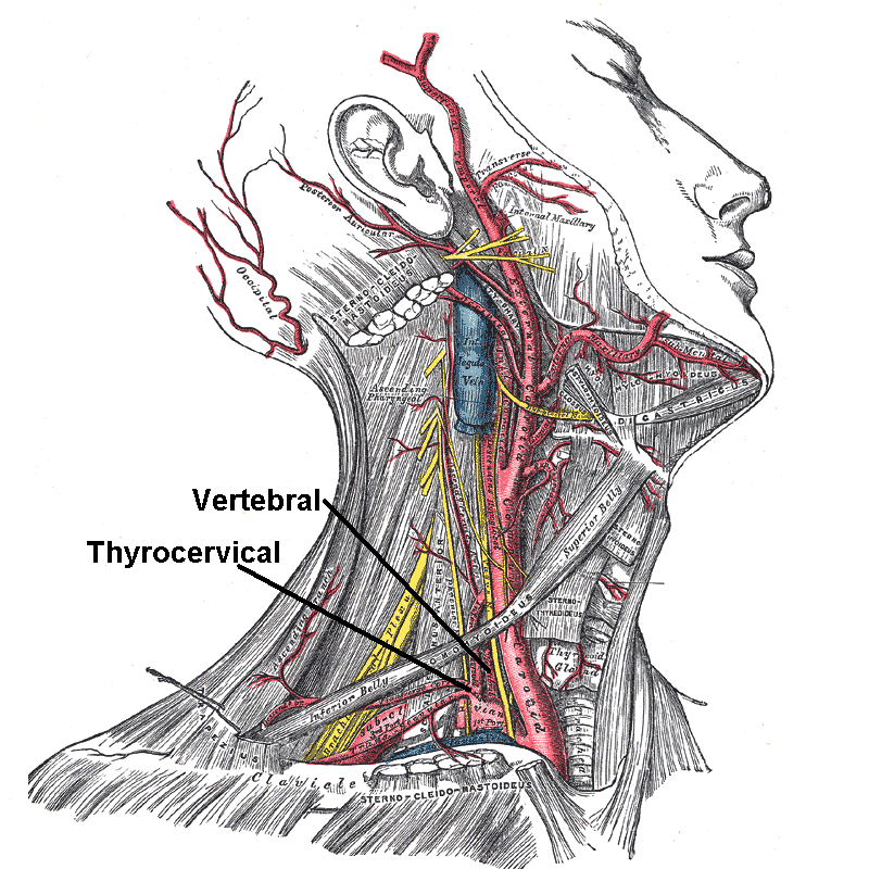
In human anatomy, the subclavian arteries are paired major arteries of the upper thorax, below the clavicle. They receive blood from the aortic arch. The left subclavian artery supplies blood to the left arm and the right subclavian artery supplies blood to the right arm, with some branches supplying the head and thorax. On the left side of the body, the subclavian comes directly off the aortic arch, while on the right side it arises from the relatively short brachiocephalic artery when it bifurcates into the subclavian and the right common carotid artery. The usual branches of the subclavian on both sides of the body are the vertebral artery, the internal thoracic artery, the thyrocervical trunk, the costocervical trunk and the dorsal scapular artery, which may branch off the transverse cervical artery, which is a branch of the thyrocervical trunk. The subclavian becomes the axillary artery at the lateral border of the first rib. Structure From its origin, the subclavian artery t ... [...More Info...] [...Related Items...] OR: [Wikipedia] [Google] [Baidu] |
David Bayford
David Bayford, FRS (c.1739 – 1790) was a London surgeon, who practised from 1761 to 1782. In later years of his life he practised as a physician. Career He was born in Hertfordshire and educated as a surgeon. He became a member of the Corporation of Surgeons, and practised as such for some years at Lewes, Sussex. In 1761, while still an apprentice surgeon, he made his discovery of the unique and bizarre cause—compression of the oesophagus by an aberrant right subclavian artery—of a fatal case of ''obstructed deglutition'' for which he coined the term dysphagia lusoria and for which he is eponymously remembered. This discovery remained unrecorded until 1787, when a paper describing the case was read on his behalf before the Medical Society of London. He was elected a Fellow of the Royal Society in 1770, when he was described as a ''Professor of Anatomy at Surgeon's Hall; and many years Lecturer in that Science and the Operations of Surgery''. He was created MD by Frederi ... [...More Info...] [...Related Items...] OR: [Wikipedia] [Google] [Baidu] |
Aortic Arch
The aortic arch, arch of the aorta, or transverse aortic arch () is the part of the aorta between the ascending and descending aorta. The arch travels backward, so that it ultimately runs to the left of the trachea. Structure The aorta begins at the level of the upper border of the second/third sternocostal articulation of the right side, behind the ventricular outflow tract and pulmonary trunk. The right atrial appendage overlaps it. The first few centimeters of the ascending aorta and pulmonary trunk lies in the same pericardial sheath. and runs at first upward, arches over the pulmonary trunk, right pulmonary artery, and right main bronchus to lie behind the right second coastal cartilage. The right lung and sternum lies anterior to the aorta at this point. The aorta then passes posteriorly and to the left, anterior to the trachea, and arches over left main bronchus and left pulmonary artery, and reaches to the left side of the T4 vertebral body. Apart from T4 vertebral body ... [...More Info...] [...Related Items...] OR: [Wikipedia] [Google] [Baidu] |
Esophagus
The esophagus (American English) or oesophagus (British English; both ), non-technically known also as the food pipe or gullet, is an organ in vertebrates through which food passes, aided by peristaltic contractions, from the pharynx to the stomach. The esophagus is a fibromuscular tube, about long in adults, that travels behind the trachea and heart, passes through the diaphragm, and empties into the uppermost region of the stomach. During swallowing, the epiglottis tilts backwards to prevent food from going down the larynx and lungs. The word ''oesophagus'' is from Ancient Greek οἰσοφάγος (oisophágos), from οἴσω (oísō), future form of φέρω (phérō, “I carry”) + ἔφαγον (éphagon, “I ate”). The wall of the esophagus from the lumen outwards consists of mucosa, submucosa (connective tissue), layers of muscle fibers between layers of fibrous tissue, and an outer layer of connective tissue. The mucosa is a stratified squamous epithel ... [...More Info...] [...Related Items...] OR: [Wikipedia] [Google] [Baidu] |
Vertebrate Trachea
The trachea, also known as the windpipe, is a cartilaginous tube that connects the larynx to the bronchi of the lungs, allowing the passage of air, and so is present in almost all air-breathing animals with lungs. The trachea extends from the larynx and branches into the two primary bronchi. At the top of the trachea the cricoid cartilage attaches it to the larynx. The trachea is formed by a number of horseshoe-shaped rings, joined together vertically by overlying ligaments, and by the trachealis muscle at their ends. The epiglottis closes the opening to the larynx during swallowing. The trachea begins to form in the second month of embryo development, becoming longer and more fixed in its position over time. It is epithelium lined with column-shaped cells that have hair-like extensions called cilia, with scattered goblet cells that produce protective mucins. The trachea can be affected by inflammation or infection, usually as a result of a viral illness affecting other parts ... [...More Info...] [...Related Items...] OR: [Wikipedia] [Google] [Baidu] |
Dysphagia
Dysphagia is difficulty in swallowing. Although classified under "symptoms and signs" in ICD-10, in some contexts it is classified as a disease#Terminology, condition in its own right. It may be a sensation that suggests difficulty in the passage of solids or liquids from the mouth to the stomach, a lack of Pharynx, pharyngeal sensation or various other inadequacies of the swallowing mechanism. Dysphagia is distinguished from other symptoms including odynophagia, which is defined as painful swallowing, and Globus Pharyngis, globus, which is the sensation of a lump in the throat. A person can have dysphagia without odynophagia (dysfunction without pain), odynophagia without dysphagia (pain without dysfunction) or both together. A psychogenic disease, psychogenic dysphagia is known as phagophobia. Classification Dysphagia is classified into the following major types: # Oropharyngeal dysphagia # Esophageal dysphagia, Esophageal and obstructive dysphagia # Neuromuscular symptom comp ... [...More Info...] [...Related Items...] OR: [Wikipedia] [Google] [Baidu] |
Johann Heinrich Ferdinand Von Autenrieth
Johann Heinrich Ferdinand von Autenrieth (20 October 1772 – 2 May 1835) was a German physician born in Stuttgart. He studied medicine at Karlsschule Stuttgart, and following graduation attended lectures by Antonio Scarpa (1752–1832) and Johann Peter Frank (1745–1821) at Pavia. Afterwards he accompanied his father to the United States, where he practiced medicine for several months in Lancaster, Pennsylvania. In 1797 he was appointed professor of anatomy, physiology, surgery and obstetrics at the University of Tübingen. In 1805 he founded an in-patient clinic at Tübingen, where in 1822 he was appointed chancellor of the university. Autenrieth specialized in forensic medicine, and was considered one of the top clinical physicians during the early part of the 19th century. One of his better written efforts was an 1806 treatise on forensics titled "''Anleitung für gerichtliche Ärzte und Wundärzte''". He died in Tübingen. Associated eponym * "Bayford-Autenrieth dyspha ... [...More Info...] [...Related Items...] OR: [Wikipedia] [Google] [Baidu] |
Aberrant Subclavian Artery
Aberrant subclavian artery, or aberrant subclavian artery syndrome, is a rare anatomical variant of the origin of the right or left subclavian artery. This abnormality is the most common congenital vascular anomaly of the aortic arch, occurring in approximately 1% of individuals. Presentation This condition is usually asymptomatic. The aberrant artery usually arises just distal to the left subclavian artery and crosses in the posterior part of the mediastinum on its way to the right upper extremity. In 80% of individuals it crosses behind the esophagus. Such course of this aberrant vessel may cause a vascular ring around the trachea and esophagus. Dysphagia due to an aberrant right subclavian artery is termed dysphagia lusoria, although this is a rare complication. In addition to dysphagia, aberrant right subclavian artery may cause stridor, dyspnoea, chest pain, or fever. An aberrant right subclavian artery may compress the recurrent laryngeal nerve causing a palsy of that ... [...More Info...] [...Related Items...] OR: [Wikipedia] [Google] [Baidu] |
Ortner's Syndrome
Ortner's syndrome is a rare cardiovocal syndrome and refers to recurrent laryngeal nerve palsy from cardiovascular disease. It was first described by Norbert Ortner (1865–1935), an Austrian physician, in 1897. Dysphagia caused by a similar mechanism is referred to as dysphagia aortica (also called Dysphagia megalatriensis), or, in the case of subclavian artery aberrancy, as dysphagia lusoria. Due to compression of the recurrent laryngeal nerve, it can cause the hoarseness of the voice, which can also be a sign of mitral stenosis. A second Ortner's syndrome, Ortner's syndrome II, refers to abdominal angina. Causes Due to its low frequency of occurrence, more common causes of hoarseness should be considered when suspecting left recurrent laryngeal nerve palsy (LRLN). When considering cardiovocal syndrome, the most common historical cause is a dilated left atrium due to mitral stenosis, but other causes, including pulmonary hypertension, thoracic aortic aneurysms, an enlarge ... [...More Info...] [...Related Items...] OR: [Wikipedia] [Google] [Baidu] |


