|
Dental Anatomy
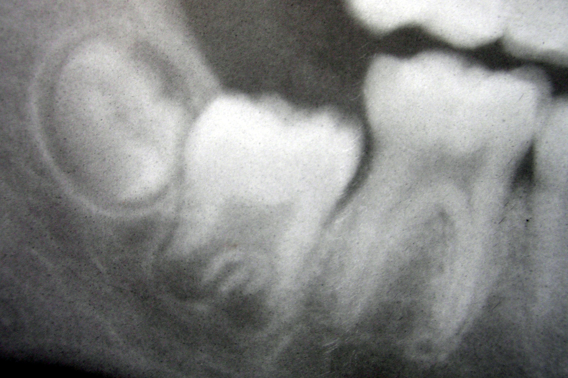
Dental anatomy is a field of anatomy dedicated to the study of human tooth structures. The development, appearance, and classification of teeth fall within its purview. (The function of teeth as they contact one another falls elsewhere, under dental occlusion.) Tooth formation begins before birth, and the teeth's eventual morphology is dictated during this time. Dental anatomy is also a taxonomical science: it is concerned with the naming of teeth and the structures of which they are made, this information serving a practical purpose in dental treatment. Usually, there are 20 primary ("baby") teeth and 32 permanent teeth, the last four being third molars or " wisdom teeth", each of which may or may not grow in. Among primary teeth, 10 usually are found in the maxilla (upper jaw) and the other 10 in the mandible (lower jaw). Among permanent teeth, 16 are found in the maxilla and the other 16 in the mandible. Each tooth has specific distinguishing features. Growing of tooth ... [...More Info...] [...Related Items...] OR: [Wikipedia] [Google] [Baidu] |
Anatomy
Anatomy () is the branch of biology concerned with the study of the structure of organisms and their parts. Anatomy is a branch of natural science that deals with the structural organization of living things. It is an old science, having its beginnings in prehistoric times. Anatomy is inherently tied to developmental biology, embryology, comparative anatomy, evolutionary biology, and phylogeny, as these are the processes by which anatomy is generated, both over immediate and long-term timescales. Anatomy and physiology, which study the structure and function of organisms and their parts respectively, make a natural pair of related disciplines, and are often studied together. Human anatomy is one of the essential basic sciences that are applied in medicine. The discipline of anatomy is divided into macroscopic and microscopic. Macroscopic anatomy, or gross anatomy, is the examination of an animal's body parts using unaided eyesight. Gross anatomy also includes the br ... [...More Info...] [...Related Items...] OR: [Wikipedia] [Google] [Baidu] |
Cementum
Cementum is a specialized calcified substance covering the root of a tooth. The cementum is the part of the periodontium that attaches the teeth to the alveolar bone by anchoring the periodontal ligament.Illustrated Dental Embryology, Histology, and Anatomy, Bath-Balogh and Fehrenbach, Elsevier, 2011, page 170. Structure The cells of cementum are the entrapped cementoblasts, the cementocytes. Each cementocyte lies in its lacuna, similar to the pattern noted in bone. These lacunae also have canaliculi or canals. Unlike those in bone, however, these canals in cementum do not contain nerves, nor do they radiate outward. Instead, the canals are oriented toward the periodontal ligament and contain cementocytic processes that exist to diffuse nutrients from the ligament because it is vascularized. After the apposition of cementum in layers, the cementoblasts that do not become entrapped in cementum line up along the cemental surface along the length of the outer covering of the per ... [...More Info...] [...Related Items...] OR: [Wikipedia] [Google] [Baidu] |
Stratum Intermedium
The stratum intermedium in a developing tooth is a layer of two or three cells between the inner enamel epithelium and the newly forming cells of the stellate reticulum. It first appears during the early bell stage of tooth development, at around the 14th week of intrauterine life. These cells are closely attached by desmosomes and gap junctions .The stratum intermedium has a notably high alkaline phosphatase activity. This layer, along with the inner enamel epithelium, is responsible for the tooth enamel formation Formation may refer to: Linguistics * Back-formation, the process of creating a new lexeme by removing or affixes * Word formation, the creation of a new word by adding affixes Mathematics and science * Cave formation or speleothem, a secondary .... It is a part of the dental (enamel) organ. Stratum intermedium stores glycogen. It is absent in the part of the tooth germ that outlines the root portions of the tooth which does not form enamel. References *Cate, A. ... [...More Info...] [...Related Items...] OR: [Wikipedia] [Google] [Baidu] |
Stellate Reticulum
The stellate reticulum is a group of cells located in the center of the enamel organ of a developing tooth. These cells are star-shaped and synthesize glycosaminoglycans. As glycosaminoglycans are produced, water is drawn in between the cells, stretching them apart. As they are moved further away from one another, the stellate reticular cells maintain contact with one another through desmosome A desmosome (; "binding body"), also known as a macula adherens (plural: maculae adherentes) (Latin for ''adhering spot''), is a cell structure specialized for cell-to-cell adhesion. A type of junctional complex, they are localized spot-like adh ...s, resulting in their unique appearance. The stellate reticulum is lost after the first layer of enamel is laid down. This brings cells in the inner enamel epithelium closer to blood vessels at the periphery. References * Orbans Oral histology and embryology – 10th ed. * Cate, A.R. Ten. Oral Histology: development, structure, and function. ... [...More Info...] [...Related Items...] OR: [Wikipedia] [Google] [Baidu] |
Inner Enamel Epithelium
The inner enamel epithelium, also known as the internal enamel epithelium, is a layer of columnar cells located on the rim nearest the dental papilla of the enamel organ in a developing tooth A tooth ( : teeth) is a hard, calcified structure found in the jaws (or mouths) of many vertebrates and used to break down food. Some animals, particularly carnivores and omnivores, also use teeth to help with capturing or wounding prey, t .... This layer is first seen during the cap stage, in which these inner enamel epithelium cells are pre-ameloblast cells. These will differentiate into Ameloblasts which are responsible for secretion of enamel during tooth development. The location of the enamel organ where the outer and inner enamel epithelium join is called the cervical loop. References *Cate, A.R. Ten. ''Oral Histology: development, structure, and function.'' 5th ed. 1998. . *Ross, Michael H., Gordon I. Kaye, and Wojciech Pawlina. ''Histology: a text and atlas.'' 4th editio ... [...More Info...] [...Related Items...] OR: [Wikipedia] [Google] [Baidu] |
Outer Enamel Epithelium
The outer enamel epithelium, also known as the external enamel epithelium, is a layer of cuboidal cells located on the periphery of the enamel organ in a developing tooth. This layer is first seen during the bell stage. The rim of the enamel organ where the outer and inner enamel epithelium join is called the cervical loop The cervical loop is the location on an enamel organ in a developing tooth where the outer enamel epithelium and the inner enamel epithelium join. The cervical loop is a histologic term indicating a specific epithelial structure at the apical s .... References *Cate, A.R. Ten. Oral Histology: development, structure, and function. 5th ed. 1998. . *Ross, Michael H., Gordon I. Kaye, and Wojciech Pawlina. Histology: a text and atlas. 4th edition. 2003. . Tooth development {{dentistry-stub ... [...More Info...] [...Related Items...] OR: [Wikipedia] [Google] [Baidu] |
Dental Follicle
The dental follicle, also known as dental sac, is made up of mesenchymal cells and fibres surrounding the enamel organ and dental papilla of a developing tooth. It is a vascular fibrous sac containing the developing tooth and its odontogenic organ. The dental follicle (DF) differentiates into the periodontal ligament. In addition, it may be the precursor of other cells of the periodontium, including osteoblasts, cementoblasts and fibroblasts. They develop into the alveolar bone, the cementum with Sharpey's fibers and the periodontal ligament fibers respectively. Similar to dental papilla, the dental follicle provides nutrition to the enamel organ and dental papilla and also have an extremely rich blood supply. Role in tooth eruption The formative role of the dental follicle starts when the crown of the tooth is fully developed and just before tooth eruption into the oral cavity. Although tooth eruption mechanisms have yet to be understood entirely, generally it can be ... [...More Info...] [...Related Items...] OR: [Wikipedia] [Google] [Baidu] |
Dental Papilla
In embryology and prenatal development, the dental papilla is a condensation of ectomesenchymal cells called odontoblasts, seen in histologic sections of a developing tooth. It lies below a cellular aggregation known as the enamel organ. The dental papilla appears after 8–10 weeks intra uteral life. The dental papilla gives rise to the dentin and pulp of a tooth. The enamel organ, dental papilla, and dental follicle together forms one unit, called the tooth germ. This is of importance because all the tissues of a tooth and its supporting structures form from these distinct cellular aggregations. Similar to dental follicle, the dental papilla has a very rich blood supply and provides nutrition to the enamel organ. Embryology Formation of dental papilla occurs in the Cap stage of Odontogenesis. The cap stage The cap stage is the second stage of tooth development and occurs during the ninth or tenth week of prenatal development. Unequal proliferation of the tooth ... [...More Info...] [...Related Items...] OR: [Wikipedia] [Google] [Baidu] |
Enamel Organ
The enamel organ, also known as the dental organ, is a cellular aggregation seen in a developing tooth and it lies above the dental papilla. The enamel organ which is differentiated from the primitive oral epithelium lining the stomodeum.The enamel organ is responsible for the formation of enamel, initiation of dentine formation, establishment of the shape of a tooth's crown, and establishment of the dentoenamel junction. The enamel organ has four layers; the inner enamel epithelium, outer enamel epithelium, stratum intermedium, and the stellate reticulum. The dental papilla, the differentiated ectomesenchyme deep to the enamel organ, will produce dentin and the dental pulp. The surrounding ectomesenchyme tissue, the dental follicle, is the primitive cementum, periodontal ligament and alveolar bone beneath the tooth root. The site where the internal enamel epithelium and external enamel epithelium coalesce is the cervical root, important in proliferation of the dental root. ... [...More Info...] [...Related Items...] OR: [Wikipedia] [Google] [Baidu] |
Branchial Arch
Branchial arches, or gill arches, are a series of bony "loops" present in fish, which support the gills. As gills are the primitive condition of vertebrates, all vertebrate embryos develop pharyngeal arches, though the eventual fate of these arches varies between taxa. In jawed fish, the first arch develops into the jaws, the second into the hyomandibular complex, with the posterior arches supporting gills. In amphibians and reptiles, many elements are lost including the gill arches, resulting in only the oral jaws and a hyoid apparatus remaining. In mammals and birds, the hyoid is still more simplified. All basal vertebrates breathe with gills. The gills are carried right behind the head, bordering the posterior margins of a series of openings from the esophagus to the exterior. Each gill is supported by a cartilaginous or bony gill arch. Bony fish have four pairs of arches, cartilaginous fish have five to seven pairs, and primitive jawless fish have seven. The verte ... [...More Info...] [...Related Items...] OR: [Wikipedia] [Google] [Baidu] |
Permanent Teeth
Permanent teeth or adult teeth are the second set of teeth formed in diphyodont mammals. In humans and old world simians, there are thirty-two permanent teeth, consisting of six maxillary and six mandibular molars, four maxillary and four mandibular premolars, two maxillary and two mandibular canines, four maxillary and four mandibular incisors. Timeline The first permanent tooth usually appears in the mouth In animal anatomy, the mouth, also known as the oral cavity, or in Latin cavum oris, is the opening through which many animals take in food and issue vocal sounds. It is also the cavity lying at the upper end of the alimentary canal, bounded on t ... at around six years of age, and the mouth will then be in a transition time with both primary (or deciduous dentition) teeth and permanent teeth during the mixed dentition period until the last primary tooth is lost or shed. The first of the permanent teeth to erupt are the permanent first molars, right behind the last ' ... [...More Info...] [...Related Items...] OR: [Wikipedia] [Google] [Baidu] |
Uterus
The uterus (from Latin ''uterus'', plural ''uteri'') or womb () is the organ in the reproductive system of most female mammals, including humans that accommodates the embryonic and fetal development of one or more embryos until birth. The uterus is a hormone-responsive sex organ that contains glands in its lining that secrete uterine milk for embryonic nourishment. In the human, the lower end of the uterus, is a narrow part known as the isthmus that connects to the cervix, leading to the vagina. The upper end, the body of the uterus, is connected to the fallopian tubes, at the uterine horns, and the rounded part above the openings to the fallopian tubes is the fundus. The connection of the uterine cavity with a fallopian tube is called the uterotubal junction. The fertilized egg is carried to the uterus along the fallopian tube. It will have divided on its journey to form a blastocyst that will implant itself into the lining of the uterus – the endometrium, ... [...More Info...] [...Related Items...] OR: [Wikipedia] [Google] [Baidu] |


.jpg)
