|
Davson–Danielli Model
The Davson–Danielli model (or paucimolecular model) was a model of the plasma membrane of a cell, proposed in 1935 by Hugh Davson and James Danielli. The model describes a phospholipid bilayer that lies between two layers of globular proteins, which is both trilaminar and lipoprotinious. The phospholipid bilayer had already been proposed by Gorter and Grendel in 1925; however, the flanking proteinaceous layers in the Davson–Danielli model were novel and intended to explain Danielli's observations on the surface tension of lipid bilayers (It is now known that the phospholipid head groups are sufficient to explain the measured surface tension). Evidence for the model included electron microscopy, in which high-resolution micrographs showed three distinct layers within a cell membrane, with an inner white core and two flanking dark layers. Since proteins usually appear dark and phospholipids white, the micrographs were interpreted as a phospholipid bilayer sandwiched between t ... [...More Info...] [...Related Items...] OR: [Wikipedia] [Google] [Baidu] |
Plasma Membrane
The cell membrane (also known as the plasma membrane (PM) or cytoplasmic membrane, and historically referred to as the plasmalemma) is a biological membrane that separates and protects the interior of all cells from the outside environment (the extracellular space). The cell membrane consists of a lipid bilayer, made up of two layers of phospholipids with cholesterols (a lipid component) interspersed between them, maintaining appropriate membrane fluidity at various temperatures. The membrane also contains membrane proteins, including integral proteins that span the membrane and serve as membrane transporters, and peripheral proteins that loosely attach to the outer (peripheral) side of the cell membrane, acting as enzymes to facilitate interaction with the cell's environment. Glycolipids embedded in the outer lipid layer serve a similar purpose. The cell membrane controls the movement of substances in and out of cells and organelles, being selectively permeable to ions an ... [...More Info...] [...Related Items...] OR: [Wikipedia] [Google] [Baidu] |
Hugh Davson
Hugh Davson, Baron Davson (25 November 1909 – 2 July 1996) was an English physiologist who worked on membrane transport and ocular fluids. Davson was born in Paddington, London, the son of physician Wilfred Maynard Davson and Mary Louisa Scott.''1911 England Census'' He attended University College School. He later studied at University College London and took a variety of research posts at institutes such as UCL, and Canada's Dalhousie University. With James Danielli he proposed a model for cell membrane structure which became known as the Davson-Danielli or "protein sandwich" model. In 1931 he married the society portrait painter Marjorie Heath with whom he had one daughter. He was a cousin of the renowned journalist and broadcaster, Alistair Cooke Alistair Cooke (born Alfred Cooke; 20 November 1908 – 30 March 2004) was a British-American writer whose work as a journalist, television personality and radio broadcaster was done primarily in the United States. [...More Info...] [...Related Items...] OR: [Wikipedia] [Google] [Baidu] |
James Danielli
James Frederic Danielli FRS (1911–1984) was an English biologist. He was famous for research on the structure and the permeability of cell membranes, developing a physical-chemical model in collaboration with the physiologist Hugh Davson Hugh Davson, Baron Davson (25 November 1909 – 2 July 1996) was an English physiologist who worked on membrane transport and ocular fluids. Davson was born in Paddington, London, the son of physician Wilfred Maynard Davson and Mary Louisa Scott. .... This became known as the Davson-Danielli or "protein sandwich" model. He also carried out studies on the chemistry of enzymes and proteins and tried to construct an artificial "cell". References English physiologists Fellows of the Royal Society 1911 births 1984 deaths {{England-med-bio-stub ... [...More Info...] [...Related Items...] OR: [Wikipedia] [Google] [Baidu] |
Phospholipid Bilayer
The lipid bilayer (or phospholipid bilayer) is a thin polar membrane made of two layers of lipid molecules. These membranes are flat sheets that form a continuous barrier around all cells. The cell membranes of almost all organisms and many viruses are made of a lipid bilayer, as are the nuclear membrane surrounding the cell nucleus, and membranes of the membrane-bound organelles in the cell. The lipid bilayer is the barrier that keeps ions, proteins and other molecules where they are needed and prevents them from diffusing into areas where they should not be. Lipid bilayers are ideally suited to this role, even though they are only a few nanometers in width, because they are impermeable to most water-soluble (hydrophilic) molecules. Bilayers are particularly impermeable to ions, which allows cells to regulate salt concentrations and pH by transporting ions across their membranes using proteins called ion pumps. Biological bilayers are usually composed of amphiphilic phospholip ... [...More Info...] [...Related Items...] OR: [Wikipedia] [Google] [Baidu] |
Globular Protein
In biochemistry, globular proteins or spheroproteins are spherical ("globe-like") proteins and are one of the common protein types (the others being fibrous, disordered and membrane proteins). Globular proteins are somewhat water-soluble (forming colloids in water), unlike the fibrous or membrane proteins. There are multiple fold classes of globular proteins, since there are many different architectures that can fold into a roughly spherical shape. The term globin can refer more specifically to proteins including the globin fold. Globular structure and solubility The term globular protein is quite old (dating probably from the 19th century) and is now somewhat archaic given the hundreds of thousands of proteins and more elegant and descriptive structural motif vocabulary. The globular nature of these proteins can be determined without the means of modern techniques, but only by using ultracentrifuges or dynamic light scattering techniques. The spherical structure is induc ... [...More Info...] [...Related Items...] OR: [Wikipedia] [Google] [Baidu] |
Electron Microscope
An electron microscope is a microscope that uses a beam of accelerated electrons as a source of illumination. As the wavelength of an electron can be up to 100,000 times shorter than that of visible light photons, electron microscopes have a higher resolving power than light microscopes and can reveal the structure of smaller objects. A scanning transmission electron microscope has achieved better than 50 pm resolution in annular dark-field imaging mode and magnifications of up to about 10,000,000× whereas most light microscopes are limited by diffraction to about 200 nm resolution and useful magnifications below 2000×. Electron microscopes use shaped magnetic fields to form electron optical lens systems that are analogous to the glass lenses of an optical light microscope. Electron microscopes are used to investigate the ultrastructure of a wide range of biological and inorganic specimens including microorganisms, cells, large molecules, biopsy samples, ... [...More Info...] [...Related Items...] OR: [Wikipedia] [Google] [Baidu] |
Micrograph
A micrograph or photomicrograph is a photograph or digital image taken through a microscope or similar device to show a magnified image of an object. This is opposed to a macrograph or photomacrograph, an image which is also taken on a microscope but is only slightly magnified, usually less than 10 times. Micrography is the practice or art of using microscopes to make photographs. A micrograph contains extensive details of microstructure. A wealth of information can be obtained from a simple micrograph like behavior of the material under different conditions, the phases found in the system, failure analysis, grain size estimation, elemental analysis and so on. Micrographs are widely used in all fields of microscopy. Types Photomicrograph A light micrograph or photomicrograph is a micrograph prepared using an optical microscope, a process referred to as ''photomicroscopy''. At a basic level, photomicroscopy may be performed simply by connecting a camera to a microscope, th ... [...More Info...] [...Related Items...] OR: [Wikipedia] [Google] [Baidu] |
Amphiphile
An amphiphile (from the Greek αμφις amphis, both, and φιλíα philia, love, friendship), or amphipath, is a chemical compound possessing both hydrophilic (''water-loving'', polar) and lipophilic (''fat-loving'') properties. Such a compound is called amphiphilic or amphipathic. Common amphiphilic substances are soaps, detergents, and lipoproteins. The phospholipid amphiphiles are the major structural component of cell membranes. Amphiphiles are the basis for a number of areas of research in chemistry and biochemistry, notably that of lipid polymorphism. Organic compounds containing hydrophilic groups at both ends of the molecule are called bolaamphiphilic. The micelles they form in the aggregate are prolate. Structure The lipophilic group is typically a large hydrocarbon moiety, such as a long chain of the form CH3(CH2)n, with n > 4. The hydrophilic group falls into one of the following categories: # charged groups #* anionic. Examples, with the lipophilic part of the m ... [...More Info...] [...Related Items...] OR: [Wikipedia] [Google] [Baidu] |
Seymour Jonathan Singer
Seymour Jonathan Singer (May 23, 1924 – February 2, 2017) was an American cell biologist and professor of biology, emeritus, at the University of California, San Diego. Biography Singer was born in New York City and attended Columbia University, where he earned his B.A. in 1943. He received his doctorate from the Polytechnic Institute of Brooklyn in 1947. He worked as a postdoctoral fellow in the lab of Linus Pauling at Caltech during 1947–1948, where he, along with Harvey Itano, co-discovered the basis of abnormal hemoglobin in sickle-cell anemia, reported in the famous paper " Sickle Cell Anemia, a Molecular Disease". He worked for the U.S. Public Health Service between 1948 and 1950. He joined the Chemistry Department at Yale University as assistant professor in 1951, and was promoted to Associate Professor in 1957 and Professor in 1960. There he developed the ferritin-antibody, which was the first electron-dense reagent used for cell staining in electron microscopy im ... [...More Info...] [...Related Items...] OR: [Wikipedia] [Google] [Baidu] |
Garth L
Garth may refer to: Places *Garth, Alberta, Canada *Garth, Bridgend, a village in south Wales :* Garth railway station (Bridgend) *Garth, Ceredigion, small village in Wales *Garth, Powys, a village in mid Wales :* Garth railway station (Powys) *Garth Hill, The Garth, Garth Hill or Garth Mountain, a mountain near Cardiff, Wales *Garth, one of many other minor place names in the United Kingdom Buildings and structures *Garth (Guilsfield), a historic house in Guilsfield, Montgomeryshire, UK *Castle Garth, a medieval fortification in Newcastle upon Tyne, England *Garth Pier, a Grade II listed structure in Bangor, Gwynedd, North Wales *Garth Castle, home to Clan Stewart of Atholl, north-west of Aberfeldy, Scotland Arts and entertainment * ''Garth'' (comic strip), published in the British newspaper ''Daily Mirror'' from 1943 to 1997 *Planet Garth, setting of David Brin's novel ''The Uplift War'' People and fictional characters *Garth (name), a list of people and fictional characters ... [...More Info...] [...Related Items...] OR: [Wikipedia] [Google] [Baidu] |
Fluid Mosaic Model
The fluid mosaic model explains various observations regarding the structure of functional cell membranes. According to this biological model, there is a lipid bilayer (two molecules thick layer consisting primarily of amphipathic phospholipids) in which protein molecules are embedded. The phospholipid bilayer gives fluidity and elasticity to the membrane. Small amounts of carbohydrates are also found in the cell membrane. The biological model, which was devised by Seymour Jonathan Singer and Garth L. Nicolson in 1972, describes the cell membrane as a two-dimensional liquid that restricts the lateral diffusion of membrane components. Such domains are defined by the existence of regions within the membrane with special lipid and protein cocoon that promote the formation of lipid rafts or protein and glycoprotein complexes. Another way to define membrane domains is the association of the lipid membrane with the cytoskeleton filaments and the extracellular matrix through membrane pr ... [...More Info...] [...Related Items...] OR: [Wikipedia] [Google] [Baidu] |
Immunofluorescence
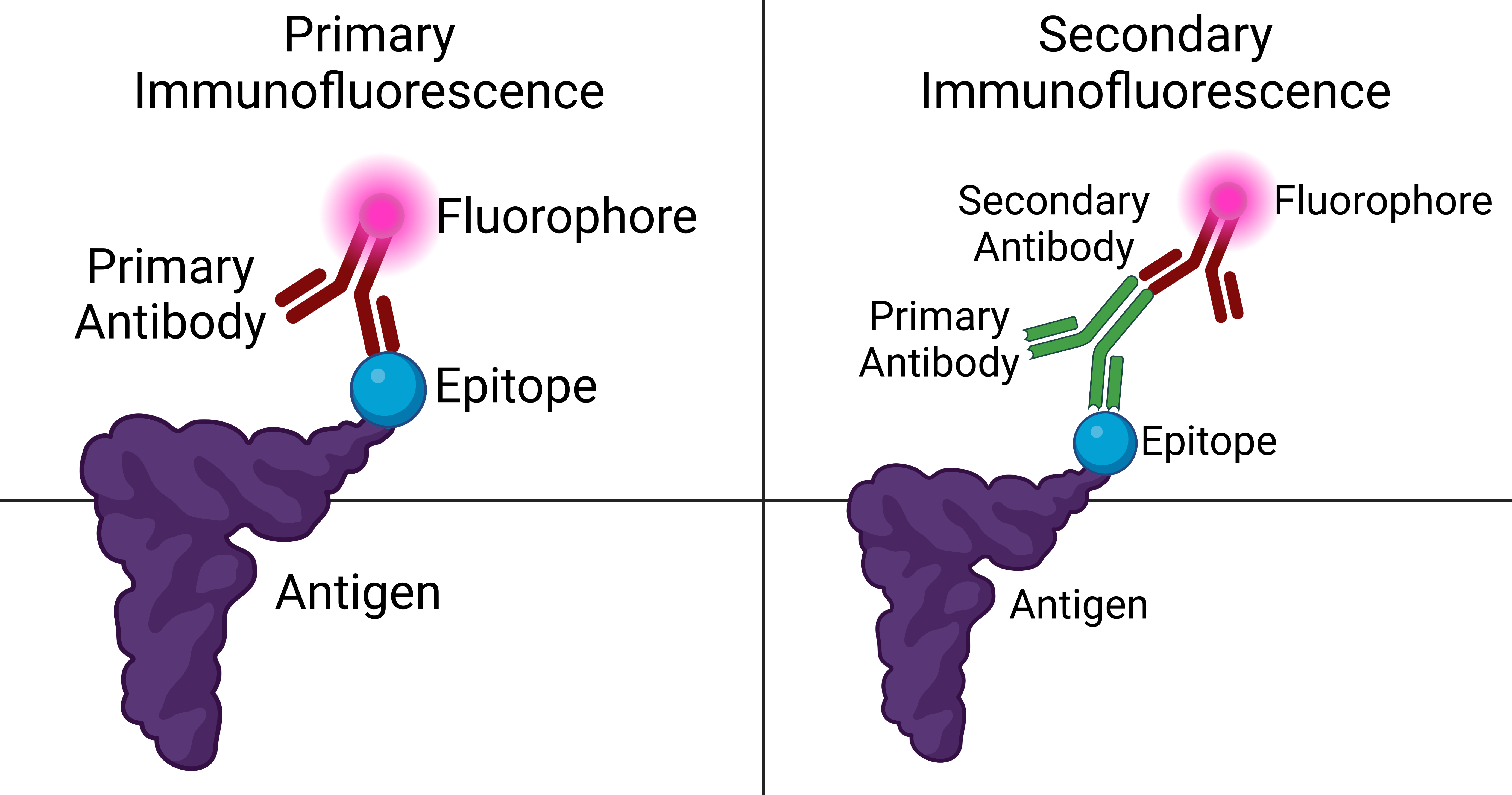
Immunofluorescence is a technique used for light microscopy with a fluorescence microscope and is used primarily on microbiological samples. This technique uses the specificity of antibodies to their antigen to target fluorescent dyes to specific biomolecule targets within a cell, and therefore allows visualization of the distribution of the target molecule through the sample. The specific region an antibody recognizes on an antigen is called an epitope. There have been efforts in epitope mapping since many antibodies can bind the same epitope and levels of binding between antibodies that recognize the same epitope can vary. Additionally, the binding of the fluorophore to the antibody itself cannot interfere with the immunological specificity of the antibody or the binding capacity of its antigen. Immunofluorescence is a widely used example of immunostaining (using antibodies to stain proteins) and is a specific example of immunohistochemistry (the use of the antibody-antigen rel ... [...More Info...] [...Related Items...] OR: [Wikipedia] [Google] [Baidu] |


