|
DNA Damage (naturally Occurring)
DNA damage is an alteration in the chemical structure of DNA, such as a break in a strand of DNA, a nucleobase missing from the backbone of DNA, or a chemically changed base such as 8-OHdG. DNA damage can occur naturally or via environmental factors, but is distinctly different from mutation, although both are types of error in DNA. DNA damage is an abnormal chemical structure in DNA, while a mutation is a change in the sequence of base pairs. DNA damages cause changes in the structure of the genetic material and prevents the replication mechanism from functioning and performing properly. The DNA damage response (DDR) is a complex signal transduction pathway which recognizes when DNA is damaged and initiates the cellular response to the damage. DNA damage and mutation have different biological consequences. While most DNA damages can undergo DNA repair, such repair is not 100% efficient. Un-repaired DNA damages accumulate in non-replicating cells, such as cells in the brains o ... [...More Info...] [...Related Items...] OR: [Wikipedia] [Google] [Baidu] |
Nucleobase
Nucleobases, also known as ''nitrogenous bases'' or often simply ''bases'', are nitrogen-containing biological compounds that form nucleosides, which, in turn, are components of nucleotides, with all of these monomers constituting the basic building blocks of nucleic acids. The ability of nucleobases to form base pairs and to stack one upon another leads directly to long-chain helical structures such as ribonucleic acid (RNA) and deoxyribonucleic acid (DNA). Five nucleobases—adenine (A), cytosine (C), guanine (G), thymine (T), and uracil (U)—are called ''primary'' or ''canonical''. They function as the fundamental units of the genetic code, with the bases A, G, C, and T being found in DNA while A, G, C, and U are found in RNA. Thymine and uracil are distinguished by merely the presence or absence of a methyl group on the fifth carbon (C5) of these heterocyclic six-membered rings. In addition, some viruses have aminoadenine (Z) instead of adenine. It differs in having an ... [...More Info...] [...Related Items...] OR: [Wikipedia] [Google] [Baidu] |
Depurination
Depurination is a chemical reaction of purine deoxyribonucleosides, deoxyadenosine and deoxyguanosine, and ribonucleosides, adenosine or guanosine, in which the β-N-glycosidic bond is hydrolytically cleaved releasing a nucleic base, adenine or guanine, respectively. The second product of depurination of deoxyribonucleosides and ribonucleosides is sugar, 2'-deoxyribose and ribose, respectively. More complex compounds containing nucleoside residues, nucleotides and nucleic acids, also suffer from depurination. Deoxyribonucleosides and their derivatives are substantially more prone to depurination than their corresponding ribonucleoside counterparts. Loss of pyrimidine bases (cytosine and thymine) occurs by a similar mechanism, but at a substantially lower rate. When depurination occurs with DNA, it leads to the formation of apurinic site and results in an alteration of the structure. Studies estimate that as many as 5,000 purines are lost this way each day in a typical human cell ... [...More Info...] [...Related Items...] OR: [Wikipedia] [Google] [Baidu] |
DNA Mismatch Repair
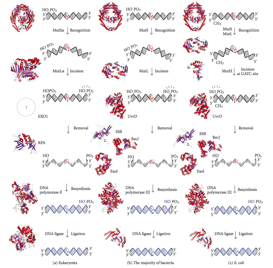
DNA mismatch repair (MMR) is a system for recognizing and repairing erroneous insertion, deletion, and mis-incorporation of nucleobase, bases that can arise during DNA replication and Genetic recombination, recombination, as well as DNA repair, repairing some forms of DNA damage. Mismatch repair is strand-specific. During DNA synthesis the newly synthesised (daughter) strand will commonly include errors. In order to begin repair, the mismatch repair machinery distinguishes the newly synthesised strand from the template (parental). In gram-negative bacteria, transient methylase, hemimethylation distinguishes the strands (the parental is methylated and daughter is not). However, in other prokaryotes and eukaryotes, the exact mechanism is not clear. It is suspected that, in eukaryotes, newly synthesized lagging-strand DNA transiently contains Nick (DNA), nicks (before being sealed by DNA ligase) and provides a signal that directs mismatch proofreading systems to the appropriate str ... [...More Info...] [...Related Items...] OR: [Wikipedia] [Google] [Baidu] |
Microhomology-mediated End Joining
Microhomology-mediated end joining (MMEJ), also known as alternative nonhomologous end-joining (Alt-NHEJ) is one of the pathways for repairing double-strand breaks in DNA. As reviewed by McVey and Lee, the foremost distinguishing property of MMEJ is the use of microhomologous sequences during the alignment of broken ends before joining, thereby resulting in deletions flanking the original break. MMEJ is frequently associated with chromosome abnormalities such as deletions, translocations, inversions and other complex rearrangements. There are multiple pathways for repairing double strand breaks, mainly non-homologous end joining (NHEJ), homologous recombination (HR), and MMEJ. NHEJ directly joins both ends of the double strand break and is relatively accurate, although small (usually less than a few nucleotides) insertions or deletions sometimes occur. HR is highly accurate and uses the sister chromatid as a template for accurate repair of the DSB. MMEJ is distinguished from these ... [...More Info...] [...Related Items...] OR: [Wikipedia] [Google] [Baidu] |
G0 Phase
The G0 phase describes a cellular state outside of the replicative cell cycle. Classically, cells were thought to enter G0 primarily due to environmental factors, like nutrient deprivation, that limited the resources necessary for proliferation. Thus it was thought of as a ''resting phase''. G0 is now known to take different forms and occur for multiple reasons. For example, most adult neuronal cells, among the most metabolically active cells in the body, are fully differentiated and reside in a terminal G0 phase. Neurons reside in this state, not because of stochastic or limited nutrient supply, but as a part of their developmental program. G0 was first suggested as a cell state based on early cell cycle studies. When the first studies defined the four phases of the cell cycle using radioactive labeling techniques, it was discovered that not all cells in a population proliferate at similar rates. A population's “growth fraction” – or the fraction of the population that w ... [...More Info...] [...Related Items...] OR: [Wikipedia] [Google] [Baidu] |
Non-homologous End Joining
Non-homologous end joining (NHEJ) is a pathway that repairs double-strand breaks in DNA. NHEJ is referred to as "non-homologous" because the break ends are directly ligated without the need for a homologous template, in contrast to homology directed repair(HDR), which requires a homologous sequence to guide repair. NHEJ is active in both non-dividing and proliferating cells, while HDR is not readily accessible in non-dividing cells. The term "non-homologous end joining" was coined in 1996 by Moore and Haber. NHEJ is typically guided by short homologous DNA sequences called microhomologies. These microhomologies are often present in single-stranded overhangs on the ends of double-strand breaks. When the overhangs are perfectly compatible, NHEJ usually repairs the break accurately. Imprecise repair leading to loss of nucleotides can also occur, but is much more common when the overhangs are not compatible. Inappropriate NHEJ can lead to translocations and telomere fusion, hallmarks ... [...More Info...] [...Related Items...] OR: [Wikipedia] [Google] [Baidu] |
S Phase
S phase (Synthesis Phase) is the phase of the cell cycle in which DNA is replicated, occurring between G1 phase and G2 phase. Since accurate duplication of the genome is critical to successful cell division, the processes that occur during S-phase are tightly regulated and widely conserved. Regulation Entry into S-phase is controlled by the G1 restriction point (R), which commits cells to the remainder of the cell-cycle if there is adequate nutrients and growth signaling. This transition is essentially irreversible; after passing the restriction point, the cell will progress through S-phase even if environmental conditions become unfavorable. Accordingly, entry into S-phase is controlled by molecular pathways that facilitate a rapid, unidirectional shift in cell state. In yeast, for instance, cell growth induces accumulation of Cln3 cyclin, which complexes with the cyclin dependent kinase CDK2. The Cln3-CDK2 complex promotes transcription of S-phase genes by inactivating ... [...More Info...] [...Related Items...] OR: [Wikipedia] [Google] [Baidu] |
Homology-directed Repair
Homology-directed repair (HDR) is a mechanism in cells to repair double-strand DNA lesions. The most common form of HDR is homologous recombination. The HDR mechanism can only be used by the cell when there is a homologous piece of DNA present in the nucleus, mostly in G2 and S phase of the cell cycle. Other examples of homology-directed repair include single-strand annealing and breakage-induced replication. When the homologous DNA is absent, another process called non-homologous end joining (NHEJ) takes place instead. Cancer suppression HDR is important for suppressing the formation of cancer. HDR maintains genomic stability by repairing broken DNA strands; it is assumed to be error free because of the use of a template. When a double strand DNA lesion is repaired by NHEJ there is no validating DNA template present so it may result in a novel DNA strand formation with loss of information. A different nucleotide sequence in the DNA strand results in a different protein expr ... [...More Info...] [...Related Items...] OR: [Wikipedia] [Google] [Baidu] |
Nucleotide Excision Repair
Nucleotide excision repair is a DNA repair mechanism. DNA damage occurs constantly because of chemicals (e.g. intercalating agents), radiation and other mutagens. Three excision repair pathways exist to repair single stranded DNA damage: Nucleotide excision repair (NER), base excision repair (BER), and DNA mismatch repair (MMR). While the BER pathway can recognize specific non-bulky lesions in DNA, it can correct only damaged bases that are removed by specific glycosylases. Similarly, the MMR pathway only targets mismatched Watson-Crick base pairs. Nucleotide excision repair (NER) is a particularly important excision mechanism that removes DNA damage induced by ultraviolet light (UV). UV DNA damage results in bulky DNA adducts - these adducts are mostly thymine dimers and 6,4-photoproducts. Recognition of the damage leads to removal of a short single-stranded DNA segment that contains the lesion. The undamaged single-stranded DNA remains and DNA polymerase uses it as a templa ... [...More Info...] [...Related Items...] OR: [Wikipedia] [Google] [Baidu] |
Base Excision Repair
Base excision repair (BER) is a cellular mechanism, studied in the fields of biochemistry and genetics, that repairs damaged DNA throughout the cell cycle. It is responsible primarily for removing small, non-helix-distorting base lesions from the genome. The related nucleotide excision repair pathway repairs bulky helix-distorting lesions. BER is important for removing damaged bases that could otherwise cause mutations by mispairing or lead to breaks in DNA during replication. BER is initiated by DNA glycosylases, which recognize and remove specific damaged or inappropriate bases, forming AP sites. These are then cleaved by an AP endonuclease. The resulting single-strand break can then be processed by either short-patch (where a single nucleotide is replaced) or long-patch BER (where 2–10 new nucleotides are synthesized). Lesions processed by BER Single bases in DNA can be chemically damaged by a variety of mechanisms, the most common ones being deamination, oxidation, ... [...More Info...] [...Related Items...] OR: [Wikipedia] [Google] [Baidu] |
Mutagen
In genetics, a mutagen is a physical or chemical agent that permanently changes nucleic acid, genetic material, usually DNA, in an organism and thus increases the frequency of mutations above the natural background level. As many mutations can cause cancer in animals, such mutagens can therefore be carcinogens, although not all necessarily are. All mutagens have characteristic mutational signatures with some chemicals becoming mutagenic through cellular processes. The process of DNA becoming modified is called mutagenesis. Not all mutations are caused by mutagens: so-called "spontaneous mutations" occur due to spontaneous hydrolysis, DNA error, errors in DNA replication, repair and Genetic recombination, recombination. Discovery The first mutagens to be identified were carcinogens, substances that were shown to be linked to cancer. Tumors were described more than 2,000 years before the discovery of chromosomes and DNA; in 500 B.C., the Greece, Greek physician Hippocrates named tu ... [...More Info...] [...Related Items...] OR: [Wikipedia] [Google] [Baidu] |
Depurination
Depurination is a chemical reaction of purine deoxyribonucleosides, deoxyadenosine and deoxyguanosine, and ribonucleosides, adenosine or guanosine, in which the β-N-glycosidic bond is hydrolytically cleaved releasing a nucleic base, adenine or guanine, respectively. The second product of depurination of deoxyribonucleosides and ribonucleosides is sugar, 2'-deoxyribose and ribose, respectively. More complex compounds containing nucleoside residues, nucleotides and nucleic acids, also suffer from depurination. Deoxyribonucleosides and their derivatives are substantially more prone to depurination than their corresponding ribonucleoside counterparts. Loss of pyrimidine bases (cytosine and thymine) occurs by a similar mechanism, but at a substantially lower rate. When depurination occurs with DNA, it leads to the formation of apurinic site and results in an alteration of the structure. Studies estimate that as many as 5,000 purines are lost this way each day in a typical human cell ... [...More Info...] [...Related Items...] OR: [Wikipedia] [Google] [Baidu] |


