|
Corollary Discharge Theory
The corollary discharge theory (CD) of motion perception helps understand how the brain can detect motion through the visual system, even though the body is not moving. When a signal is sent from the motor cortex of the brain to the eye muscles, a copy of that signal (see efference copy) is sent through the brain as well. The brain does this in order to distinguish real movements in the visual world from our own body and eye movement. The original signal and copy signal are then believed to be compared somewhere in the brain. Such a structure has not yet been identified, but it is believed to be the Medial superior temporal area, Medial Superior Temporal Area (MST). The original signal and copy need to be compared in order to determine if the change in vision was caused by eye movement or movement in the world. If the two signals cancel then no motion is perceived, but if they do not cancel then the residual signal is perceived as motion in the real world. Without a corollary dischar ... [...More Info...] [...Related Items...] OR: [Wikipedia] [Google] [Baidu] |
Efference Copy
In physiology, an efference copy or efferent copy is an internal copy of an outflowing ('' efferent''), movement-producing signal generated by an organism's motor system.Jeannerod, Marc (2003): "Action Monitoring and Forward Control of Movements". In: Michael Arbib (Ed.), ''The Handbook of Brain Theory and Neural Networks.'' Second Edition. Cambridge, Mass.: MIT Press, pp. 83–85, here: p. 83. It can be collated with the (reafferent) sensory input that results from the agent's movement, enabling a comparison of actual movement with desired movement, and a shielding of perception from particular self-induced effects on the sensory input to achieve perceptual stability. Together with internal models, efference copies can serve to enable the brain to predict the effects of an action. An equal term with a different history is ''corollary discharge''. Efference copies are important in enabling motor adaptation such as to enhance gaze stability. They have a role in the perception of se ... [...More Info...] [...Related Items...] OR: [Wikipedia] [Google] [Baidu] |
Medial Superior Temporal Area
The medial superior temporal (MST) area is a part of the cerebral cortex, which lies in the dorsal stream of the visual area of the primate Primates are a diverse order of mammals. They are divided into the strepsirrhines, which include the lemurs, galagos, and lorisids, and the haplorhines, which include the tarsiers and the simians (monkeys and apes, the latter including huma ... brain. The MST receives most of its inputs from the middle temporal (MT) area, which is involved primarily in the detection of motion. The MST uses the incoming information to compute things such as optic flow. References Cerebral cortex {{Neuroanatomy-stub ... [...More Info...] [...Related Items...] OR: [Wikipedia] [Google] [Baidu] |
René Descartes
René Descartes ( or ; ; Latinized: Renatus Cartesius; 31 March 1596 – 11 February 1650) was a French philosopher, scientist, and mathematician, widely considered a seminal figure in the emergence of modern philosophy and science. Mathematics was central to his method of inquiry, and he connected the previously separate fields of geometry and algebra into analytic geometry. Descartes spent much of his working life in the Dutch Republic, initially serving the Dutch States Army, later becoming a central intellectual of the Dutch Golden Age. Although he served a Protestant state and was later counted as a deist by critics, Descartes considered himself a devout Catholic. Many elements of Descartes' philosophy have precedents in late Aristotelianism, the revived Stoicism of the 16th century, or in earlier philosophers like Augustine. In his natural philosophy, he differed from the schools on two major points: first, he rejected the splitting of corporeal substance into mat ... [...More Info...] [...Related Items...] OR: [Wikipedia] [Google] [Baidu] |
Superior Colliculus
In neuroanatomy, the superior colliculus () is a structure lying on the roof of the mammalian midbrain. In non-mammalian vertebrates, the homologous structure is known as the optic tectum, or optic lobe. The adjective form ''tectal'' is commonly used for both structures. In mammals, the superior colliculus forms a major component of the midbrain. It is a paired structure and together with the paired inferior colliculi forms the corpora quadrigemina. The superior colliculus is a layered structure, with a pattern that is similar to all mammals. The layers can be grouped into the superficial layers ( stratum opticum and above) and the deeper remaining layers. Neurons in the superficial layers receive direct input from the retina and respond almost exclusively to visual stimuli. Many neurons in the deeper layers also respond to other modalities, and some respond to stimuli in multiple modalities. The deeper layers also contain a population of motor-related neurons, capable of activat ... [...More Info...] [...Related Items...] OR: [Wikipedia] [Google] [Baidu] |
Retina
The retina (from la, rete "net") is the innermost, light-sensitive layer of tissue of the eye of most vertebrates and some molluscs. The optics of the eye create a focused two-dimensional image of the visual world on the retina, which then processes that image within the retina and sends nerve impulses along the optic nerve to the visual cortex to create visual perception. The retina serves a function which is in many ways analogous to that of the film or image sensor in a camera. The neural retina consists of several layers of neurons interconnected by synapses and is supported by an outer layer of pigmented epithelial cells. The primary light-sensing cells in the retina are the photoreceptor cells, which are of two types: rods and cones. Rods function mainly in dim light and provide monochromatic vision. Cones function in well-lit conditions and are responsible for the perception of colour through the use of a range of opsins, as well as high-acuity vision used for task ... [...More Info...] [...Related Items...] OR: [Wikipedia] [Google] [Baidu] |
Visual Field
The visual field is the "spatial array of visual sensations available to observation in introspectionist psychological experiments". Or simply, visual field can be defined as the entire area that can be seen when an eye is fixed straight at a point. The equivalent concept for optical instruments and image sensors is the field of view (FOV). In optometry, ophthalmology, and neurology, a visual field test is used to determine whether the visual field is affected by diseases that cause local scotoma or a more extensive loss of vision or a reduction in sensitivity (increase in threshold). Normal limits The normal (monocular) human visual field extends to approximately 60 degrees nasally (toward the nose, or inward) from the vertical meridian in each eye, to 107 degrees temporally (away from the nose, or outwards) from the vertical meridian, and approximately 70 degrees above and 80 below the horizontal meridian. The binocular visual field is the superimposition of the two monocular ... [...More Info...] [...Related Items...] OR: [Wikipedia] [Google] [Baidu] |
Frontal Eye Fields
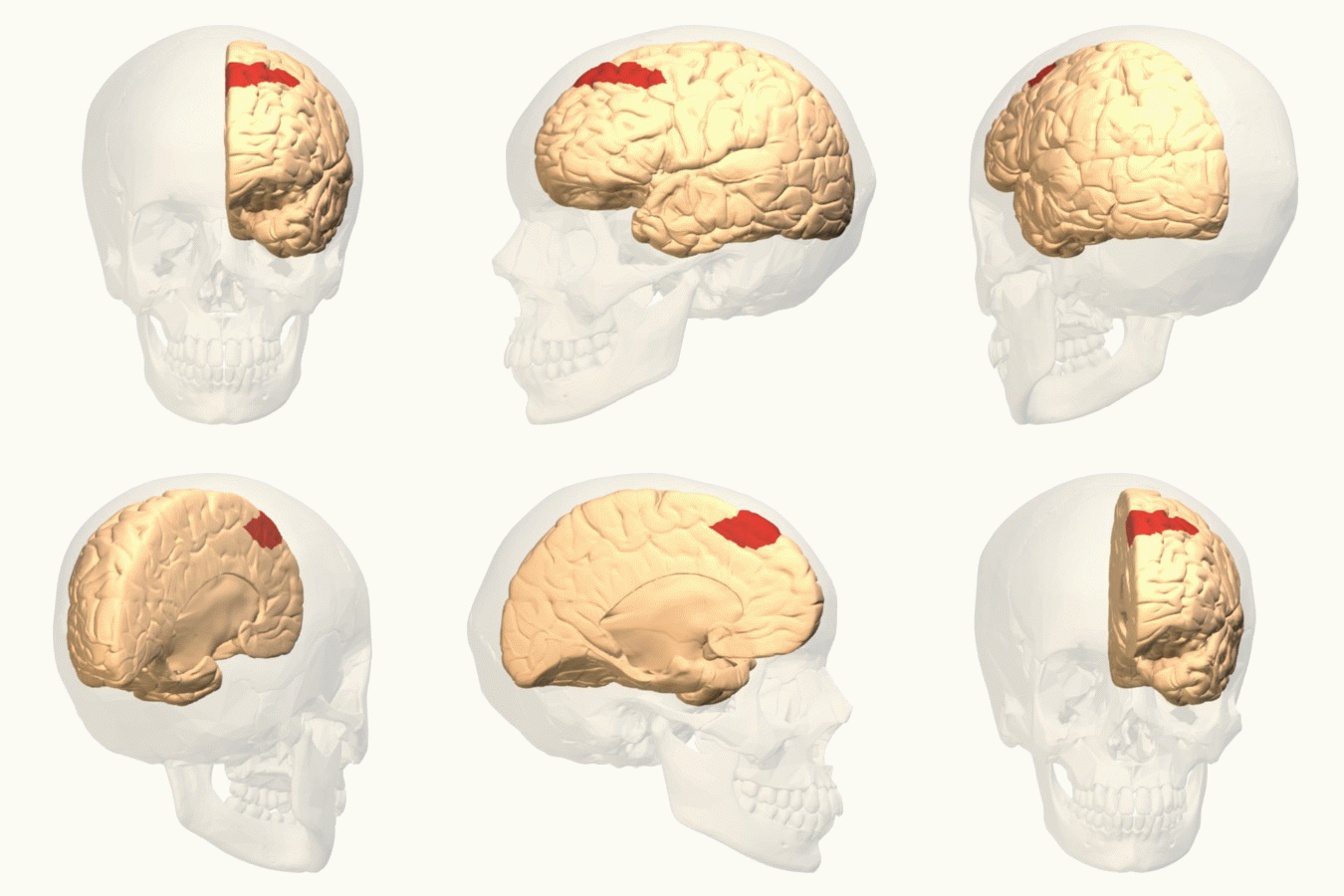
The frontal eye fields (FEF) are a region located in the frontal cortex, more specifically in Brodmann area 8 or BA8, of the primate brain. In humans, it can be more accurately said to lie in a region around the intersection of the middle frontal gyrus with the precentral gyrus, consisting of a frontal and parietal portion. The FEF is responsible for saccadic eye movements for the purpose of visual field perception and awareness, as well as for voluntary eye movement. The FEF communicates with extraocular muscles indirectly via the paramedian pontine reticular formation. Destruction of the FEF causes deviation of the eyes to the ipsilateral side. Function The cortical area called frontal eye field (FEF) plays an important role in the control of visual attention and eye movements. Electrical stimulation in the FEF elicits saccadic eye movements. The FEF have a topographic structure and represents saccade targets in retinotopic coordinates. The frontal eye field is reported to b ... [...More Info...] [...Related Items...] OR: [Wikipedia] [Google] [Baidu] |
Medial Dorsal Nucleus
The medial dorsal nucleus (or dorsomedial nucleus of thalamus) is a large nucleus in the thalamus. It is believed to play a role in memory. Structure It relays inputs from the amygdala and olfactory cortex and projects to the prefrontal cortex and the limbic system and in turn relays them to the prefrontal association cortex. As a result, it plays a crucial role in attention, planning, organization, abstract thinking, multi-tasking, and active memory. The connections of the medial dorsal nucleus have even been used to delineate the prefrontal cortex of the Göttingen minipig brain. By stereology the number of brain cells in the region has been estimated to around 6.43 million neurons in the adult human brain and 36.3 million glial cell Glia, also called glial cells (gliocytes) or neuroglia, are non-neuronal cells in the central nervous system (brain and spinal cord) and the peripheral nervous system that do not produce electrical impulses. They maintain homeostasis, form m ... [...More Info...] [...Related Items...] OR: [Wikipedia] [Google] [Baidu] |
Saccade
A saccade ( , French for ''jerk'') is a quick, simultaneous movement of both eyes between two or more phases of fixation in the same direction.Cassin, B. and Solomon, S. ''Dictionary of Eye Terminology''. Gainesville, Florida: Triad Publishing Company, 1990. In contrast, in smooth pursuit movements, the eyes move smoothly instead of in jumps. The phenomenon can be associated with a shift in frequency of an emitted signal or a movement of a body part or device. Controlled cortically by the frontal eye fields (FEF), or subcortically by the superior colliculus, saccades serve as a mechanism for fixation, rapid eye movement, and the fast phase of optokinetic nystagmus. The word appears to have been coined in the 1880s by French ophthalmologist Émile Javal, who used a mirror on one side of a page to observe eye movement in silent reading, and found that it involves a succession of discontinuous individual movements. Function Humans and many animals do not look at a scene in f ... [...More Info...] [...Related Items...] OR: [Wikipedia] [Google] [Baidu] |
Occipital Lobe
The occipital lobe is one of the four major lobes of the cerebral cortex in the brain of mammals. The name derives from its position at the back of the head, from the Latin ''ob'', "behind", and ''caput'', "head". The occipital lobe is the visual processing center of the mammalian brain containing most of the anatomical region of the visual cortex. The primary visual cortex is Brodmann area 17, commonly called V1 (visual one). Human V1 is located on the medial side of the occipital lobe within the calcarine sulcus; the full extent of V1 often continues onto the occipital pole. V1 is often also called striate cortex because it can be identified by a large stripe of myelin, the Stria of Gennari. Visually driven regions outside V1 are called extrastriate cortex. There are many extrastriate regions, and these are specialized for different visual tasks, such as visuospatial processing, color differentiation, and motion perception. Bilateral lesions of the occipital lobe can lead ... [...More Info...] [...Related Items...] OR: [Wikipedia] [Google] [Baidu] |


