|
Congenital Red–green Color Blindness
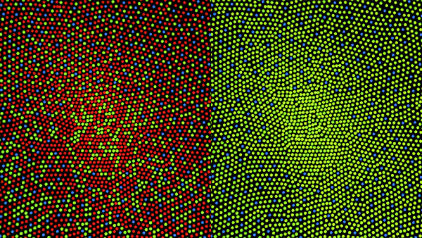
Congenital red–green color blindness is an inherited condition that is the root cause of the majority of cases of color blindness. It has no significant symptoms aside from its minor to moderate effect on color vision. It is caused by variation in the functionality of the red and/or green opsin proteins, which are the photosensitive pigment in the cone cells of the retina, which mediate color vision. Males are more likely to inherit red–green color blindness than females, because the genes for the relevant opsins are on the X chromosome. Screening for congenital red–green color blindness is typically performed with the Ishihara or similar color vision test. There is no cure for color blindness. This form of colorblindness is sometimes referred to historically as daltonism after John Dalton, who had congenital red–green color blindness and was the first to scientifically study it. In other languages, ''daltonism'' is still used to describe red–green color blindness, ... [...More Info...] [...Related Items...] OR: [Wikipedia] [Google] [Baidu] |
Ishihara Test
The Ishihara test is a color vision test for detection of red-green color deficiencies. It was named after its designer, Shinobu Ishihara, a professor at the University of Tokyo, who first published his tests in 1917.S. Ishihara, Tests for color-blindness (Handaya, Tokyo, Hongo Harukicho, 1917). The test consists of a number of Ishihara plates, which are a type of pseudoisochromatic plate. Each plate depicts a solid circle of colored dots appearing randomized in color and size. Within the pattern are dots which form a number or shape clearly visible to those with normal color vision, and invisible, or difficult to see, to those with a red-green color vision defect. Other plates are intentionally designed to reveal numbers only to those with a red-green color vision deficiency, and be invisible to those with normal red-green color vision. The full test consists of 38 plates, but the existence of a severe deficiency is usually apparent after only a few plates. There are also Ish ... [...More Info...] [...Related Items...] OR: [Wikipedia] [Google] [Baidu] |
Photopsin
Vertebrate visual opsins are a subclass of ciliary opsins and mediate vision in vertebrates. They include the opsins in human rod and cone cells. They are often abbreviated to ''opsin'', as they were the first opsins discovered and are still the most widely studied opsins. Opsins Opsin refers strictly to the apoprotein (without bound retinal). When an opsin binds retinal to form a holoprotein, it is referred to as Retinylidene protein. However, the distinction is often ignored, and opsin may refer loosely to both (regardless of whether retinal is bound). Opsins are G-protein-coupled receptors (GPCRs) and must bind retinal — typically 11-''cis''-retinal — in order to be photosensitive, since the retinal acts as the chromophore. When the Retinylidene protein absorbs a photon, the retinal isomerizes and is released by the opsin. The process that follows the isomerization and renewal of retinal is known as the visual cycle. Free 11-''cis''-retinal is photosensitive an ... [...More Info...] [...Related Items...] OR: [Wikipedia] [Google] [Baidu] |
Luminous Efficiency Function
A luminous efficiency function or luminosity function represents the average spectral sensitivity of human visual perception of light. It is based on subjective judgements of which of a pair of different-colored lights is brighter, to describe relative sensitivity to light of different wavelengths. It is not an absolute reference to any particular individual, but is a standard observer representation of visual sensitivity of theoretical human eye. It is valuable as a baseline for experimental purposes, and in colorimetry. Different luminous efficiency functions apply under different lighting conditions, varying from photopic in brightly lit conditions through mesopic to scotopic under low lighting conditions. When not specified, ''the luminous efficiency function'' generally refers to the photopic luminous efficiency function. The CIE photopic luminous efficiency function or is a standard function established by the Commission Internationale de l'Éclairage (CIE) and may b ... [...More Info...] [...Related Items...] OR: [Wikipedia] [Google] [Baidu] |
Opponent Process
The opponent process is a color theory that states that the human visual system interprets information about color by processing signals from photoreceptor cells in an antagonistic manner. The opponent-process theory suggests that there are three opponent channels, each comprising an opposing color pair: red versus green, blue versus yellow, and black versus white (luminance). The theory was first proposed in 1892 by the German physiologist Ewald Hering. Color theory Complementary colors When staring at a bright color for awhile (e.g. red), then looking away at a white field, an afterimage is perceived, such that the original color will evoke its complementary color (green, in the case of red input). When complementary colors are combined or mixed, they "cancel each other out" and become neutral (white or gray). That is, complementary colors are never perceived as a mixture; there is no "greenish red" or "yellowish blue", despite claims to the contrary. The strongest color con ... [...More Info...] [...Related Items...] OR: [Wikipedia] [Google] [Baidu] |
OPN1MW
Green-sensitive opsin is a protein that in humans is encoded by the ''OPN1MW'' gene. OPN1MW2 OPN1MW2 is a duplication of the OPN1MW gene, which encodes the medium wavelength sensitive (MWS) photopsin. The gene duplication is present in about 50% of X-chromosomes, so is present in 50% of males and at least once 75% of females. It caused by ... is a similar opsin. See also * Opsin References Further reading * * * * * * * * * * * * * External links GeneReviews/NIH/NCBI/UW entry on Red-Green Color Vision Defects G protein-coupled receptors Color vision {{transmembranereceptor-stub ... [...More Info...] [...Related Items...] OR: [Wikipedia] [Google] [Baidu] |
OPN1LW
OPN1LW is a gene on the X chromosome that encodes for long wave sensitive (LWS) opsin, or red cone photopigment. It is responsible for perception of visible light in the yellow-green range on the visible spectrum (around 500-570nm). The gene contains 6 exons with variability that induces shifts in the spectral range. OPN1LW is subject to homologous recombination with OPN1MW, as the two have very similar sequences. These recombinations can lead to various vision problems, such as red-green colourblindness and blue monochromacy. The protein encoded is a G-protein coupled receptor with embedded 11-''cis''-retinal, whose light excitation causes a cis-trans conformational change that begins the process of chemical signalling to the brain. Gene OPN1LW produces red-sensitive opsin, while its counterparts, OPN1MW and OPN1SW, produce green-sensitive and blue-sensitive opsin respectively. OPN1LW and OPN1MW are on the X chromosome at position Xq28. They are in a tandem array, composed of a ... [...More Info...] [...Related Items...] OR: [Wikipedia] [Google] [Baidu] |
Dynamic Range
Dynamic range (abbreviated DR, DNR, or DYR) is the ratio between the largest and smallest values that a certain quantity can assume. It is often used in the context of signals, like sound and light. It is measured either as a ratio or as a base-10 ( decibel) or base-2 (doublings, bits or stops) logarithmic value of the difference between the smallest and largest signal values. Electronically reproduced audio and video is often processed to fit the original material with a wide dynamic range into a narrower recorded dynamic range that can more easily be stored and reproduced; this processing is called dynamic range compression. Human perception The human senses of sight and hearing have a relatively high dynamic range. However, a human cannot perform these feats of perception at both extremes of the scale at the same time. The human eye takes time to adjust to different light levels, and its dynamic range in a given scene is actually quite limited due to optical glare. The ins ... [...More Info...] [...Related Items...] OR: [Wikipedia] [Google] [Baidu] |
Spectral Sensitivity
Spectral sensitivity is the relative efficiency of detection, of light or other signal, as a function of the frequency or wavelength of the signal. In visual neuroscience, spectral sensitivity is used to describe the different characteristics of the photopigments in the rod cells and cone cells in the retina of the eye. It is known that the rod cells are more suited to scotopic vision and cone cells to photopic vision, and that they differ in their sensitivity to different wavelengths of light. It has been established that the maximum spectral sensitivity of the human eye under daylight conditions is at a wavelength of 555 nm, while at night the peak shifts to 507 nm. In photography, film and sensors are often described in terms of their spectral sensitivity, to supplement their characteristic curves that describe their responsivity. A database of camera spectral sensitivity is created and its space analyzed. For X-ray films, the spectral sensitivity is chosen to b ... [...More Info...] [...Related Items...] OR: [Wikipedia] [Google] [Baidu] |
Gamut
In color reproduction, including computer graphics and photography, the gamut, or color gamut , is a certain ''complete subset'' of colors. The most common usage refers to the subset of colors which can be accurately represented in a given circumstance, such as within a given color space or by a certain output device. Another sense, less frequently used but still correct, refers to the complete set of colors found within an image at a given time. In this context, digitizing a photograph, converting a digitized image to a different color space, or outputting it to a given medium using a certain output device generally alters its gamut, in the sense that some of the colors in the original are lost in the process. Introduction The term ''gamut'' was adopted from the field of music, where in middle age Latin "gamut" meant the entire range of musical notes of which musical melodies are composed; Shakespeare's use of the term in ''The Taming of the Shrew'' is sometimes attributed to ... [...More Info...] [...Related Items...] OR: [Wikipedia] [Google] [Baidu] |
Trichromacy
Trichromacy or trichromatism is the possessing of three independent channels for conveying color information, derived from the three different types of cone cells in the eye. Organisms with trichromacy are called trichromats. The normal explanation of trichromacy is that the organism's retina contains three types of color receptors (called cone cells in vertebrates) with different Absorption spectroscopy, absorption spectra. In actuality the number of such receptor types may be greater than three, since different types may be active at different light intensities. In vertebrates with three types of cone cells, at low light intensities the rod cells may contribute to color vision. Humans and other animals that are trichromats Humans and some other mammals have Evolution, evolved trichromacy based partly on pigments inherited from early vertebrates. In fish and birds, for example, Tetrachromacy, four pigments are used for vision. These extra cone receptor visual pigments detect ... [...More Info...] [...Related Items...] OR: [Wikipedia] [Google] [Baidu] |


