|
Complex Cell
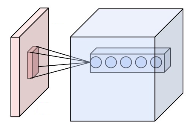
Complex cells can be found in the primary visual cortex (V1), the secondary visual cortex (V2), and Brodmann area 19 ( V3). Like a simple cell, a complex cell will respond primarily to oriented edges and gratings, however it has a degree of spatial invariance. This means that its receptive field cannot be mapped into fixed excitatory and inhibitory zones. Rather, it will respond to patterns of light in a certain orientation within a large receptive field, regardless of the exact location. Some complex cells respond optimally only to movement in a certain direction. These cells were discovered by Torsten Wiesel and David Hubel in the early 1960s. They refrained from reporting on the complex cells in (Hubel 1959) because they did not feel that they understood them well enough at the time. In Hubel and Wiesel (1962), they reported that complex cells were intermixed with simple cells and when excitatory and inhibitory regions could be established, the summation and mutual antagonis ... [...More Info...] [...Related Items...] OR: [Wikipedia] [Google] [Baidu] |
Visual Cortex
The visual cortex of the brain is the area of the cerebral cortex that processes visual information. It is located in the occipital lobe. Sensory input originating from the eyes travels through the lateral geniculate nucleus in the thalamus and then reaches the visual cortex. The area of the visual cortex that receives the sensory input from the lateral geniculate nucleus is the primary visual cortex, also known as visual area 1 ( V1), Brodmann area 17, or the striate cortex. The extrastriate areas consist of visual areas 2, 3, 4, and 5 (also known as V2, V3, V4, and V5, or Brodmann area 18 and all Brodmann area 19). Both hemispheres of the brain include a visual cortex; the visual cortex in the left hemisphere receives signals from the right visual field, and the visual cortex in the right hemisphere receives signals from the left visual field. Introduction The primary visual cortex (V1) is located in and around the calcarine fissure in the occipital lobe. Each hemisphere's V1 ... [...More Info...] [...Related Items...] OR: [Wikipedia] [Google] [Baidu] |
Brodmann Area 19
Brodmann area 19, or BA 19, is part of the occipital lobe cortex in the human brain. Along with area 18, it comprises the extrastriate (or peristriate) cortex. In humans with normal sight, extrastriate cortex is a visual association area, with feature-extracting, shape recognition, attentional, and multimodal integrating functions. This area is also known as peristriate area 19, and it refers to a subdivision of the cytoarchitecturally defined occipital region of cerebral cortex. In the human it is located in parts of the lingual gyrus, the cuneus, the lateral occipital gyrus (H) and the superior occipital gyrus (H) of the occipital lobe where it is bounded approximately by the parieto-occipital sulcus. It is bounded on one side by the parastriate area 18, which it surrounds. It is bounded rostrally by the angular area 39 (H) and the occipitotemporal area 37 (H) (Brodmann-1909). In animals Brodmann area 19-1909 is a subdivision of the cerebral cortex of the guenon defined on t ... [...More Info...] [...Related Items...] OR: [Wikipedia] [Google] [Baidu] |
Simple Cell
A simple cell in the visual cortex, primary visual cortex is a cell that responds primarily to oriented edges and gratings (bars of particular orientations). These cells were discovered by Torsten Wiesel and David Hubel in the late 1950s. Such cells are tuned to different frequencies and orientations, even with different phase relationships, possibly for extracting disparity (depth) information and to attribute depth to detected lines and edges. This may result in a 3D 'wire-frame' representation as used in computer graphics. The fact that input from the left and right eyes is very close in the so-called cortical hypercolumns is an indication that depth processing occurs at a very early stage, aiding recognition of 3D objects. Later, many other cells with specific functions have been discovered: (a) end-stopped cells which are thought to detect singularities like line and edge crossings, vertices and line endings; (b) bar and grating cells. The latter are not linear operators be ... [...More Info...] [...Related Items...] OR: [Wikipedia] [Google] [Baidu] |
Translational Invariance
In geometry, to translate a geometric figure is to move it from one place to another without rotating it. A translation "slides" a thing by . In physics and mathematics, continuous translational symmetry is the invariance of a system of equations under any translation. Discrete translational symmetry is invariant under discrete translation. Analogously an operator on functions is said to be translationally invariant with respect to a translation operator T_\delta if the result after applying doesn't change if the argument function is translated. More precisely it must hold that \forall \delta \ A f = A (T_\delta f). Laws of physics are translationally invariant under a spatial translation if they do not distinguish different points in space. According to Noether's theorem, space translational symmetry of a physical system is equivalent to the momentum conservation law. Translational symmetry of an object means that a particular translation does not change the object. For ... [...More Info...] [...Related Items...] OR: [Wikipedia] [Google] [Baidu] |
Receptive Field
The receptive field, or sensory space, is a delimited medium where some physiological stimuli can evoke a sensory neuronal response in specific organisms. Complexity of the receptive field ranges from the unidimensional chemical structure of odorants to the multidimensional spacetime of human visual field, through the bidimensional skin surface, being a receptive field for touch perception. Receptive fields can positively or negatively alter the membrane potential with or without affecting the rate of action potentials. A sensory space can be dependent of an animal's location. For a particular sound wave traveling in an appropriate transmission medium, by means of sound localization, an auditory space would amount to a reference system that continuously shifts as the animal moves (taking into consideration the space inside the ears as well). Conversely, receptive fields can be largely independent of the animal's location, as in the case of place cells. A sensory space can also m ... [...More Info...] [...Related Items...] OR: [Wikipedia] [Google] [Baidu] |
Torsten Wiesel
Torsten Nils Wiesel (born 3 June 1924) is a Swedish neurophysiologist. With David H. Hubel, he received the 1981 Nobel Prize in Physiology or Medicine, for their discoveries concerning information processing in the visual system; the prize was shared with Roger W. Sperry for his independent research on the cerebral hemispheres. Career Wiesel was born in Uppsala, Sweden in 1924, the youngest of five children. In 1947, he began his scientific career in Carl Gustaf Bernhard's laboratory at the Karolinska Institute, where he received his medical degree in 1954. He went on to teach in the Institute's department of physiology and worked in the child psychiatry unit of the Karolinska Hospital. In 1955 he moved to the United States to work at Johns Hopkins School of Medicine under Stephen Kuffler. Wiesel began a fellowship in ophthalmology, and in 1958 he became an assistant professor. That same year, he met David Hubel, beginning a collaboration that would last over twenty years. In ... [...More Info...] [...Related Items...] OR: [Wikipedia] [Google] [Baidu] |
David Hubel
David Hunter Hubel (February 27, 1926 – September 22, 2013) was a Canadian American neurophysiologist noted for his studies of the structure and function of the visual cortex. He was co-recipient with Torsten Wiesel of the 1981 Nobel Prize in Physiology or Medicine (shared with Roger W. Sperry), for their discoveries concerning information processing in the visual system. For much of his career, Hubel worked as the Professor of Neurobiology at Johns Hopkins University and Harvard Medical School. In 1978, Hubel and Wiesel were awarded the Louisa Gross Horwitz Prize from Columbia University. In 1983, Hubel received the Golden Plate Award of the American Academy of Achievement. Early life and education David H. Hubel was born in Windsor, Ontario, Canada, to American parents in 1926. His grandfather emigrated as a child to the United States from the Bavarian town of Nördlingen. In 1929, his family moved to Montreal, where he spent his formative years. His father was a che ... [...More Info...] [...Related Items...] OR: [Wikipedia] [Google] [Baidu] |
The MIT Press
The MIT Press is a university press affiliated with the Massachusetts Institute of Technology (MIT) in Cambridge, Massachusetts (United States). It was established in 1962. History The MIT Press traces its origins back to 1926 when MIT published under its own name a lecture series entitled ''Problems of Atomic Dynamics'' given by the visiting German physicist and later Nobel Prize winner, Max Born. Six years later, MIT's publishing operations were first formally instituted by the creation of an imprint called Technology Press in 1932. This imprint was founded by James R. Killian, Jr., at the time editor of MIT's alumni magazine and later to become MIT president. Technology Press published eight titles independently, then in 1937 entered into an arrangement with John Wiley & Sons in which Wiley took over marketing and editorial responsibilities. In 1962 the association with Wiley came to an end after a further 125 titles had been published. The press acquired its modern name afte ... [...More Info...] [...Related Items...] OR: [Wikipedia] [Google] [Baidu] |
Stephen Kuffler
Stephen William Kuffler (August 24 Táp, Austria-Hungary, 1913 – October 11, 1980) was a pre-eminent Hungarian-American neurophysiologist. He is often referred to as the "Father of Modern Neuroscience". Kuffler, alongside noted Nobel Laureates Sir John Eccles and Sir Bernard Katz gave research lectures at the University of Sydney, strongly influencing its intellectual environment while working at Sydney Hospital. He founded the Harvard Neurobiology department in 1966, and made numerous seminal contributions to our understanding of vision, neural coding, and the neural implementation of behavior. He is known for his research on neuromuscular junctions in frogs, presynaptic inhibition, and the neurotransmitter GABA. In 1972, he was awarded the Louisa Gross Horwitz Prize from Columbia University. Honors and awards Kuffler was widely recognized as a truly original and creative neuroscientist. In addition to numerous prizes, honorary degrees, and special lectureships from co ... [...More Info...] [...Related Items...] OR: [Wikipedia] [Google] [Baidu] |
Christina Enroth-Cugell
Christina Alma Elisabeth Enroth-Cugell (1919 – June 15, 2016), was a vision scientist who was a professor at Northwestern University for 31 years, was a founding faculty member and one of the first women to teach at the McCormick School of Engineering and chaired the Department of Neurobiology at the Weinberg College of Arts and Sciences from 1984 to 1986. Her husband David Cugell was a professor at the Feinberg School of Medicine for 58 years, the longest tenure in the school’s history. Enroth-Cugell was born in Helsinki, Finland. She earned a joint M.D./Ph.D. from the Karolinska Institute in Sweden and did post-doctorate work at Harvard University Harvard University is a private Ivy League research university in Cambridge, Massachusetts. Founded in 1636 as Harvard College and named for its first benefactor, the Puritan clergyman John Harvard, it is the oldest institution of higher le .... Enroth-Cugell was one of the first female interns at Passavant Memorial Hosp ... [...More Info...] [...Related Items...] OR: [Wikipedia] [Google] [Baidu] |
Cerebrum
The cerebrum, telencephalon or endbrain is the largest part of the brain containing the cerebral cortex (of the two cerebral hemispheres), as well as several subcortical structures, including the hippocampus, basal ganglia, and olfactory bulb. In the human brain, the cerebrum is the uppermost region of the central nervous system. The cerebrum prenatal development, develops prenatally from the forebrain (prosencephalon). In mammals, the Dorsum (biology), dorsal telencephalon, or Pallium (neuroanatomy), pallium, develops into the cerebral cortex, and the ventral telencephalon, or Pallium (neuroanatomy), subpallium, becomes the basal ganglia. The cerebrum is also divided into approximately symmetric Lateralization of brain function, left and right cerebral hemispheres. With the assistance of the cerebellum, the cerebrum controls all voluntary actions in the human body. Structure The cerebrum is the largest part of the brain. Depending upon the position of the animal it lies eithe ... [...More Info...] [...Related Items...] OR: [Wikipedia] [Google] [Baidu] |

