|
Companion Shadow
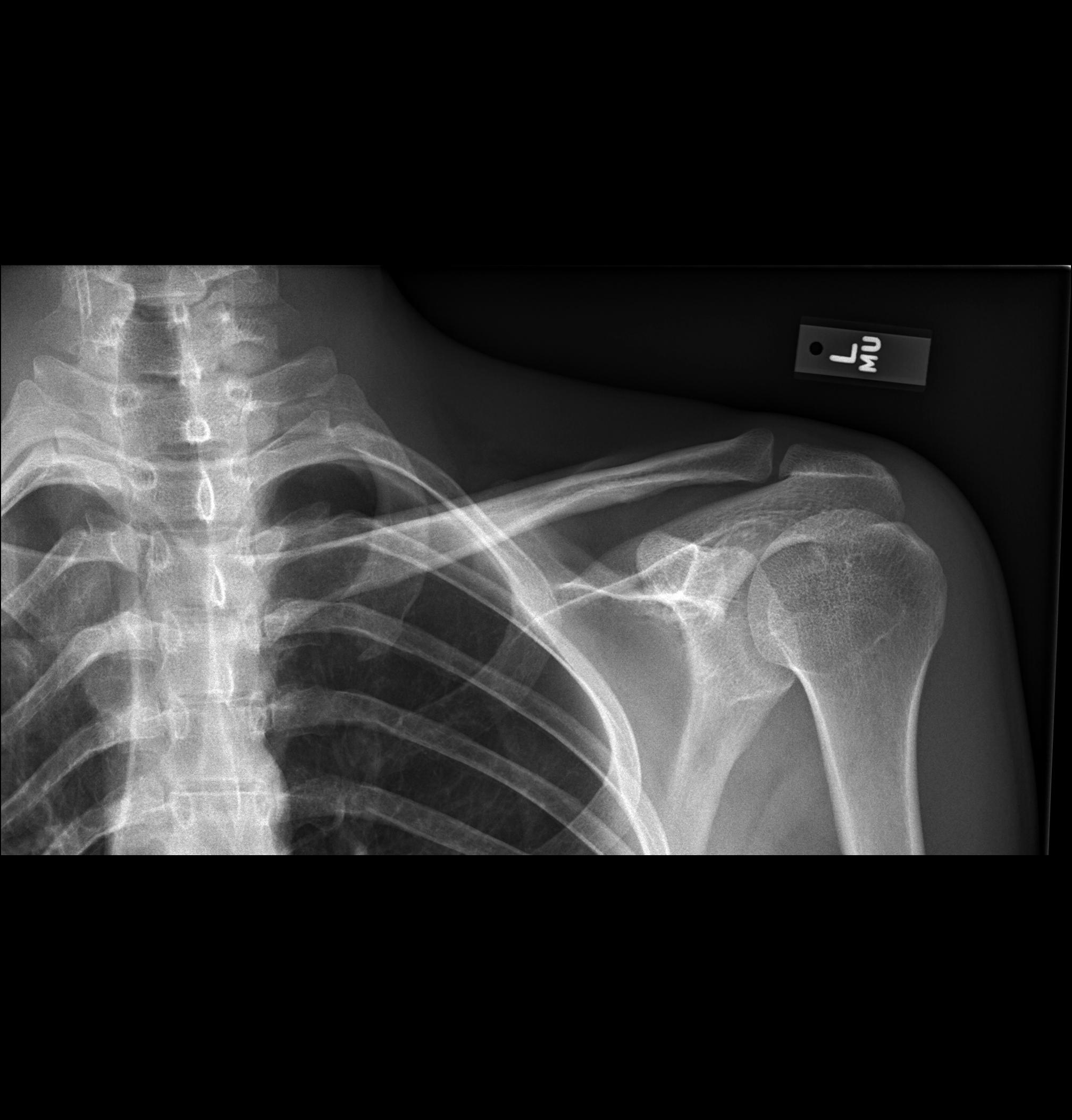
Companion shadow is a term used in describing radiographs that denotes the appearance of a smooth, homogenous, radiodensity with a well-defined margin that runs parallel with a bony landmark. Companion shadows represent soft tissue that overlies the respective bony landmark in profile. They are not seen in every radiograph and can be misinterpreted as pathology. Types of companion shadow * Clavicular companion shadow is a thin soft-tissue stripe along the upper edge of the clavicle. Lange S., Walsh G. Radiology of chest diseases, 2nd Edition, 1998 * Rib companion shadows parallel the and measure 1–5 mm in diameter project adjacent to the inferior and inferolateral margi ... [...More Info...] [...Related Items...] OR: [Wikipedia] [Google] [Baidu] |
Radiographs
Radiography is an imaging technology, imaging technique using X-rays, gamma rays, or similar ionizing radiation and non-ionizing radiation to view the internal form of an object. Applications of radiography include medical radiography ("diagnostic" and "therapeutic") and industrial radiography. Similar techniques are used in airport security (where "body scanners" generally use backscatter X-ray). To create an image in conventional radiography, a beam of X-rays is produced by an X-ray generator and is projected toward the object. A certain amount of the X-rays or other radiation is absorbed by the object, dependent on the object's density and structural composition. The X-rays that pass through the object are captured behind the object by a X-ray detector, detector (either photographic film or a digital detector). The generation of flat two dimensional images by this technique is called projectional radiography. In computed tomography (CT scanning) an X-ray source and its associa ... [...More Info...] [...Related Items...] OR: [Wikipedia] [Google] [Baidu] |
Soft Tissue
Soft tissue is all the tissue in the body that is not hardened by the processes of ossification or calcification such as bones and teeth. Soft tissue connects, surrounds or supports internal organs and bones, and includes muscle, tendons, ligaments, fat, fibrous tissue, lymph and blood vessels, fasciae, and synovial membranes. with :q=a_E_E_ \qquad Q=b_E_E_ quadratic forms of Green-Lagrange strains E_ and a_, b_ and c material constants. W is the strain energy function per volume unit, which is the mechanical strain energy for a given temperature. Isotropic simplification The Fung-model, simplified with isotropic hypothesis (same mechanical properties in all directions). This written in respect of the principal stretches (\lambda_i): :W = \frac\left (\lambda_1^2 + \lambda_2^2 + \lambda_3^2 - 3) + b\left( e^ -1 \right) \right/math> , where a, b and c are constants. Simplification for small and big stretches For small strains, the exponential term is very small, thus neg ... [...More Info...] [...Related Items...] OR: [Wikipedia] [Google] [Baidu] |
Pathology
Pathology is the study of the causes and effects of disease or injury. The word ''pathology'' also refers to the study of disease in general, incorporating a wide range of biology research fields and medical practices. However, when used in the context of modern medical treatment, the term is often used in a narrower fashion to refer to processes and tests that fall within the contemporary medical field of "general pathology", an area which includes a number of distinct but inter-related medical specialties that diagnose disease, mostly through analysis of tissue, cell, and body fluid samples. Idiomatically, "a pathology" may also refer to the predicted or actual progression of particular diseases (as in the statement "the many different forms of cancer have diverse pathologies", in which case a more proper choice of word would be " pathophysiologies"), and the affix ''pathy'' is sometimes used to indicate a state of disease in cases of both physical ailment (as in cardiomy ... [...More Info...] [...Related Items...] OR: [Wikipedia] [Google] [Baidu] |
Clavicle
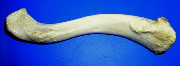
The clavicle, or collarbone, is a slender, S-shaped long bone approximately 6 inches (15 cm) long that serves as a strut between the shoulder blade and the sternum (breastbone). There are two clavicles, one on the left and one on the right. The clavicle is the only long bone in the body that lies horizontally. Together with the shoulder blade, it makes up the shoulder girdle. It is a palpable bone and, in people who have less fat in this region, the location of the bone is clearly visible. It receives its name from the Latin ''clavicula'' ("little key"), because the bone rotates along its axis like a key when the shoulder is abducted. The clavicle is the most commonly fractured bone. It can easily be fractured by impacts to the shoulder from the force of falling on outstretched arms or by a direct hit. Structure The collarbone is a thin doubly curved long bone that connects the arm to the trunk of the body. Located directly above the first rib, it acts as a strut to k ... [...More Info...] [...Related Items...] OR: [Wikipedia] [Google] [Baidu] |
Ribs
The rib cage, as an enclosure that comprises the ribs, vertebral column and sternum in the thorax of most vertebrates, protects vital organs such as the heart, lungs and great vessels. The sternum, together known as the thoracic cage, is a semi-rigid bony and cartilaginous structure which surrounds the thoracic cavity and supports the shoulder girdle to form the core part of the human skeleton. A typical human thoracic cage consists of 12 pairs of ribs and the adjoining costal cartilages, the sternum (along with the manubrium and xiphoid process), and the 12 thoracic vertebrae articulating with the ribs. Together with the skin and associated fascia and muscles, the thoracic cage makes up the thoracic wall and provides attachments for extrinsic skeletal muscles of the neck, upper limbs, upper abdomen and back. The rib cage intrinsically holds the muscles of respiration ( diaphragm, intercostal muscles, etc.) that are crucial for active inhalation and forced exhalation, and t ... [...More Info...] [...Related Items...] OR: [Wikipedia] [Google] [Baidu] |
Muscles
Skeletal muscles (commonly referred to as muscles) are organs of the vertebrate muscular system and typically are attached by tendons to bones of a skeleton. The muscle cells of skeletal muscles are much longer than in the other types of muscle tissue, and are often known as muscle fibers. The muscle tissue of a skeletal muscle is striated – having a striped appearance due to the arrangement of the sarcomeres. Skeletal muscles are voluntary muscles under the control of the somatic nervous system. The other types of muscle are cardiac muscle which is also striated and smooth muscle which is non-striated; both of these types of muscle tissue are classified as involuntary, or, under the control of the autonomic nervous system. A skeletal muscle contains multiple fascicles – bundles of muscle fibers. Each individual fiber, and each muscle is surrounded by a type of connective tissue layer of fascia. Muscle fibers are formed from the fusion of developmental myoblasts in a p ... [...More Info...] [...Related Items...] OR: [Wikipedia] [Google] [Baidu] |
Intercostal Space
The intercostal space (ICS) is the anatomic space between two ribs (Lat. costa). Since there are 12 ribs on each side, there are 11 intercostal spaces, each numbered for the rib superior to it. Structures in intercostal space * several kinds of intercostal muscle * intercostal arteries and intercostal veins * intercostal lymph nodes * intercostal nerves Order of components Muscles There are 3 muscular layers in each intercostal space, consisting of the external intercostal muscle, the internal intercostal muscle, and the thinner innermost intercostal muscle. These muscles help to move the ribs during breathing. Neurovascular bundles Neurovascular bundles are located between the internal intercostal muscle and the innermost intercostal muscle. The neurovascular bundle has a strict order of vein-artery- nerve (VAN), from top to bottom. This neurovascular bundle runs high in the intercostal space, and the smaller collateral neurovascular bundle runs just superior ... [...More Info...] [...Related Items...] OR: [Wikipedia] [Google] [Baidu] |
Pleural Disease
Pleural disease occurs in the pleural space, which is the thin fluid-filled area in between the two pulmonary pleurae in the human body. There are several disorders and complications that can occur within the pleural area, and the surrounding tissues in the lung. Pleural content anomalies * Pneumothorax: a collection of air within the pleural cavity, arising either from the outside or from the lung. Pneumothoraces may be traumatic, iatrogenic, or spontaneous. A tension pneumothorax is a particular type of pneumothorax where the air may enter (though a defect of the chest wall, lung, or airways) on inspiration, but cannot exit on expiration. Each breath increases the amount of trapped air in the chest cavity, leading to further lung compression. This is often an urgent situation and may progress to a medical emergency if there is compromise of the venous return to the heart causing hypotension and rarely shock. * Pleural effusion: a fluid accumulation within the pleural space. A ... [...More Info...] [...Related Items...] OR: [Wikipedia] [Google] [Baidu] |
Lung Disease
The lungs are the primary organs of the respiratory system in humans and most other animals, including some snails and a small number of fish. In mammals and most other vertebrates, two lungs are located near the backbone on either side of the heart. Their function in the respiratory system is to extract oxygen from the air and transfer it into the bloodstream, and to release carbon dioxide from the bloodstream into the atmosphere, in a process of gas exchange. Respiration is driven by different muscular systems in different species. Mammals, reptiles and birds use their different muscles to support and foster breathing. In earlier tetrapods, air was driven into the lungs by the pharyngeal muscles via buccal pumping, a mechanism still seen in amphibians. In humans, the main muscle of respiration that drives breathing is the diaphragm. The lungs also provide airflow that makes vocal sounds including human speech possible. Humans have two lungs, one on the left and on ... [...More Info...] [...Related Items...] OR: [Wikipedia] [Google] [Baidu] |
Scapula
The scapula (plural scapulae or scapulas), also known as the shoulder blade, is the bone that connects the humerus (upper arm bone) with the clavicle (collar bone). Like their connected bones, the scapulae are paired, with each scapula on either side of the body being roughly a mirror image of the other. The name derives from the Classical Latin word for trowel or small shovel, which it was thought to resemble. In compound terms, the prefix omo- is used for the shoulder blade in medical terminology. This prefix is derived from ὦμος (ōmos), the Ancient Greek word for shoulder, and is cognate with the Latin , which in Latin signifies either the shoulder or the upper arm bone. The scapula forms the back of the shoulder girdle. In humans, it is a flat bone, roughly triangular in shape, placed on a posterolateral aspect of the thoracic cage. Structure The scapula is a thick, flat bone lying on the thoracic wall that provides an attachment for three groups of muscles: intrin ... [...More Info...] [...Related Items...] OR: [Wikipedia] [Google] [Baidu] |

.jpg)


