|
Circumvallate Placenta
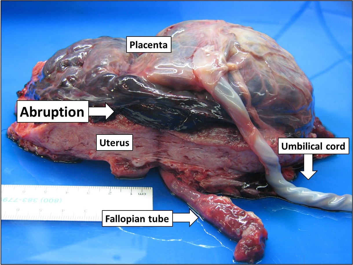
Circumvallate placenta is a rare condition affecting about 1-2% of pregnancies, in which the amnion and chorion fetal membranes essentially "double back" on the fetal side around the edges of the placenta. After delivery, a circumvallate placenta has a thick ring of membranes on its fetal surface. Circumvallate placenta is a placental morphological abnormality associated with increased fetal morbidity and mortality due to the restricted availability of nutrients and oxygen to the developing fetus. Physicians may be able to detect a circumvallate placenta during pregnancy by using an ultrasound. However, in other cases, a circumvallate placenta is not identified until delivery of the baby. Circumvallate placenta can increase the risk of associated complications such as preterm delivery and placental abruption. Occasionally, a circumvallate placenta can also increase the risk of neonatal death and emergency caesarean section. Although there is no existing treatment for circumval ... [...More Info...] [...Related Items...] OR: [Wikipedia] [Google] [Baidu] |
Pregnancy
Pregnancy is the time during which one or more offspring develops ( gestates) inside a woman's uterus (womb). A multiple pregnancy involves more than one offspring, such as with twins. Pregnancy usually occurs by sexual intercourse, but can also occur through assisted reproductive technology procedures. A pregnancy may end in a live birth, a miscarriage, an induced abortion, or a stillbirth. Childbirth typically occurs around 40 weeks from the start of the last menstrual period (LMP), a span known as the gestational age. This is just over nine months. Counting by fertilization age, the length is about 38 weeks. Pregnancy is "the presence of an implanted human embryo or fetus in the uterus"; implantation occurs on average 8–9 days after fertilization. An '' embryo'' is the term for the developing offspring during the first seven weeks following implantation (i.e. ten weeks' gestational age), after which the term ''fetus'' is used until birth. Signs an ... [...More Info...] [...Related Items...] OR: [Wikipedia] [Google] [Baidu] |
Placental Abruption
Placental abruption is when the placenta separates early from the uterus, in other words separates before childbirth. It occurs most commonly around 25 weeks of pregnancy. Symptoms may include vaginal bleeding, lower abdominal pain, and dangerously low blood pressure. Complications for the mother can include disseminated intravascular coagulopathy and kidney failure. Complications for the baby can include fetal distress, low birthweight, preterm delivery, and stillbirth. The cause of placental abruption is not entirely clear. Risk factors include smoking, pre-eclampsia, prior abruption (most important and predictive risk factor), trauma during pregnancy, cocaine use, and previous cesarean section. Diagnosis is based on symptoms and supported by ultrasound. It is classified as a complication of pregnancy. For small abruption, bed rest may be recommended, while for more significant abruptions or those that occur near term, delivery may be recommended. If everything is stable, ... [...More Info...] [...Related Items...] OR: [Wikipedia] [Google] [Baidu] |
Umbilical Cord
In placental mammals, the umbilical cord (also called the navel string, birth cord or ''funiculus umbilicalis'') is a conduit between the developing embryo or fetus and the placenta. During prenatal development, the umbilical cord is physiologically and genetically part of the fetus and (in humans) normally contains two arteries (the umbilical arteries) and one vein (the umbilical vein), buried within Wharton's jelly. The umbilical vein supplies the fetus with oxygenated, nutrient-rich blood from the placenta. Conversely, the fetal heart pumps low-oxygen, nutrient-depleted blood through the umbilical arteries back to the placenta. Structure and development The umbilical cord develops from and contains remnants of the yolk sac and allantois. It forms by the fifth week of development, replacing the yolk sac as the source of nutrients for the embryo. The cord is not directly connected to the mother's circulatory system, but instead joins the placenta, which transfers materials t ... [...More Info...] [...Related Items...] OR: [Wikipedia] [Google] [Baidu] |
Blood Vessel
The blood vessels are the components of the circulatory system that transport blood throughout the human body. These vessels transport blood cells, nutrients, and oxygen to the tissues of the body. They also take waste and carbon dioxide away from the tissues. Blood vessels are needed to sustain life, because all of the body's tissues rely on their functionality. There are five types of blood vessels: the arteries, which carry the blood away from the heart; the arterioles; the capillaries, where the exchange of water and chemicals between the blood and the tissues occurs; the venules; and the veins, which carry blood from the capillaries back towards the heart. The word ''vascular'', meaning relating to the blood vessels, is derived from the Latin ''vas'', meaning vessel. Some structures – such as cartilage, the epithelium, and the lens and cornea of the eye – do not contain blood vessels and are labeled ''avascular''. Etymology * artery: late Middle English; from Latin ... [...More Info...] [...Related Items...] OR: [Wikipedia] [Google] [Baidu] |
Fibrin
Fibrin (also called Factor Ia) is a fibrous, non-globular protein involved in the clotting of blood. It is formed by the action of the protease thrombin on fibrinogen, which causes it to polymerize. The polymerized fibrin, together with platelets, forms a hemostatic plug or clot over a wound site. When the lining of a blood vessel is broken, platelets are attracted, forming a platelet plug. These platelets have thrombin receptors on their surfaces that bind serum thrombin molecules, which in turn convert soluble fibrinogen in the serum into fibrin at the wound site. Fibrin forms long strands of tough insoluble protein that are bound to the platelets. Factor XIII completes the cross-linking of fibrin so that it hardens and contracts. The cross-linked fibrin forms a mesh atop the platelet plug that completes the clot. Fibrin was discovered by Marcello Malpighi in 1666. Role in disease Excessive generation of fibrin due to activation of the coagulation cascade leads to thrombos ... [...More Info...] [...Related Items...] OR: [Wikipedia] [Google] [Baidu] |
Decidua
The decidua is the modified mucosal lining of the uterus (that is, modified endometrium) that forms every month, in preparation for pregnancy. It is shed off each month when there is no fertilised egg to support. The decidua is under the influence of progesterone. Endometrial cells become highly characteristic. The decidua forms the maternal part of the placenta and remains for the duration of the pregnancy. After birth the decidua is shed together with the placenta. Structure The part of the decidua that interacts with the trophoblast is the ''decidua basalis'' (also called ''decidua placentalis''), while the ''decidua capsularis'' grows over the embryo on the luminal side, enclosing it into the endometrium. The remainder of the decidua is termed the ''decidua parietalis'' or ''decidua vera'', and it will fuse with the decidua capsularis by the fourth month of gestation. Three morphologically distinct layers of the decidua basalis can then be described: * Compact outer laye ... [...More Info...] [...Related Items...] OR: [Wikipedia] [Google] [Baidu] |
Placenta
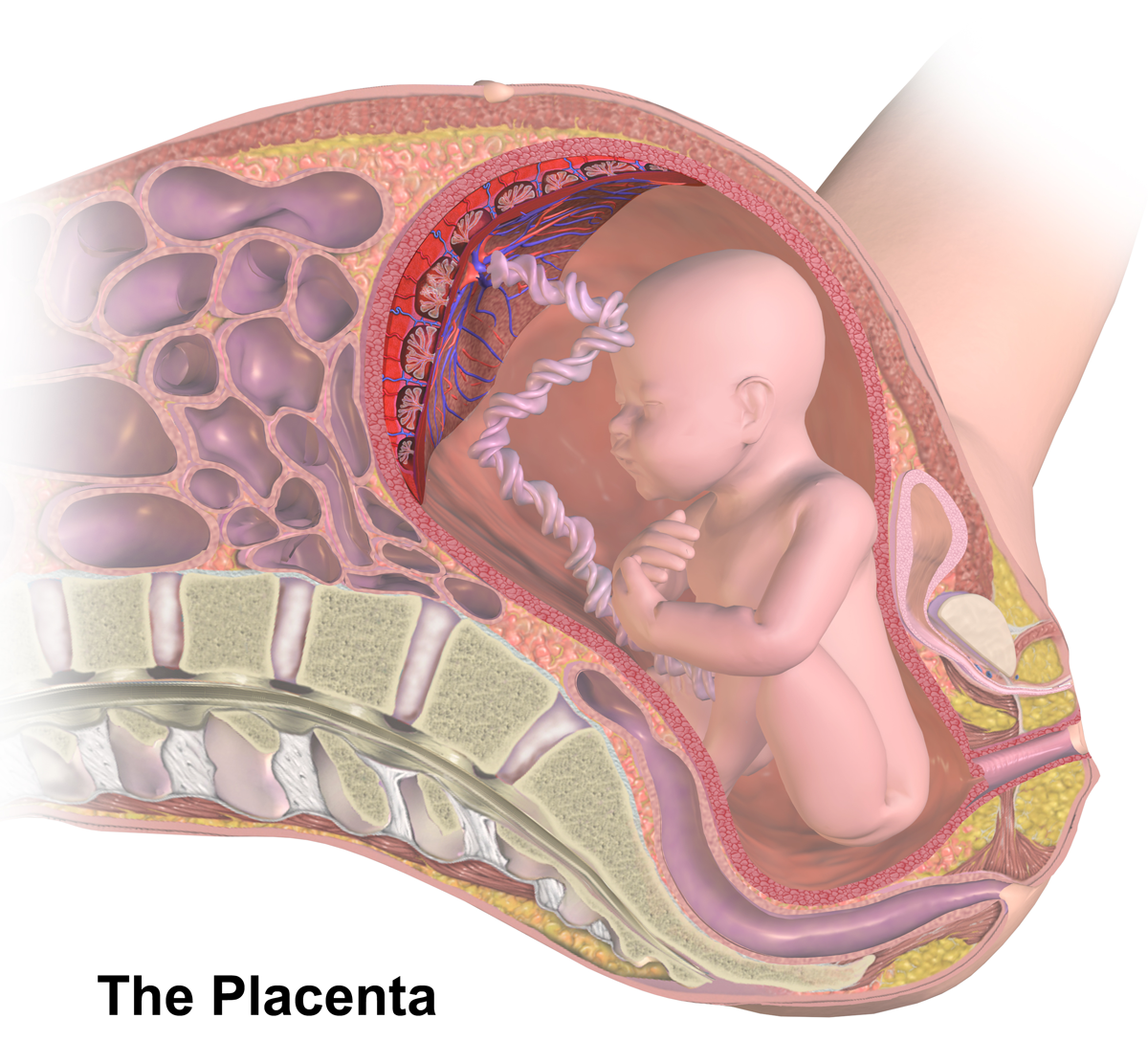
The placenta is a temporary embryonic and later fetal organ that begins developing from the blastocyst shortly after implantation. It plays critical roles in facilitating nutrient, gas and waste exchange between the physically separate maternal and fetal circulations, and is an important endocrine organ, producing hormones that regulate both maternal and fetal physiology during pregnancy. The placenta connects to the fetus via the umbilical cord, and on the opposite aspect to the maternal uterus in a species-dependent manner. In humans, a thin layer of maternal decidual (endometrial) tissue comes away with the placenta when it is expelled from the uterus following birth (sometimes incorrectly referred to as the 'maternal part' of the placenta). Placentas are a defining characteristic of placental mammals, but are also found in marsupials and some non-mammals with varying levels of development. Mammalian placentas probably first evolved about 150 million to 200 million years ... [...More Info...] [...Related Items...] OR: [Wikipedia] [Google] [Baidu] |
Mother
] A mother is the female parent of a child. A woman may be considered a mother by virtue of having given childbirth, birth, by raising a child who may or may not be her biological offspring, or by supplying her ovum for fertilisation in the case of gestational surrogacy. An adoptive mother is a female who has become the child's parent through the legal process of adoption. A biological mother is the female genetic contributor to the creation of the infant, through sexual intercourse or egg donation. A biological mother may have legal obligations to a child not raised by her, such as an obligation of monetary support. A putative mother is a female whose biological relationship to a child is alleged but has not been established. A stepmother is a woman who is married to a child's father and they may form a family unit, but who generally does not have the legal rights and responsibilities of a parent in relation to the child. A father is the male counterpart of a mother. Women who ... [...More Info...] [...Related Items...] OR: [Wikipedia] [Google] [Baidu] |
Basal Plate (placenta)
During pregnancy changes in the placenta involve the disappearance of the greater portion of the stratum compactum, but the deeper part of this layer persists and is condensed to form what is known as the basal plate. Between this plate and the uterine muscular fibres are the stratum spongiosum and the boundary layer; through these and the basal plate the uterine arteries and veins pass to and from the intervillous space In the placenta, the intervillous space is the space between chorionic villi, and contains maternal blood. The trophoblast, which is a collection of cells that invades the maternal endometrium to gain access to nutrition for the fetus, prolif .... Decidual septum is one of the structures derived from basal plate. References External links Slide at uottawa.ca Embryology {{Portal bar, Anatomy ... [...More Info...] [...Related Items...] OR: [Wikipedia] [Google] [Baidu] |
Chorion
The chorion is the outermost fetal membrane around the embryo in mammals, birds and reptiles (amniotes). It develops from an outer fold on the surface of the yolk sac, which lies outside the zona pellucida (in mammals), known as the vitelline membrane in other animals. In insects it is developed by the follicle cells while the egg is in the ovary.Chapman, R.F. (1998) "The insects: structure and function", Section ''The egg and embryology''. Previewed in Google Bookon 26 Sep 2009. Structure In humans and other mammals (excluding monotremes), the chorion is one of the fetal membranes that exist during pregnancy between the developing fetus and mother. The chorion and the amnion together form the amniotic sac. In humans it is formed by extraembryonic mesoderm and the two layers of trophoblast that surround the embryo and other membranes; the chorionic villi emerge from the chorion, invade the endometrium, and allow the transfer of nutrients from maternal blood to fetal blood. ... [...More Info...] [...Related Items...] OR: [Wikipedia] [Google] [Baidu] |
Caesarean Section
Caesarean section, also known as C-section or caesarean delivery, is the surgical procedure by which one or more babies are delivered through an incision in the mother's abdomen, often performed because vaginal delivery would put the baby or mother at risk. Reasons for the operation include obstructed labor, twin pregnancy, high blood pressure in the mother, breech birth, and problems with the placenta or umbilical cord. A caesarean delivery may be performed based upon the shape of the mother's pelvis or history of a previous C-section. A trial of vaginal birth after C-section may be possible. The World Health Organization recommends that caesarean section be performed only when medically necessary. Most C-sections are performed without a medical reason, upon request by someone, usually the mother. A C-section typically takes 45 minutes to an hour. It may be done with a spinal block, where the woman is awake, or under general anesthesia. A urinary catheter is used to drain ... [...More Info...] [...Related Items...] OR: [Wikipedia] [Google] [Baidu] |
Pregnancy Ultrasound
Obstetric ultrasonography, or prenatal ultrasound, is the use of medical ultrasonography in pregnancy, in which sound waves are used to create real-time visual images of the developing embryo or fetus in the uterus (womb). The procedure is a standard part of prenatal care in many countries, as it can provide a variety of information about the health of the mother, the timing and progress of the pregnancy, and the health and development of the embryo or fetus. The International Society of Ultrasound in Obstetrics and Gynecology (ISUOG) recommends that pregnant women have routine obstetric ultrasounds between 18 weeks' and 22 weeks' gestational age (the anatomy scan) in order to confirm pregnancy dating, to measure the fetus so that growth abnormalities can be recognized quickly later in pregnancy, and to assess for congenital malformations and multiple pregnancies (twins, etc). Additionally, the ISUOG recommends that pregnant patients who desire genetic testing have obstetric ultra ... [...More Info...] [...Related Items...] OR: [Wikipedia] [Google] [Baidu] |


.jpg)

