|
Choroidal Nevus
Choroidal nevus (plural: nevi) is a type of eye neoplasm that is classified under choroidal tumors as a type of benign (non-cancerous) melanocytic tumor. A choroidal nevus can be described as an unambiguous pigmented blue or green-gray choroidal lesion, found at the front of the eye, around the iris, or the rear end of the eye. Nevi are usually darkly pigmented tumors because they comprise melanocytes. J. Donald M. Gass, Dr. Gass, one of the leading specialists on eye diseases, speculates that a choroidal nevus grows from small cells resting as Hyperplasia, hyperplastic lesions, and exhibits growth primarily. In most cases, choroidal nevus is an asymptomatic disease, however, in serious conditions, adverse symptoms can be observed. Choroidal nevus is usually diagnosed through an ophthalmic eye examination, or more specialized technologies such as photographic imaging, ophthalmoscopy, ultrasonography and Optical coherence tomography, ocular coherence tomography (OCT). Choroidal n ... [...More Info...] [...Related Items...] OR: [Wikipedia] [Google] [Baidu] |
Eye Neoplasm
Eye neoplasms can affect all parts of the eye, and can be a benign tumor or a malignant tumor (cancer). Eye cancers can be primary (starts within the eye) or metastatic cancer (spread to the eye from another organ). The two most common cancers that spread to the eye from another organ are breast cancer and lung cancer. Other less common sites of origin include the prostate, kidney, thyroid, skin, colon and blood or bone marrow. Types Tumors in the eye and orbit can be benign like dermoid cysts, or malignant like rhabdomyosarcoma and retinoblastoma. Malignant The most common eyelid tumor is called basal cell carcinoma. This tumor can grow around the eye but rarely spreads to other parts of the body. Other types of common eyelid cancers include squamous carcinoma, sebaceous carcinoma and malignant melanoma. The most common orbital malignancy is ''orbital lymphoma''. This tumor can be diagnosed by biopsy with histopathologic and immunohistochemical analysis. Most patients with o ... [...More Info...] [...Related Items...] OR: [Wikipedia] [Google] [Baidu] |
Uveal Melanoma
Uveal melanoma is a type of eye cancer in the uvea of the eye. It is traditionally classed as originating in the Iris (anatomy), iris, choroid, and ciliary body, but can also be divided into class I (low metastatic risk) and class II (high metastatic risk). Symptoms include blurred vision, loss of vision or photopsia, but there may be no symptoms. Tumors arise from the pigment cells (melanocytes) that reside within the uvea and give color to the eye. These melanocytes are distinct from the retinal pigment epithelium cells underlying the retina that do not form melanomas. When eye melanoma is spread to distant parts of the body, the five-year survival rate is about 15%.Eye Cancer Survival Rates American Cancer Society, Last Medical Review: December 9, 2014 Last Revised: February 5, 2016 [...More Info...] [...Related Items...] OR: [Wikipedia] [Google] [Baidu] |
Optomap
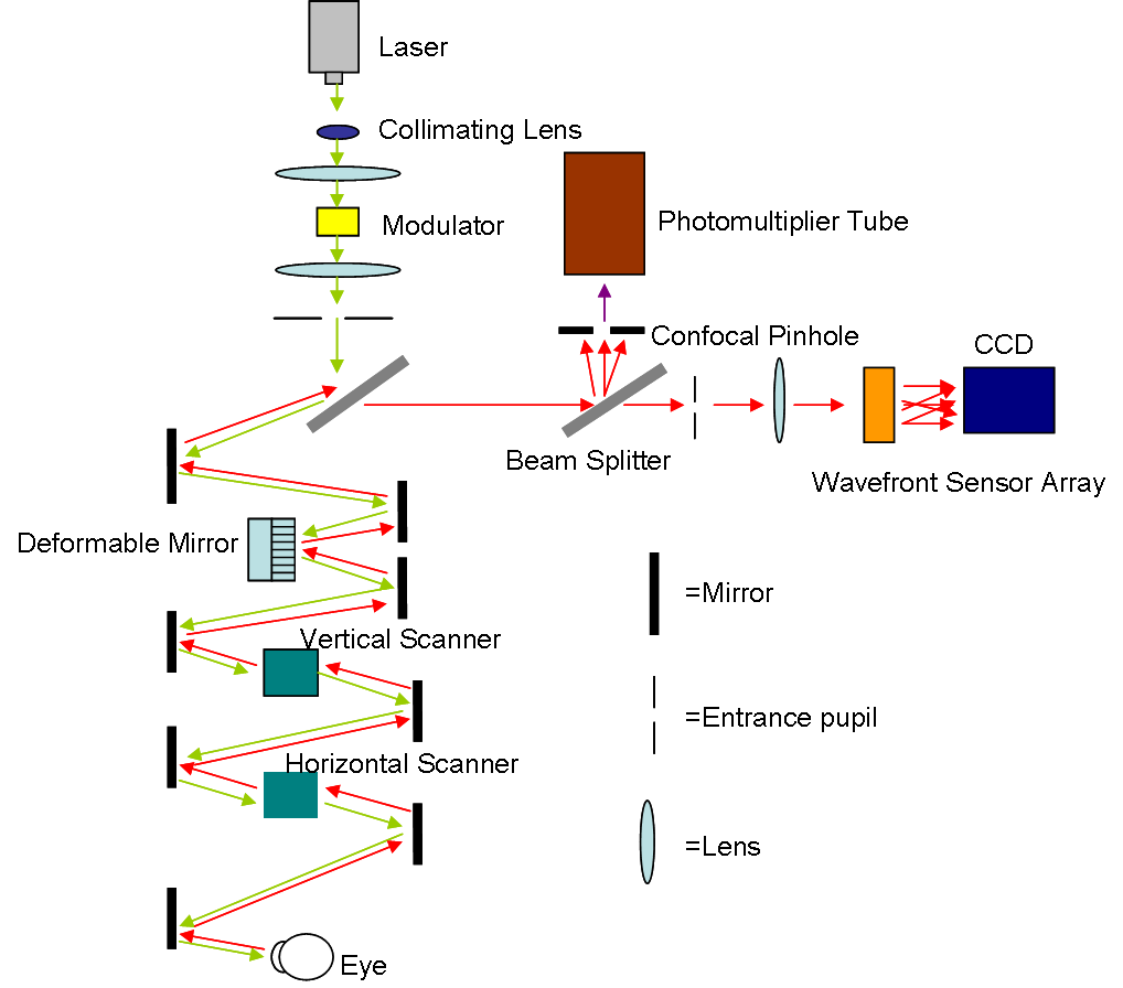
Scanning laser ophthalmoscopy (SLO) is a method of examination of the eye. It uses the technique of confocal laser scanning microscopy for diagnostic imaging of the retina or cornea of the human eye. As a method used to image the retina with a high degree of spatial sensitivity, it is helpful in the diagnosis of glaucoma, macular degeneration, and other retinal disorders. It has further been combined with adaptive optics technology to provide sharper images of the retina."Roorda Lab" — (last accessed: 9 December 2006) Published on October 25, 2006—(last accessed: 9 December 2006) Scanning laser ophthalmoscopy SLO utilizes horizontal and vertical scanning mirrors to scan a specific regi ...[...More Info...] [...Related Items...] OR: [Wikipedia] [Google] [Baidu] |
A-scan Ultrasound Biometry
A-scan ultrasound biometry, commonly referred to as an A-scan (short for Amplitude scan), is a routine type of diagnostic test used in optometry or ophthalmology. The A-scan provides data on the length of the eye, which is a major determinant in common sight disorders. The most common use of the A-scan is to determine eye length for calculation of intraocular lens power. Briefly, the total refractive power of the emmetropic eye is approximately 60. Of this power, the cornea provides roughly 40 diopters, and the crystalline lens 20 diopters. When a cataract is removed, the lens is replaced by an artificial lens implant. By measuring both the length of the eye (A-scan) and the power of the cornea (keratometry), a simple formula can be used to calculate the power of the intraocular lens needed. There are several different formulas that can be used depending on the actual characteristics of the eye. All the information here is not valid for medical purposes. The other major use ... [...More Info...] [...Related Items...] OR: [Wikipedia] [Google] [Baidu] |
B-scan
Medical ultrasound includes diagnostic techniques (mainly imaging techniques) using ultrasound, as well as therapeutic applications of ultrasound. In diagnosis, it is used to create an image of internal body structures such as tendons, muscles, joints, blood vessels, and internal organs, to measure some characteristics (e.g. distances and velocities) or to generate an informative audible sound. Its aim is usually to find a source of disease or to exclude pathology. The usage of ultrasound to produce visual images for medicine is called medical ultrasonography or simply sonography. The practice of examining pregnant women using ultrasound is called obstetric ultrasonography, and was an early development of clinical ultrasonography. Ultrasound is composed of sound waves with frequencies which are significantly higher than the range of human hearing (>20,000 Hz). Ultrasonic images, also known as sonograms, are created by sending pulses of ultrasound into tissue using a pro ... [...More Info...] [...Related Items...] OR: [Wikipedia] [Google] [Baidu] |
Lipofuscin
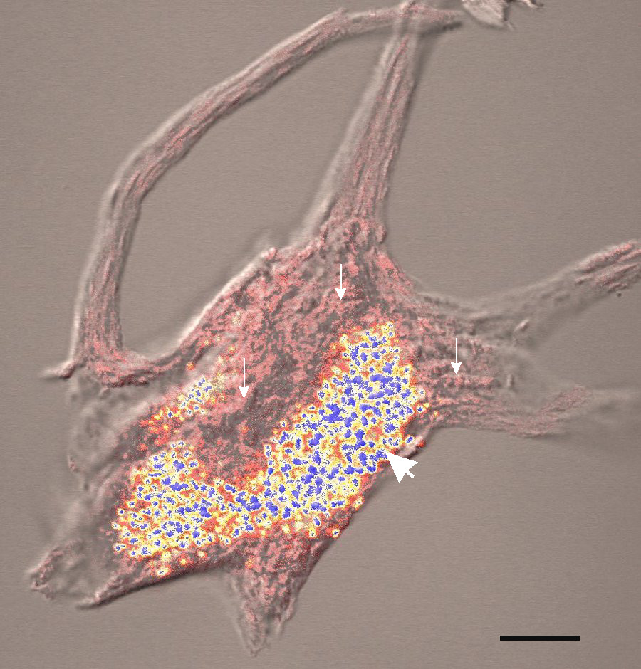
Lipofuscin is the name given to fine yellow-brown pigment granules composed of lipid-containing residues of lysosomal digestion. It is considered to be one of the aging or "wear-and-tear" pigments, found in the liver, kidney, heart muscle, retina, adrenals, nerve cells, and ganglion cells. Formation and turnover Lipofuscin appears to be the product of the oxidation of unsaturated fatty acids and may be symptomatic of membrane damage, or damage to mitochondria and lysosomes. Aside from a large lipid content, lipofuscin is known to contain sugars and metals, including mercury, aluminium, iron, copper and zinc.Chris Gaugler,Lipofuscin", ''Stanislaus Journal of Biochemical Reviews'' May 1997 Lipofuscin is also accepted as consisting of oxidized proteins (30–70%) as well as lipids (20–50%). It is a type of lipochrome and is specifically arranged around the nucleus. The accumulation of lipofuscin-like material may be the result of an imbalance between formation and disposal mech ... [...More Info...] [...Related Items...] OR: [Wikipedia] [Google] [Baidu] |
Autofluorescence
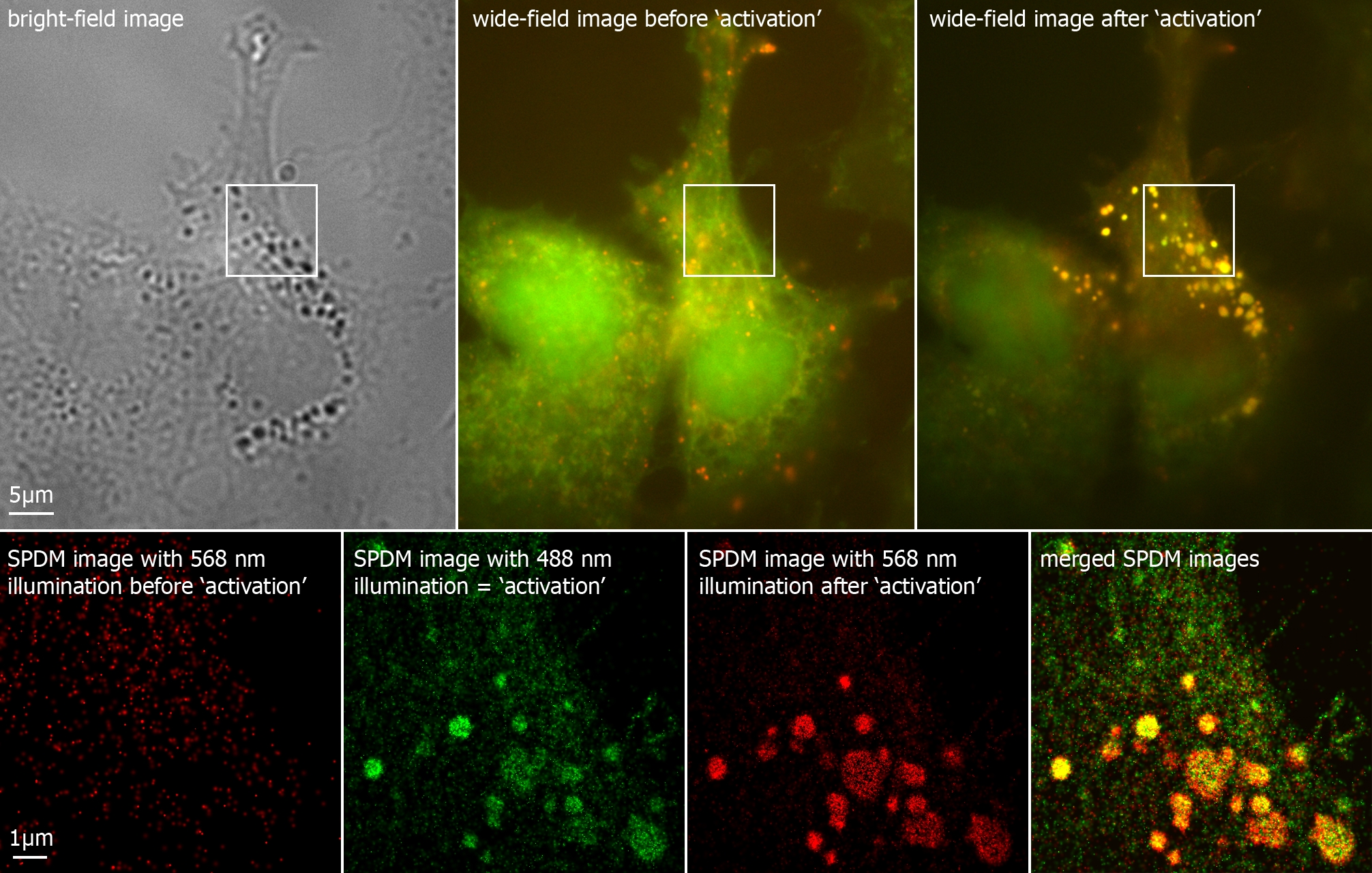
Autofluorescence is the natural emission of light by biological structures such as mitochondria and lysosomes when they have absorbed light, and is used to distinguish the light originating from artificially added fluorescent markers (fluorophores). The most commonly observed autofluorescencing molecules are NADPH and flavins; the extracellular matrix can also contribute to autofluorescence because of the intrinsic properties of collagen and elastin. Generally, proteins containing an increased amount of the amino acids tryptophan, tyrosine, and phenylalanine show some degree of autofluorescence. Autofluorescence also occurs in non-biological materials found in many papers and textiles. Autofluorescence from U.S. paper money has been demonstrated as a means for discerning counterfeit currency from authentic currency. Microscopy Autofluorescence can be problematic in fluorescence microscopy. Light-emitting stains (such as fluorescently labelled antibodies) are applied to sampl ... [...More Info...] [...Related Items...] OR: [Wikipedia] [Google] [Baidu] |
Retinal Pigment Epithelium
The pigmented layer of retina or retinal pigment epithelium (RPE) is the pigmented cell layer just outside the neurosensory retina that nourishes retinal visual cells, and is firmly attached to the underlying choroid and overlying retinal visual cells. History The RPE was known in the 18th and 19th centuries as the pigmentum nigrum, referring to the observation that the RPE is dark (black in many animals, brown in humans); and as the tapetum nigrum, referring to the observation that in animals with a tapetum lucidum, in the region of the tapetum lucidum the RPE is not pigmented. Anatomy The RPE is composed of a single layer of hexagonal cells that are densely packed with pigment granules. When viewed from the outer surface, these cells are smooth and hexagonal in shape. When seen in section, each cell consists of an outer non-pigmented part containing a large oval nucleus and an inner pigmented portion which extends as a series of straight thread-like processes between the rods, ... [...More Info...] [...Related Items...] OR: [Wikipedia] [Google] [Baidu] |
Fluorescein Angiography
Fluorescein angiography (FA), fluorescent angiography (FAG), or fundus fluorescein angiography (FFA) is a technique for examining the circulation of the retina and choroid (parts of the fundus) using a fluorescent dye and a specialized camera. Sodium fluorescein is added into the systemic circulation, the retina is illuminated with blue light at a wavelength of 490 nanometers, and an angiogram is obtained by photographing the fluorescent green light that is emitted by the dye. The fluorescein is administered intravenously in intravenous fluorescein angiography (IVFA) and orally in oral fluorescein angiography (OFA). The test is a dye tracing method. The fluorescein dye also reappears in the patient urine, causing the urine to appear darker, and sometimes orange. It can also cause discolouration of the saliva. Fluorescein angiography is one of several health care applications of this dye, all of which have a risk of severe adverse effects. See fluorescein safety in health care a ... [...More Info...] [...Related Items...] OR: [Wikipedia] [Google] [Baidu] |
Dysplastic Nevus Syndrome
Dysplastic nevus syndrome, also known as familial atypical multiple mole–melanoma (FAMMM) syndrome, is an inherited cutaneous condition described in certain families, and characterized by unusual nevi and multiple inherited melanomas. First described in 1820, the condition is inherited in an autosomal dominant pattern, and caused by mutations in the CDKN2A gene. In addition to melanoma, individuals with the condition are at increased risk for pancreatic cancer. The diagnosis of dysplastic nevus syndrome is based on clinical presentation and family history. Treatment consists of resection of malignant skin lesions (melanoma). Screening for pancreatic cancer may be considered, particularly if there is a family history. Signs and symptoms Dysplastic nevus syndrome is characterized by unusual nevi and multiple inherited melanomas.Czajkowski, R., ''FAMMM Syndrome: Pathogenesis and Management'', Dermatologic Surgery, Vol. 30 Issue 2 p2, p291-296, 2004. Genetics The CDKN2A gene ... [...More Info...] [...Related Items...] OR: [Wikipedia] [Google] [Baidu] |
Welding
Welding is a fabrication (metal), fabrication process that joins materials, usually metals or thermoplastics, by using high heat to melt the parts together and allowing them to cool, causing Fusion welding, fusion. Welding is distinct from lower temperature techniques such as brazing and soldering, which do not melting, melt the base metal (parent metal). In addition to melting the base metal, a filler material is typically added to the joint to form a pool of molten material (the weld pool) that cools to form a joint that, based on weld configuration (butt, full penetration, fillet, etc.), can be stronger than the base material. Pressure may also be used in conjunction with heat or by itself to produce a weld. Welding also requires a form of shield to protect the filler metals or melted metals from being contaminated or Oxidation, oxidized. Many different energy sources can be used for welding, including a gas flame (chemical), an electric arc (electrical), a laser, an electron ... [...More Info...] [...Related Items...] OR: [Wikipedia] [Google] [Baidu] |


