|
Centric Relation
In dentistry, centric relation is the mandibular jaw position in which the head of the condyle is situated as far anterior and superior as it possibly can within the mandibular fossa/glenoid fossa. It is defined as, ''"The maxillo-mandibular relationship in which the condyles articulate with the thinnest avascular portion of their respective discs with the complex in the anterior-superior position against the slopes of the articular eminences. This position is independent of tooth contact. This position is clinically discernible when the mandible is directed superiorly and anteriorly. It is restricted to a purely rotary movement about the transverse horizontal axis". — GPT.'' This position is used when restoring edentulous patients with removable or either implant-supported hybrid or fixed prostheses. Because the dentist wants to be able to reproducibly relate the patient's maxilla and mandible, but the patient does not have teeth with which to establish his or her own ... [...More Info...] [...Related Items...] OR: [Wikipedia] [Google] [Baidu] |
Human Mandible
In anatomy, the mandible, lower jaw or jawbone is the largest, strongest and lowest bone in the human facial skeleton. It forms the lower jaw and holds the lower tooth, teeth in place. The mandible sits beneath the maxilla. It is the only movable bone of the skull (discounting the ossicles of the middle ear). It is connected to the temporal bones by the temporomandibular joints. The bone is formed prenatal development, in the fetus from a fusion of the left and right mandibular prominences, and the point where these sides join, the mandibular symphysis, is still visible as a faint ridge in the midline. Like other symphyses in the body, this is a midline articulation where the bones are joined by fibrocartilage, but this articulation fuses together in early childhood.Illustrated Anatomy of the Head and Neck, Fehrenbach and Herring, Elsevier, 2012, p. 59 The word "mandible" derives from the Latin word ''mandibula'', "jawbone" (literally "one used for chewing"), from ''wikt:mandere ... [...More Info...] [...Related Items...] OR: [Wikipedia] [Google] [Baidu] |
Mandibular Condyle
The condyloid process or condylar process is the process on the human and other mammalian species' mandibles that ends in a condyle, the mandibular condyle. It is thicker than the coronoid process of the mandible and consists of two portions: the condyle and the constricted portion which supports it, the neck. Condyle The most superior part of the mandible, the condyle presents an articular surface for articulation with the articular disk of the temporomandibular joint; it is convex from before backward and from side to side, and extends farther on the posterior than on the anterior surface. Its long axis is directed medialward and slightly backward, and if prolonged to the middle line will meet that of the opposite condyle near the anterior margin of the foramen magnum. At the lateral extremity of the condyle is a small tubercle for the attachment of the temporomandibular ligament. The articular surface of the condyle is covered by fibrous tissue, and interfaces with an articu ... [...More Info...] [...Related Items...] OR: [Wikipedia] [Google] [Baidu] |
Mandibular Fossa
The mandibular fossa, also known as the glenoid fossa in some dental literature, is the depression in the temporal bone that articulates with the mandible. Structure In the temporal bone, the mandibular fossa is bounded anteriorly by the articular tubercle and posteriorly by the tympanic portion of the temporal bone, which separates it from the external acoustic meatus. The fossa is divided into two parts by a narrow slit, the petrotympanic fissure (Glaserian fissure). It is concave in shape to receive the condyloid process of the mandible. Development The mandibular fossa develops from condylar cartilage. This may be stimulated by SOX9 or ALK2, as has been seen in mouse models. Function The condyloid process of the mandible articulates with the temporal bone of the skull at the mandibular fossa. Clinical significance Problems with morphogenesis during embryonic development can lead to the mandibular fossa not forming. This may be caused by mutations to SOX9 or ... [...More Info...] [...Related Items...] OR: [Wikipedia] [Google] [Baidu] |
Dentures
Dentures (also known as false teeth) are prosthetic devices constructed to replace missing teeth, and are supported by the surrounding soft and hard tissues of the oral cavity. Conventional dentures are removable (removable partial denture or complete denture). However, there are many denture designs, some which rely on bonding or clasping onto teeth or dental implants (fixed prosthodontics). There are two main categories of dentures, the distinction being whether they are used to replace missing teeth on the mandibular arch or on the maxillary arch. Medical uses Dentures do not feel like real teeth, nor do they function like real teeth. Dentures can help people through: * Mastication or chewing ability is improved by replacing edentulous areas with denture teeth. * Aesthetics, because the presence of teeth gives a natural appearance to the face, and wearing a denture to replace missing teeth provides support for the lips and cheeks and corrects the collapsed appearance that ... [...More Info...] [...Related Items...] OR: [Wikipedia] [Google] [Baidu] |
Dental Implant
A dental implant (also known as an endosseous implant or fixture) is a prosthesis that interfaces with the bone of the jaw or skull to support a dental prosthesis such as a crown, bridge, denture, or facial prosthesis or to act as an orthodontic anchor. The basis for modern dental implants is a biologic process called osseointegration, in which materials such as titanium or zirconia form an intimate bond to bone. The implant fixture is first placed so that it is likely to osseointegrate, then a dental prosthetic is added. A variable amount of healing time is required for osseointegration before either the dental prosthetic (a tooth, bridge or denture) is attached to the implant or an abutment is placed which will hold a dental prosthetic/crown. Success or failure of implants depends on the health of the person receiving the treatment, drugs which affect the chances of osseointegration, and the health of the tissues in the mouth. The amount of stress that will be put on the impla ... [...More Info...] [...Related Items...] OR: [Wikipedia] [Google] [Baidu] |
Fixed Prosthodontics
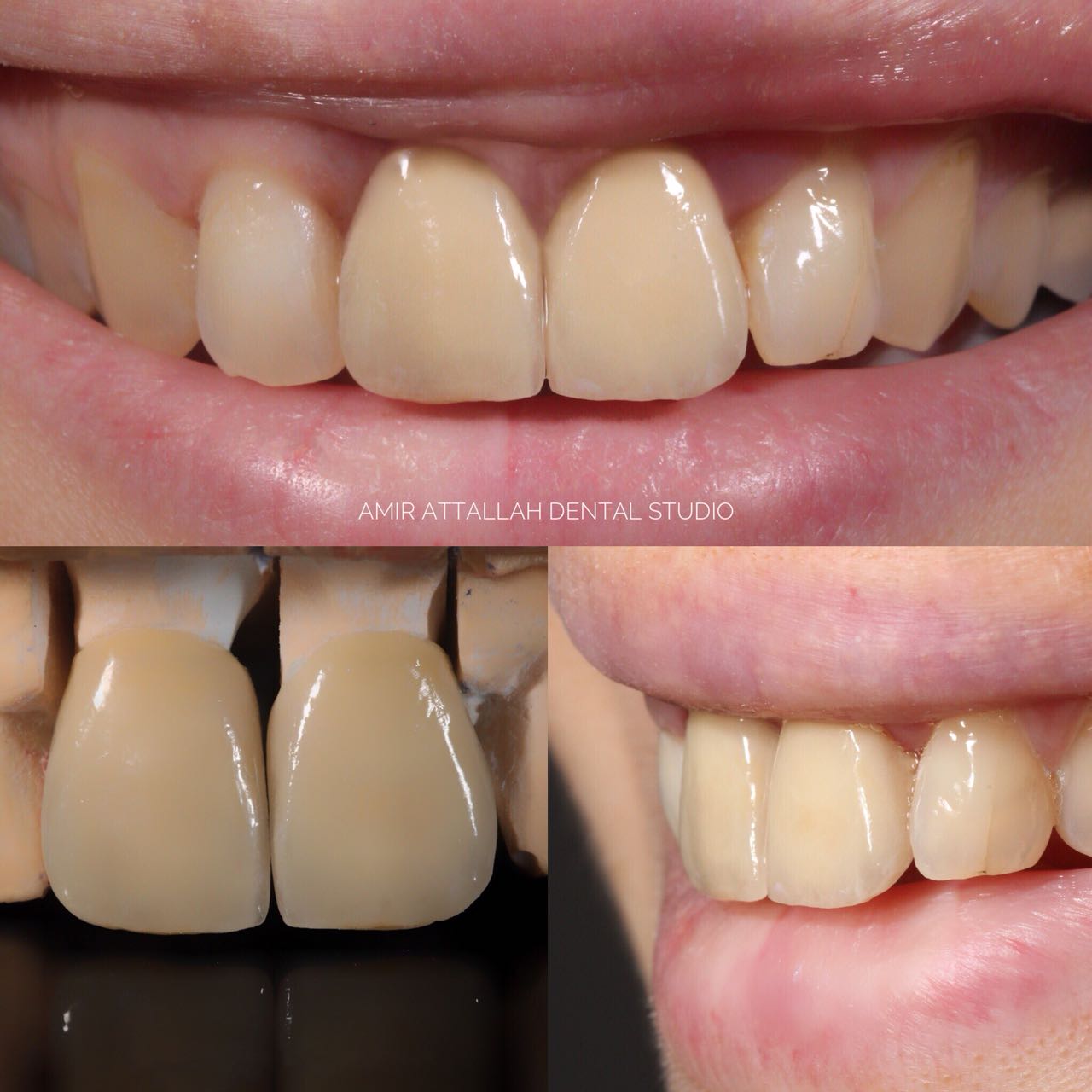
Fixed prosthodontics is the area of prosthodontics focused on permanently attached (fixed) dental prostheses. Such dental restorations, also referred to as indirect restorations, include crowns, bridges (fixed dentures), inlays, onlays, and veneers. Prosthodontists are specialist dentists who have undertaken training recognized by academic institutions in this field. Fixed prosthodontics can be used to restore single or multiple teeth, spanning areas where teeth have been lost. In general, the main advantages of fixed prosthodontics when compared to direct restorations is the superior strength when used in large restorations, and the ability to create an aesthetic looking tooth. As with any dental restoration, principles used to determine the appropriate restoration involves consideration of the materials to be used, extent of tooth destruction, orientation and location of tooth, and condition of neighboring teeth. A good source of information about this subject can be found at ... [...More Info...] [...Related Items...] OR: [Wikipedia] [Google] [Baidu] |
Maxilla
The maxilla (plural: ''maxillae'' ) in vertebrates is the upper fixed (not fixed in Neopterygii) bone of the jaw formed from the fusion of two maxillary bones. In humans, the upper jaw includes the hard palate in the front of the mouth. The two maxillary bones are fused at the intermaxillary suture, forming the anterior nasal spine. This is similar to the mandible (lower jaw), which is also a fusion of two mandibular bones at the mandibular symphysis. The mandible is the movable part of the jaw. Structure In humans, the maxilla consists of: * The body of the maxilla * Four processes ** the zygomatic process ** the frontal process of maxilla ** the alveolar process ** the palatine process * three surfaces – anterior, posterior, medial * the Infraorbital foramen * the maxillary sinus * the incisive foramen Articulations Each maxilla articulates with nine bones: * two of the cranium: the frontal and ethmoid * seven of the face: the nasal, zygomatic, lacrimal, inferior n ... [...More Info...] [...Related Items...] OR: [Wikipedia] [Google] [Baidu] |
Vertical Dimension Of Occlusion
Vertical dimension of occlusion, or VDO, also known as occlusal vertical dimension (OVD), is a term used in dentistry to indicate the superior-inferior relationship of the maxilla and the mandible when the teeth are occluded in maximum intercuspation. A VDO is not only possessed by people who have teeth, however; for completely edentulous individuals who do not have any teeth with which to position themselves in maximum intercuspation, VDO can be measured based on subjective signs related to esthetics and phonetics. In terms of esthetics, an appropriately measured VDO will appear to a layman's eye as an ordinary configuration of the patient's nose, lips and chin. An excessive VDO will appear as though the patient has something stuffed into their mouth, and the patient may not even be able to close their lips. A telltale indication of an excessive VDO is a patient straining to close their lips around the wax rims during VDO determination. Conversely, a deficient VDO will appea ... [...More Info...] [...Related Items...] OR: [Wikipedia] [Google] [Baidu] |
Dental Anatomy
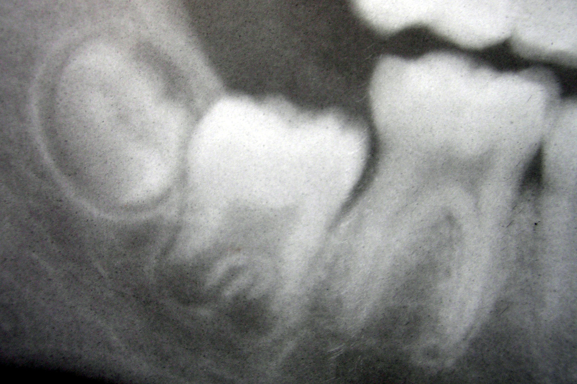
Dental anatomy is a field of anatomy dedicated to the study of human tooth structures. The development, appearance, and classification of teeth fall within its purview. (The function of teeth as they contact one another falls elsewhere, under dental occlusion.) Tooth formation begins before birth, and the teeth's eventual morphology is dictated during this time. Dental anatomy is also a taxonomical science: it is concerned with the naming of teeth and the structures of which they are made, this information serving a practical purpose in dental treatment. Usually, there are 20 primary ("baby") teeth and 32 permanent teeth, the last four being third molars or "wisdom teeth", each of which may or may not grow in. Among primary teeth, 10 usually are found in the maxilla (upper jaw) and the other 10 in the mandible (lower jaw). Among permanent teeth, 16 are found in the maxilla and the other 16 in the mandible. Each tooth has specific distinguishing features. Growing of tooth ... [...More Info...] [...Related Items...] OR: [Wikipedia] [Google] [Baidu] |


