|
Cation Channels Of Sperm
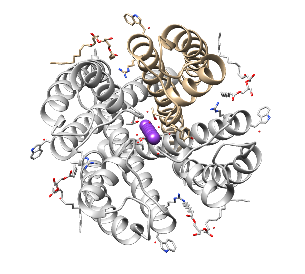
The cation channels of sperm also known as Catsper channels or CatSper, are ion channels that are related to the two-pore channels and distantly related to TRP channels. The four members of this family form voltage-gated Ca2+ channels that seem to be specific to sperm. As sperm encounter the more alkaline environment of the female reproductive tract, CatSper channels become activated by the altered ion concentration. These channels are required for proper fertilization. The study of these channels has been slow because they do not traffic to the cell membrane in many heterologous systems. There are several factors that can activate the CatSper calcium channel, depending on species. In the human, the channel is activated by progesterone released by the oocyte. Progesterone binds to the protein ABHD2 which is present in the sperm plasma membrane, which causes ABHD2 to cleave an inhibitor of CatSper (2-arachidonoylglycerol) into arachidonic acid and glycerol. The human CatSp ... [...More Info...] [...Related Items...] OR: [Wikipedia] [Google] [Baidu] |
CatSper1
CatSper1, is a protein which in humans is encoded by the ''CATSPER1'' gene. CatSper1 is a member of the cation channels of sperm family of protein. The four proteins in this family together form a Ca2+-permeant ion channel specific essential for the correct function of sperm cells. Function Calcium ions play a primary role in the regulation of sperm motility. This gene belongs to a family of putative cation channels that are specific to spermatozoa and localize to the flagellum A flagellum (; ) is a hairlike appendage that protrudes from certain plant and animal sperm cells, and from a wide range of microorganisms to provide motility. Many protists with flagella are termed as flagellates. A microorganism may have f .... The protein family features a single repeat with six membrane-spanning segments and a predicted calcium-selective pore region. References Further reading * * * * * * * * * * Ion channels {{membrane-protein-stub ... [...More Info...] [...Related Items...] OR: [Wikipedia] [Google] [Baidu] |
Arachidonic Acid
Arachidonic acid (AA, sometimes ARA) is a polyunsaturated omega-6 fatty acid 20:4(ω-6), or 20:4(5,8,11,14). It is structurally related to the saturated arachidic acid found in cupuaçu butter. Its name derives from the New Latin word ''arachis'' (peanut), but peanut oil does not contain any arachidonic acid. Chemistry In chemical structure, arachidonic acid is a carboxylic acid with a 20-carbon chain and four ''cis''-double bonds; the first double bond is located at the sixth carbon from the omega end. Some chemistry sources define 'arachidonic acid' to designate any of the eicosatetraenoic acids. However, almost all writings in biology, medicine, and nutrition limit the term to ''all cis''-5,8,11,14-eicosatetraenoic acid. Biology Arachidonic acid is a polyunsaturated fatty acid present in the phospholipids (especially phosphatidylethanolamine, phosphatidylcholine, and phosphatidylinositides) of membranes of the body's cells, and is abundant in the brain, muscles, an ... [...More Info...] [...Related Items...] OR: [Wikipedia] [Google] [Baidu] |
Transmembrane Proteins
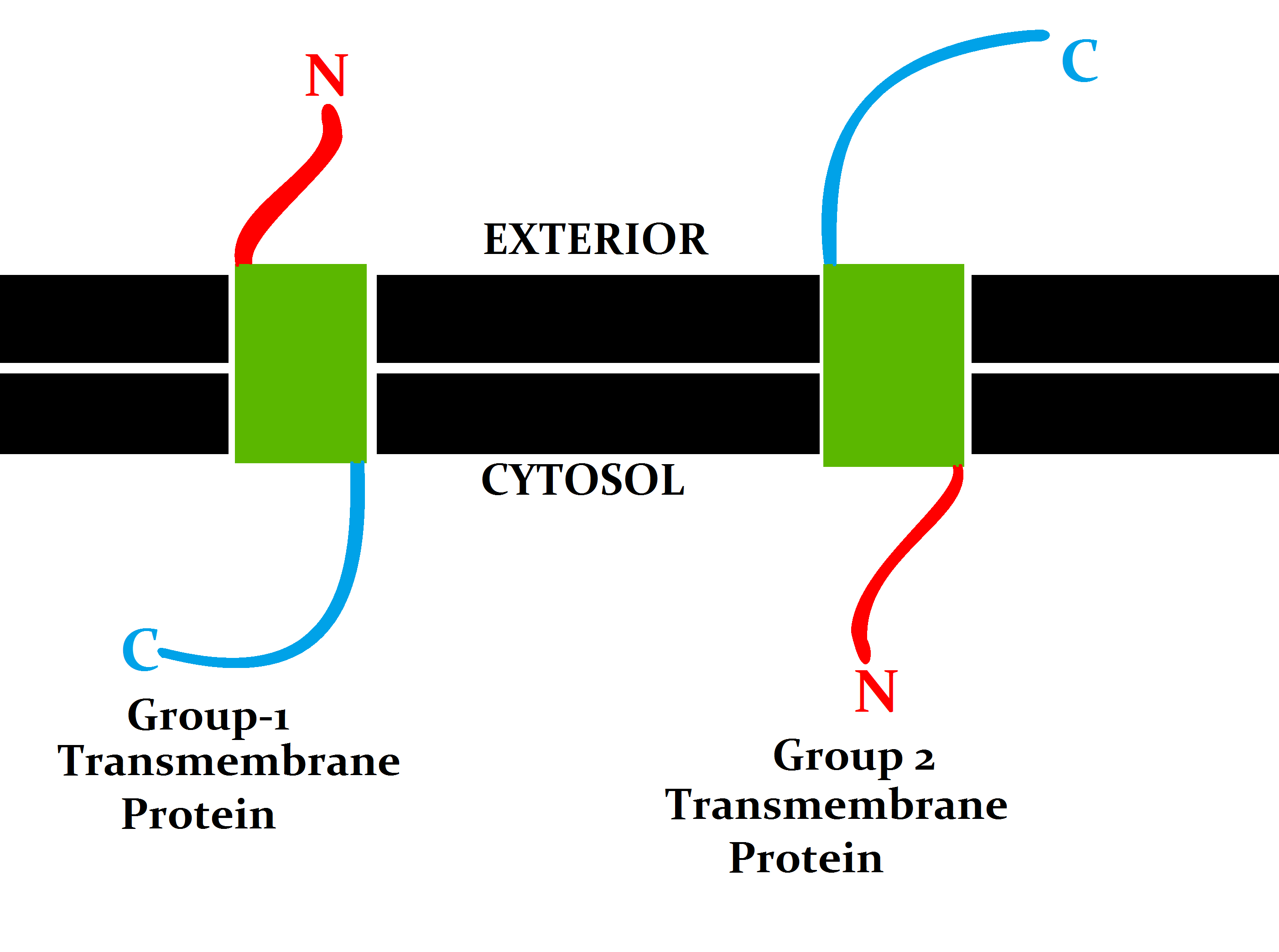
A transmembrane protein (TP) is a type of integral membrane protein that spans the entirety of the cell membrane. Many transmembrane proteins function as gateways to permit the transport of specific substances across the membrane. They frequently undergo significant conformational changes to move a substance through the membrane. They are usually highly hydrophobic and aggregate and precipitate in water. They require detergents or nonpolar solvents for extraction, although some of them (beta-barrels) can be also extracted using denaturing agents. The peptide sequence that spans the membrane, or the transmembrane segment, is largely hydrophobic and can be visualized using the hydropathy plot. Depending on the number of transmembrane segments, transmembrane proteins can be classified as single-span (or bitopic) or multi-span (polytopic). Some other integral membrane proteins are called monotopic, meaning that they are also permanently attached to the membrane, but do not pass t ... [...More Info...] [...Related Items...] OR: [Wikipedia] [Google] [Baidu] |
Voltage-gated Ion Channels
Voltage-gated ion channels are a class of transmembrane proteins that form ion channels that are activated by changes in the electrical membrane potential near the channel. The membrane potential alters the conformation of the channel proteins, regulating their opening and closing. Cell membranes are generally impermeable to ions, thus they must diffuse through the membrane through transmembrane protein channels. They have a crucial role in excitable cells such as neuronal and muscle tissues, allowing a rapid and co-ordinated depolarization in response to triggering voltage change. Found along the axon and at the synapse, voltage-gated ion channels directionally propagate electrical signals. Voltage-gated ion-channels are usually ion-specific, and channels specific to sodium (Na+), potassium (K+), calcium (Ca2+), and chloride (Cl−) ions have been identified. The opening and closing of the channels are triggered by changing ion concentration, and hence charge gradient, between t ... [...More Info...] [...Related Items...] OR: [Wikipedia] [Google] [Baidu] |
Ion Channels
Ion channels are pore-forming membrane proteins that allow ions to pass through the channel pore. Their functions include establishing a resting membrane potential, shaping action potentials and other electrical signals by gating the flow of ions across the cell membrane, controlling the flow of ions across secretory and epithelial cells, and regulating cell volume. Ion channels are present in the membranes of all cells. Ion channels are one of the two classes of ionophoric proteins, the other being ion transporters. The study of ion channels often involves biophysics, electrophysiology, and pharmacology, while using techniques including voltage clamp, patch clamp, immunohistochemistry, X-ray crystallography, fluoroscopy, and RT-PCR. Their classification as molecules is referred to as channelomics. Basic features There are two distinctive features of ion channels that differentiate them from other types of ion transporter proteins: #The rate of ion transport through the ... [...More Info...] [...Related Items...] OR: [Wikipedia] [Google] [Baidu] |
Spermatozoon
A spermatozoon (; also spelled spermatozoön; ; ) is a motile sperm cell, or moving form of the haploid cell that is the male gamete. A spermatozoon joins an ovum to form a zygote. (A zygote is a single cell, with a complete set of chromosomes, that normally develops into an embryo.) Sperm cells contribute approximately half of the nuclear genetic information to the diploid offspring (excluding, in most cases, mitochondrial DNA). In mammals, the sex of the offspring is determined by the sperm cell: a spermatozoon bearing an X chromosome will lead to a female (XX) offspring, while one bearing a Y chromosome will lead to a male (XY) offspring. Sperm cells were first observed in Antonie van Leeuwenhoek's laboratory in 1677. Mammalian spermatozoon structure, function, and size Humans The human sperm cell is the reproductive cell in males and will only survive in warm environments; once it leaves the male body the sperm's survival likelihood is reduced and it may die, thereby dec ... [...More Info...] [...Related Items...] OR: [Wikipedia] [Google] [Baidu] |
Spermatogenesis
Spermatogenesis is the process by which haploid spermatozoa develop from germ cells in the seminiferous tubules of the testis. This process starts with the mitotic division of the stem cells located close to the basement membrane of the tubules. These cells are called spermatogonial stem cells. The mitotic division of these produces two types of cells. Type A cells replenish the stem cells, and type B cells differentiate into primary spermatocytes. The primary spermatocyte divides meiotically (Meiosis I) into two secondary spermatocytes; each secondary spermatocyte divides into two equal haploid spermatids by Meiosis II. The spermatids are transformed into spermatozoa (sperm) by the process of spermiogenesis. These develop into mature spermatozoa, also known as sperm cells. Thus, the primary spermatocyte gives rise to two cells, the secondary spermatocytes, and the two secondary spermatocytes by their subdivision produce four spermatozoa and four haploid cells. Spermatozoa are ... [...More Info...] [...Related Items...] OR: [Wikipedia] [Google] [Baidu] |
Hyperactivation
Hyperactivation is a type of sperm motility. Hyperactivated sperm motility is characterised by a high amplitude, asymmetrical beating pattern of the sperm tail (flagellum). This type of motility may aid in sperm penetration of the zona pellucida, which encloses the ovum. Hyperactivation could then be followed by the acrosome reaction where the cap-like structure on the head of the cell releases the enzymes it contains. This facilitates the penetration of the ovum and fertilisation. Some definitions consider sperm activation to consist of these two processes of hyperactivation and the acrosome reaction Hyperactivation is a term also used to express an X chromosome gene dosage compensation mechanism and is seen in Drosophila. Here, a complex of proteins bind to the X-linked genes to effectively double their genetic activity. This allows males (XY) to have equal genetic activity as females (XX), whose X's are not hyperactivated. Mechanisms Mammalian sperm cells become more act ... [...More Info...] [...Related Items...] OR: [Wikipedia] [Google] [Baidu] |
Lupeol
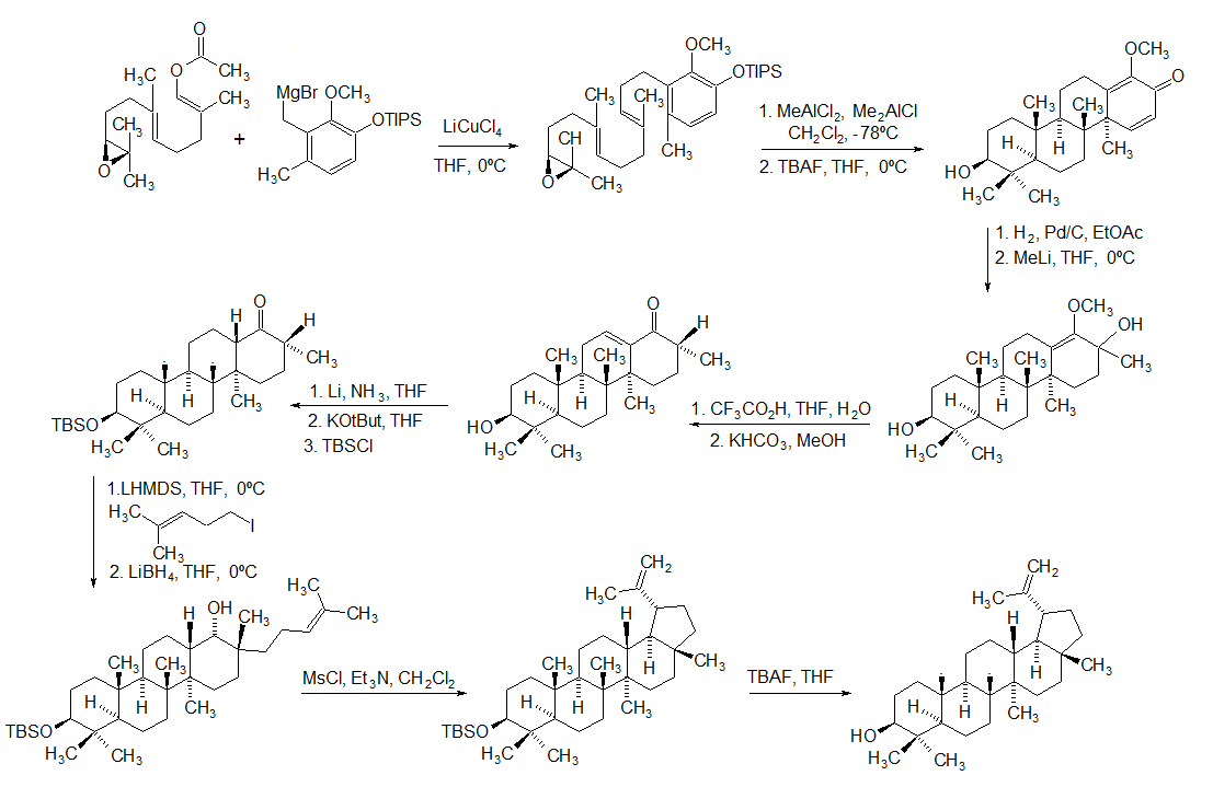
Lupeol is a pharmacologically active pentacyclic triterpenoid. It has several potential medicinal properties, like anticancer and anti-inflammatory activity. Natural occurrences Lupeol is found in a variety of plants, including mango, '' Acacia visco'' and '' Abronia villosa''. It is also found in dandelion coffee. Lupeol is present as a major component in ''Camellia japonica'' leaf. Total synthesis The first total synthesis of lupeol was reported by Gilbert Stork ''et al''. In 2009, Surendra and Corey reported a more efficient and enantioselective total synthesis of lupeol, starting from (''1E,5E'')-8- ''2S'')-3,3-dimethyloxiran-2-yl2,6-dimethylocta-1,5-dienyl acetate by use of a polycyclization. Biosynthesis Lupeol is produced by several organisms from squalene epoxide. Dammarane and baccharane skeletons are formed as intermediates. The reactions are catalyzed by the enzyme lupeol synthase. A recent study on the metabolomics of ''Camellia japonica'' leaf revealed t ... [...More Info...] [...Related Items...] OR: [Wikipedia] [Google] [Baidu] |
Pregnenolone Sulfate
Pregnenolone sulfate (PS, PREGS) is an endogenous excitatory neurosteroid that is synthesized from pregnenolone. It is known to have cognitive and memory-enhancing, antidepressant, anxiogenic, and proconvulsant effects. Biological activity Pregnenolone sulfate is a neurosteroid with excitatory effects in the brain, acting as a potent negative allosteric modulator of the GABAA receptor and a weak positive allosteric modulator of the NMDA receptor. To a lesser extent, it also acts as a negative allosteric modulator of the AMPA, kainate, and glycine receptors, and may interact with the nACh receptors as well. In addition to its effects on ligand-gated ion channels, pregnenolone sulfate is an agonist of the sigma receptor, as well as an activator of the TRPM1 and TRPM3 channels. It may also interact with potassium channels and voltage-gated sodium channels and has been found to inhibit voltage-gated calcium channels. Biochemistry Biosynthesis Pregnenolone sulfate is synthes ... [...More Info...] [...Related Items...] OR: [Wikipedia] [Google] [Baidu] |
Microvillus
Microvilli (singular: microvillus) are microscopic cellular membrane protrusions that increase the surface area for diffusion and minimize any increase in volume, and are involved in a wide variety of functions, including absorption, secretion, cellular adhesion, and mechanotransduction. Structure Microvilli are covered in plasma membrane, which encloses cytoplasm and microfilaments. Though these are cellular extensions, there are little or no cellular organelles present in the microvilli. Each microvillus has a dense bundle of cross-linked actin filaments, which serves as its structural core. 20 to 30 tightly bundled actin filaments are cross-linked by bundling proteins fimbrin (or plastin-1), villin and espin to form the core of the microvilli. In the enterocyte microvillus, the structural core is attached to the plasma membrane along its length by lateral arms made of myosin 1a and Ca2+ binding protein calmodulin. Myosin 1a functions through a binding site for filamentous ac ... [...More Info...] [...Related Items...] OR: [Wikipedia] [Google] [Baidu] |


