|
Carotid Canal
The carotid canal is a passageway in the temporal bone of the skull through which the internal carotid artery enters the middle cranial fossa from the neck. Structure The carotid canal is located within the middle cranial fossa, at the petrous part of the temporal bone. Anteriorly, it is limited by posterior margin of the greater wing of sphenoid bone. Posteromedially, it is limited by basilar part of occipital bone. It is divided in three parts, namely, ascending petrous, transverse petrous, and ascending cavernous parts. The carotid canal has two openings, namely internal and external openings. The internal opening is situated laterally to foramen lacerum. The external opening of the carotid canal is located posterolaterally to the foramen lacerum. Both internal and external openings of the carotid canal lies anterior to the jugular foramen, where the latter is located inside the posterior cranial fossa. The carotid canal is separated from middle ear and inner ear by a thin pla ... [...More Info...] [...Related Items...] OR: [Wikipedia] [Google] [Baidu] |
Temporal Bone
The temporal bones are situated at the sides and base of the skull, and lateral to the temporal lobes of the cerebral cortex. The temporal bones are overlaid by the sides of the head known as the temples, and house the structures of the ears. The lower seven cranial nerves and the major vessels to and from the brain traverse the temporal bone. Structure The temporal bone consists of four parts— the squamous, mastoid, petrous and tympanic parts. The squamous part is the largest and most superiorly positioned relative to the rest of the bone. The zygomatic process is a long, arched process projecting from the lower region of the squamous part and it articulates with the zygomatic bone. Posteroinferior to the squamous is the mastoid part. Fused with the squamous and mastoid parts and between the sphenoid and occipital bones lies the petrous part, which is shaped like a pyramid. The tympanic part is relatively small and lies inferior to the squamous part, anterior to the mast ... [...More Info...] [...Related Items...] OR: [Wikipedia] [Google] [Baidu] |
Angiography
Angiography or arteriography is a medical imaging technique used to visualize the inside, or lumen, of blood vessels and organs of the body, with particular interest in the arteries, veins, and the heart chambers. Modern angiography is performed by injecting a radio-opaque contrast agent into the blood vessel and imaging using X-ray based techniques such as fluoroscopy. The word itself comes from the Greek words ἀγγεῖον ''angeion'' 'vessel' and γράφειν ''graphein'' 'to write, record'. The film or image of the blood vessels is called an ''angiograph'', or more commonly an ''angiogram''. Though the word can describe both an arteriogram and a venogram, in everyday usage the terms angiogram and arteriogram are often used synonymously, whereas the term venogram is used more precisely. The term angiography has been applied to radionuclide angiography and newer vascular imaging techniques such as CO2 angiography, CT angiography and MR angiography. The term ''isotope a ... [...More Info...] [...Related Items...] OR: [Wikipedia] [Google] [Baidu] |
Scalp
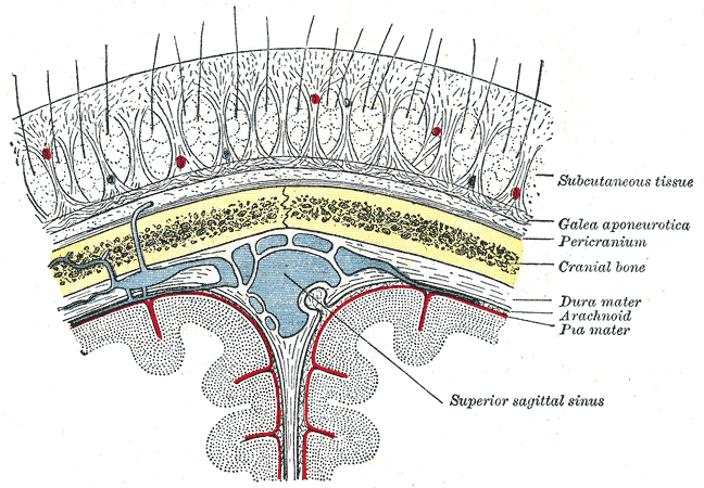
The scalp is the anatomical area bordered by the human face at the front, and by the neck at the sides and back. Structure The scalp is usually described as having five layers, which can conveniently be remembered as a mnemonic: * S: The skin on the head from which head hair grows. It contains numerous sebaceous glands and hair follicles. * C: Connective tissue. A dense subcutaneous layer of fat and fibrous tissue that lies beneath the skin, containing the nerves and vessels of the scalp. * A: The aponeurosis called epicranial aponeurosis (or galea aponeurotica) is the next layer. It is a tough layer of dense fibrous tissue which runs from the frontalis muscle anteriorly to the occipitalis posteriorly. * L: The loose areolar connective tissue layer provides an easy plane of separation between the upper three layers and the pericranium. In scalping the scalp is torn off through this layer. It also provides a plane of access in craniofacial surgery and neurosurgery. This layer i ... [...More Info...] [...Related Items...] OR: [Wikipedia] [Google] [Baidu] |
Sweat Glands
Sweat glands, also known as sudoriferous or sudoriparous glands, , are small tubular structures of the skin that produce sweat. Sweat glands are a type of exocrine gland, which are glands that produce and secrete substances onto an epithelial surface by way of a duct. There are two main types of sweat glands that differ in their structure, function, secretory product, mechanism of excretion, anatomic distribution, and distribution across species: * Eccrine sweat glands are distributed almost all over the human body, in varying densities, with the highest density in palms and soles, then on the head, but much less on the trunk and the extremities. Its water-based secretion represents a primary form of cooling in humans. * Apocrine sweat glands are mostly limited to the axillae (armpits) and perineal area in humans. They are not significant for cooling in humans, but are the sole effective sweat glands in hoofed animals, such as the camels, donkeys, horses, and cattle. Cerumin ... [...More Info...] [...Related Items...] OR: [Wikipedia] [Google] [Baidu] |
Pupil
The pupil is a black hole located in the center of the iris of the eye that allows light to strike the retina.Cassin, B. and Solomon, S. (1990) ''Dictionary of Eye Terminology''. Gainesville, Florida: Triad Publishing Company. It appears black because light rays entering the pupil are either absorbed by the tissues inside the eye directly, or absorbed after diffuse reflections within the eye that mostly miss exiting the narrow pupil. The term "pupil" was coined by Gerard of Cremona. In humans, the pupil is round, but its shape varies between species; some cats, reptiles, and foxes have vertical slit pupils, goats have horizontally oriented pupils, and some catfish have annular types. In optical terms, the anatomical pupil is the eye's aperture and the iris is the aperture stop. The image of the pupil as seen from outside the eye is the entrance pupil, which does not exactly correspond to the location and size of the physical pupil because it is magnified by the cornea. On the ... [...More Info...] [...Related Items...] OR: [Wikipedia] [Google] [Baidu] |
Superior Tarsal Muscle
The superior tarsal muscle is a smooth muscle adjoining the levator palpebrae superioris muscle that helps to raise the upper eyelid. Structure The superior tarsal muscle originates on the underside of levator palpebrae superioris and inserts on the superior tarsal plate of the eyelid. Nerve supply The superior tarsal muscle receives its innervation from the sympathetic nervous system. Postganglionic sympathetic fibers originate in the superior cervical ganglion, and travel via the internal carotid plexus, where small branches communicate with the oculomotor nerve as it passes through the cavernous sinus. The sympathetic fibres continue to the superior division of the oculomotor nerve, where they enter the superior tarsal muscle on its inferior aspect. Function Its role is not fully clear, but may be an accessory muscle to raise the upper eyelid. Clinical significance Damage to some elements of the sympathetic nervous system can inhibit this muscle, causing a drooping ey ... [...More Info...] [...Related Items...] OR: [Wikipedia] [Google] [Baidu] |
Eyelid
An eyelid is a thin fold of skin that covers and protects an eye. The levator palpebrae superioris muscle retracts the eyelid, exposing the cornea to the outside, giving vision. This can be either voluntarily or involuntarily. The human eyelid features a row of eyelashes along the eyelid margin, which serve to heighten the protection of the eye from dust and foreign debris, as well as from perspiration. "Palpebral" (and "blepharal") means relating to the eyelids. Its key function is to regularly spread the tears and other secretions on the eye surface to keep it moist, since the cornea must be continuously moist. They keep the eyes from drying out when asleep. Moreover, the blink reflex protects the eye from foreign bodies. The appearance of the human upper eyelid often varies between different populations. The prevalence of an epicanthic fold covering the inner corner of the eye account for the majority of East Asian and Southeast Asian populations, and is also found i ... [...More Info...] [...Related Items...] OR: [Wikipedia] [Google] [Baidu] |
Superior Cervical Ganglion
The superior cervical ganglion (SCG) is part of the autonomic nervous system (ANS); more specifically, it is part of the sympathetic nervous system, a division of the ANS most commonly associated with the fight or flight response. The ANS is composed of pathways that lead to and from ganglia, groups of nerve cells. A ganglion allows a large amount of divergence in a neuronal pathway and also enables a more localized circuitry for control of the innervated targets. The SCG is the only ganglion in the sympathetic nervous system that innervates the head and neck. It is the largest and most rostral (superior) of the three cervical ganglia. The SCG innervates many organs, glands and parts of the carotid system in the head. Structure Location The SCG is located opposite the second and third cervical vertebrae. It lies deep to the sheath of the internal carotid artery and internal jugular vein, and anterior to the Longus capitis muscle. The SCG contains neurons that supply sympathetic ... [...More Info...] [...Related Items...] OR: [Wikipedia] [Google] [Baidu] |
Sympathetics
The sympathetic nervous system (SNS) is one of the three divisions of the autonomic nervous system, the others being the parasympathetic nervous system and the enteric nervous system. The enteric nervous system is sometimes considered part of the autonomic nervous system, and sometimes considered an independent system. The autonomic nervous system functions to regulate the body's unconscious actions. The sympathetic nervous system's primary process is to stimulate the body's fight or flight response. It is, however, constantly active at a basic level to maintain homeostasis. The sympathetic nervous system is described as being antagonistic to the parasympathetic nervous system which stimulates the body to "feed and breed" and to (then) "rest-and-digest". Structure There are two kinds of neurons involved in the transmission of any signal through the sympathetic system: pre-ganglionic and post-ganglionic. The shorter preganglionic neurons originate in the thoracolumbar division ... [...More Info...] [...Related Items...] OR: [Wikipedia] [Google] [Baidu] |
Internal Carotid Plexus
The internal carotid plexus is situated on the lateral side of the internal carotid artery, and in the plexus there occasionally exists a small gangliform swelling, the ''carotid ganglion'', on the under surface of the artery. Postganglionic sympathetic fibres ascend from the superior cervical ganglion, along the walls of the internal carotid artery, to enter the internal carotid plexus. These fibres then distribute to deep structures, which include the Superior Tarsal Muscle and pupillary dilator muscles. Some of the fibres from the internal carotid plexus converge to form the deep petrosal nerve.Richard L. Drake, Wayne Vogel & Adam W M Mitchell, "Gray's Anatomy for Students", Elsevier inc., 2005 The internal carotid plexus communicates with the trigeminal ganglion, the abducent nerve, and the pterygopalatine ganglion (also named sphenopalatine); it distributes filaments to the wall of the internal carotid artery, and also communicates with the tympanic branch of the glossopha ... [...More Info...] [...Related Items...] OR: [Wikipedia] [Google] [Baidu] |
Academic Press
Academic Press (AP) is an academic book publisher founded in 1941. It was acquired by Harcourt, Brace & World in 1969. Reed Elsevier bought Harcourt in 2000, and Academic Press is now an imprint of Elsevier. Academic Press publishes reference books, serials and online products in the subject areas of: * Communications engineering * Economics * Environmental science * Finance * Food science and nutrition * Geophysics * Life sciences * Mathematics and statistics * Neuroscience * Physical sciences * Psychology Well-known products include the ''Methods in Enzymology'' series and encyclopedias such as ''The International Encyclopedia of Public Health'' and the ''Encyclopedia of Neuroscience''. See also * Akademische Verlagsgesellschaft (AVG) — the German predecessor, founded in 1906 by Leo Jolowicz (1868–1940), the father of Walter Jolowicz Walter may refer to: People * Walter (name), both a surname and a given name * Little Walter, American blues harmonica player Marion Wa ... [...More Info...] [...Related Items...] OR: [Wikipedia] [Google] [Baidu] |
Human Cranium
The skull is a bone protective cavity for the brain. The skull is composed of four types of bone i.e., cranial bones, facial bones, ear ossicles and hyoid bone. However two parts are more prominent: the cranium and the mandible. In humans, these two parts are the neurocranium and the viscerocranium (facial skeleton) that includes the mandible as its largest bone. The skull forms the anterior-most portion of the skeleton and is a product of cephalisation—housing the brain, and several sensory structures such as the eyes, ears, nose, and mouth. In humans these sensory structures are part of the facial skeleton. Functions of the skull include protection of the brain, fixing the distance between the eyes to allow stereoscopic vision, and fixing the position of the ears to enable sound localisation of the direction and distance of sounds. In some animals, such as horned ungulates (mammals with hooves), the skull also has a defensive function by providing the mount (on the fronta ... [...More Info...] [...Related Items...] OR: [Wikipedia] [Google] [Baidu] |


