|
Cardiopulmonary Nerves
Cardiopulmonary nerves are splanchnic nerves that are postsynaptic and sympathetic. They originate in cervical and upper thoracic ganglia and innervate the thoracic cavity. All major sympathetic cardiopulmonary nerves arise from the stellate ganglia and the caudal halves of the cervical sympathetic trunks below the level of the cricoid cartilage. Parasympathetic cardiopulmonary nerves arise from the recurrent laryngeal nerve The recurrent laryngeal nerve (RLN) is a branch of the vagus nerve ( cranial nerve X) that supplies all the intrinsic muscles of the larynx, with the exception of the cricothyroid muscles. There are two recurrent laryngeal nerves, right and ...s and the thoracic vagus immediately distal to them. These interconnect with the sympathetic cardiopulmonary nerves to form the ventral and dorsal cardiopulmonary plexuses. References Sympathetic nervous system {{Neuroanatomy-stub ... [...More Info...] [...Related Items...] OR: [Wikipedia] [Google] [Baidu] |
Splanchnic Nerves
The splanchnic nerves are paired visceral nerves (nerves that contribute to the innervation of the viscera, innervation of the internal organs), carrying fibers of the autonomic nervous system (visceral efferent fibers) as well as sensory fibers from the organs (visceral afferent fibers). All carry Sympathetic nervous system, sympathetic fibers except for the pelvic splanchnic nerves, which carry parasympathetic fibers. Types The term ''splanchnic nerves'' can refer to: * Cardiopulmonary nervesEssential Clinical Anatomy. K.L. Moore & A.M. Agur. Lippincott, 3 ed. 2007. Page 181 * Thoracic splanchnic nerves (greater, lesser, and least) * Lumbar splanchnic nerves * Sacral splanchnic nerves * Pelvic splanchnic nerves References {{Autonomic Autonomic nervous system ... [...More Info...] [...Related Items...] OR: [Wikipedia] [Google] [Baidu] |
Postsynaptic
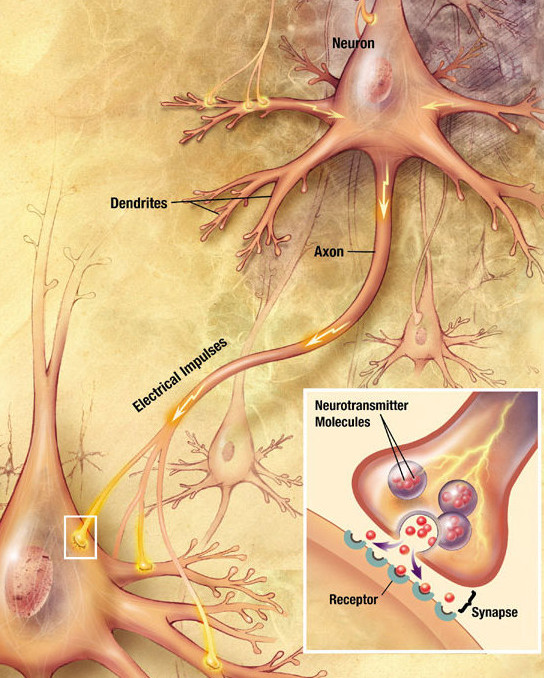
Chemical synapses are biological junctions through which neurons' signals can be sent to each other and to non-neuronal cells such as those in muscles or glands. Chemical synapses allow neurons to form circuits within the central nervous system. They are crucial to the biological computations that underlie perception and thought. They allow the nervous system to connect to and control other systems of the body. At a chemical synapse, one neuron releases neurotransmitter molecules into a small space (the synaptic cleft) that is adjacent to another neuron. The neurotransmitters are contained within small sacs called synaptic vesicles, and are released into the synaptic cleft by exocytosis. These molecules then bind to neurotransmitter receptors on the postsynaptic cell. Finally, the neurotransmitters are cleared from the synapse through one of several potential mechanisms including enzymatic degradation or re-uptake by specific transporters either on the presynaptic cel ... [...More Info...] [...Related Items...] OR: [Wikipedia] [Google] [Baidu] |
Sympathetic Nervous System
The sympathetic nervous system (SNS) is one of the three divisions of the autonomic nervous system, the others being the parasympathetic nervous system and the enteric nervous system. The enteric nervous system is sometimes considered part of the autonomic nervous system, and sometimes considered an independent system. The autonomic nervous system functions to regulate the body's unconscious actions. The sympathetic nervous system's primary process is to stimulate the body's fight or flight response. It is, however, constantly active at a basic level to maintain homeostasis. The sympathetic nervous system is described as being antagonistic to the parasympathetic nervous system which stimulates the body to "feed and breed" and to (then) "rest-and-digest". Structure There are two kinds of neurons involved in the transmission of any signal through the sympathetic system: pre-ganglionic and post-ganglionic. The shorter preganglionic neurons originate in the thoracolumbar division o ... [...More Info...] [...Related Items...] OR: [Wikipedia] [Google] [Baidu] |
Cervical Ganglia
The cervical ganglia are paravertebral ganglia of the sympathetic nervous system. Preganglionic nerves from the thoracic spinal cord enter into the cervical ganglions and synapse with its postganglionic fibers or nerves. The cervical ganglion has three paravertebral ganglia: * superior cervical ganglion (largest) - adjacent to C2 & C3; postganglionic axon projects to target: (heart, head, neck) via "hitchhiking" on the carotid arteries * middle cervical ganglion (smallest) - adjacent to C6; target: heart, neck * inferior cervical ganglion. The inferior ganglion may be fused with the first thoracic ganglion to form a single structure, the stellate ganglion. - adjacent to C7; target: heart, lower neck, arm, posterior cranial arteries Nerves emerging from cervical sympathetic ganglia contribute to the cardiac plexus The cardiac plexus is a plexus of nerves situated at the base of the heart that innervates the heart. Structure The cardiac plexus is divided into a superficial p ... [...More Info...] [...Related Items...] OR: [Wikipedia] [Google] [Baidu] |
Thoracic Ganglia
The thoracic ganglia are paravertebral ganglia. The thoracic portion of the sympathetic trunk typically has 12 thoracic ganglia. Emerging from the ganglia are thoracic splanchnic nerves (the cardiopulmonary, the greater, lesser, and least splanchnic nerves) that help provide sympathetic innervation to thoracic and abdominal structures. The thoracic part of sympathetic trunk lies posterior to the costovertebral pleura and is hence not a content of the posterior mediastinum Also, the ganglia of the thoracic sympathetic trunk have both white and gray rami communicantes. The white rami communicantes carry sympathetic fibers arising in the spinal cord into the sympathetic trunk, while the gray rami communicantes carry postganglionic nerve fibers of the sympathetic nervous system back to the spinal nerves A spinal nerve is a mixed nerve, which carries motor, sensory, and autonomic signals between the spinal cord and the body. In the human body there are 31 pairs of spinal nerves, on ... [...More Info...] [...Related Items...] OR: [Wikipedia] [Google] [Baidu] |
Thoracic Cavity
The thoracic cavity (or chest cavity) is the chamber of the body of vertebrates that is protected by the thoracic wall (rib cage and associated skin, muscle, and fascia). The central compartment of the thoracic cavity is the mediastinum. There are two openings of the thoracic cavity, a superior thoracic aperture known as the thoracic inlet and a lower inferior thoracic aperture known as the thoracic outlet. The thoracic cavity includes the tendons as well as the cardiovascular system which could be damaged from injury to the back, spine or the neck. Structure Structures within the thoracic cavity include: * structures of the cardiovascular system, including the heart and great vessels, which include the thoracic aorta, the pulmonary artery and all its branches, the superior and inferior vena cava, the pulmonary veins, and the azygos vein * structures of the respiratory system, including the diaphragm, trachea, bronchi and lungs * structures of the digestive system, including ... [...More Info...] [...Related Items...] OR: [Wikipedia] [Google] [Baidu] |
Stellate Ganglion
The stellate ganglion (or cervicothoracic ganglion) is a sympathetic ganglion formed by the fusion of the inferior cervical ganglion and the first thoracic (superior thoracic sympathetic) ganglion, which exists in 80% of people. Sometimes, the second and the third thoracic ganglia are included in this fusion. The stellate ganglion is relatively big (10–12 x 8–20 mm) compared to much smaller thoracic, lumbar and sacral ganglia, and is polygonal in shape (). Stellate ganglion is located at the level of C7, anterior to the transverse process of C7 and the neck of the first rib, superior to the cervical pleura and just below the subclavian artery. It is superiorly covered by the prevertebral lamina of the cervical fascia and anteriorly in relation with common carotid artery, subclavian artery and the beginning of vertebral artery which sometimes leaves a groove at the apex of this ganglion (this groove can sometimes even separate the stellate ganglion into so called vertebral gangli ... [...More Info...] [...Related Items...] OR: [Wikipedia] [Google] [Baidu] |
Cricoid Cartilage
The cricoid cartilage , or simply cricoid (from the Greek ''krikoeides'' meaning "ring-shaped") or cricoid ring, is the only complete ring of cartilage around the trachea. It forms the back part of the voice box and functions as an attachment site for muscles, cartilages, and ligaments involved in opening and closing the airway and in producing speech. Structure The cricoid cartilage sits just inferior to the thyroid cartilage in the neck, at the level of the C6 vertebra, and is joined to it medially by the median cricothyroid ligament and postero-laterally by the cricothyroid joints. Inferior to it are the rings of cartilage around the trachea (which are not continuous – rather they are C-shaped with a gap posteriorly). The cricoid is joined to the first tracheal ring by the cricotracheal ligament, and this can be felt as a more yielding area between the firm thyroid cartilage and firmer cricoid. It is also anatomically related to the thyroid gland; although the ... [...More Info...] [...Related Items...] OR: [Wikipedia] [Google] [Baidu] |
Recurrent Laryngeal Nerve
The recurrent laryngeal nerve (RLN) is a branch of the vagus nerve (cranial nerve X) that supplies all the intrinsic muscles of the larynx, with the exception of the cricothyroid muscles. There are two recurrent laryngeal nerves, right and left. The right and left nerves are not symmetrical, with the left nerve looping under the aortic arch, and the right nerve looping under the right subclavian artery then traveling upwards. They both travel alongside the trachea. Additionally, the nerves are among the few nerves that follow a ''recurrent'' course, moving in the opposite direction to the nerve they branch from, a fact from which they gain their name. The recurrent laryngeal nerves supply sensation to the larynx below the vocal cords, give cardiac branches to the deep cardiac plexus, and branch to the trachea, esophagus and the inferior constrictor muscles. The posterior cricoarytenoid muscles, the only muscles that can open the vocal folds, are innervated by this nerve. Th ... [...More Info...] [...Related Items...] OR: [Wikipedia] [Google] [Baidu] |


