|
Canal Of Nuck
__NOTOC__ The canal of Nuck, first described by Anton Nuck ( de) in 1691, is an abnormal patent (open) pouch of peritoneum extending into the labia majora of women. It is analogous to a patent processus vaginalis in males (see hydrocele testis, inguinal hernia). In rare cases, it may give rise to a cyst or a hydrocele in women and has potential to develop into an indirect inguinal hernia. The pouch accompanies the gubernaculum during development of the urinary and reproductive organs, more specifically during the descent of the ovaries, and normally obliterates. See also *List of homologues of the human reproductive system This list of related male and female reproductive organs shows how the male and female reproductive organs and the development of the reproductive system are related, sharing a common developmental path. This makes them biological homologues. T ... References Further reading * * * * * * * {{DEFAULTSORT:Canal Of Nuck Mammal female reproductive system< ... [...More Info...] [...Related Items...] OR: [Wikipedia] [Google] [Baidu] |
Peritoneum
The peritoneum is the serous membrane forming the lining of the abdominal cavity or coelom in amniotes and some invertebrates, such as annelids. It covers most of the intra-abdominal (or coelomic) organs, and is composed of a layer of mesothelium supported by a thin layer of connective tissue. This peritoneal lining of the cavity supports many of the abdominal organs and serves as a conduit for their blood vessels, lymphatic vessels, and nerves. The abdominal cavity (the space bounded by the vertebrae, abdominal muscles, diaphragm, and pelvic floor) is different from the intraperitoneal space (located within the abdominal cavity but wrapped in peritoneum). The structures within the intraperitoneal space are called "intraperitoneal" (e.g., the stomach and intestines), the structures in the abdominal cavity that are located behind the intraperitoneal space are called "retroperitoneal" (e.g., the kidneys), and those structures below the intraperitoneal space are called "subp ... [...More Info...] [...Related Items...] OR: [Wikipedia] [Google] [Baidu] |
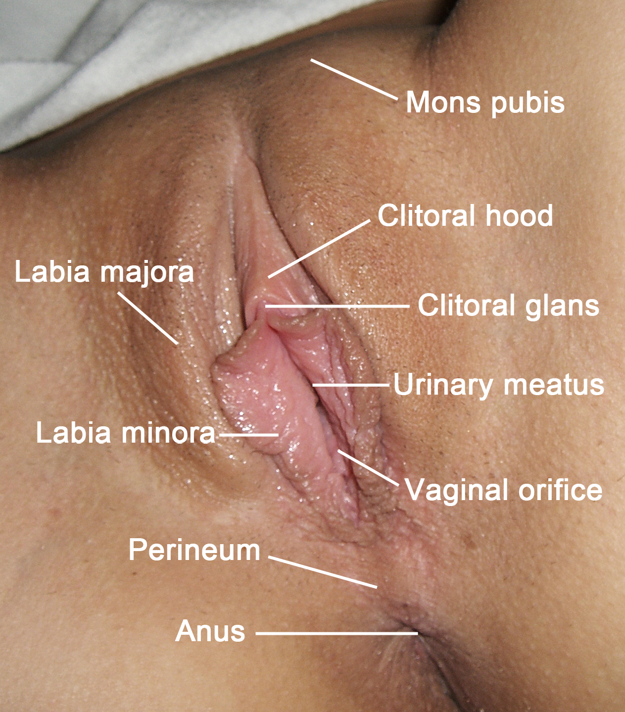
Labia Majora
The labia majora (singular: ''labium majus'') are two prominent longitudinal cutaneous folds that extend downward and backward from the mons pubis to the perineum. Together with the labia minora they form the labia of the vulva. The labia majora are homologous to the male scrotum. Etymology ''Labia majora'' is the Latin plural for big ("major") lips; the singular is ''labium majus.'' The Latin term ''labium/labia'' is used in anatomy for a number of usually paired parallel structures, but in English it is mostly applied to two pairs of parts of female external genitals (vulva)—labia majora and labia minora. Labia majora are commonly known as the outer lips, while labia minora (Latin for ''small lips''), which run alongside between them, are referred to as the inner lips. Traditionally, to avoid confusion with other lip-like structures of the body, the labia of female genitals were termed by anatomists in Latin as ''labia majora (''or ''minora) pudendi.'' Embryology Embryolo ... [...More Info...] [...Related Items...] OR: [Wikipedia] [Google] [Baidu] |
Processus Vaginalis
The vaginal process (or processus vaginalis) is an embryonic developmental outpouching of the parietal peritoneum. It is present from around the 12th week of gestation, and commences as a peritoneal outpouching. Sex differences In males, it precedes the testes in their descent down within the gubernaculum, and closes. This closure (also called ''fusion'') occurs at any point from a few weeks before birth, to a few weeks after birth. The remaining portion around the testes becomes the tunica vaginalis. If it does not close in females, it forms the canal of Nuck. Clinical significance Failure of closure of the vaginal process leads to the propensity to develop a number of abnormalities. Peritoneal fluid can travel down a patent vaginal process leading to the formation of a hydrocele. Persistent patent processus vaginalis is more common on the right than the left. Accumulation of blood in a persistent processus vaginalis could result in a hematocele. There is the potential for an ... [...More Info...] [...Related Items...] OR: [Wikipedia] [Google] [Baidu] |
Hydrocele Testis
A hydrocele testis is an accumulation of clear fluid within the cavum vaginale, the potential space between the layers of the tunica vaginalis of the testicle. It is the most common form of hydrocele and is often referred to simply as a "hydrocele". A primary hydrocele testis causes a painless enlargement in the scrotum on the affected side and is thought to be due to the defective absorption of fluid secreted between the two layers of the tunica vaginalis (investing membrane). A secondary hydrocele is secondary to either inflammation or a neoplasm in the testis. A hydrocele testis usually occurs on one side, but can also affect both sides. The accumulation can be a marker of physical trauma, infection, tumor or varicocele surgery, but the cause is generally unknown. Indirect inguinal hernia indicates increased risk of hydrocele testis. Signs and symptoms A hydrocele testis feels like a small fluid-filled balloon inside the scrotum. It is smooth, and is mainly in front of the test ... [...More Info...] [...Related Items...] OR: [Wikipedia] [Google] [Baidu] |
Inguinal Hernia
An inguinal hernia is a hernia (protrusion) of abdominal-cavity contents through the inguinal canal. Symptoms, which may include pain or discomfort especially with or following coughing, exercise, or bowel movements, are absent in about a third of patients. Symptoms often get worse throughout the day and improve when lying down. A bulging area may occur that becomes larger when bearing down. Inguinal hernias occur more often on the right than left side. The main concern is strangulation, where the blood supply to part of the intestine is blocked. This usually produces severe pain and tenderness of the area. Risk factors for the development of a hernia include: smoking, chronic obstructive pulmonary disease, obesity, pregnancy, peritoneal dialysis, collagen vascular disease, and previous open appendectomy, among others. Predisposition to hernias is Genetic predisposition, genetic and they occur more often in certain families. Deleterious mutations causing predisposition to he ... [...More Info...] [...Related Items...] OR: [Wikipedia] [Google] [Baidu] |
Cyst
A cyst is a closed sac, having a distinct envelope and cell division, division compared with the nearby Biological tissue, tissue. Hence, it is a cluster of Cell (biology), cells that have grouped together to form a sac (like the manner in which water molecules group together to form a bubble); however, the distinguishing aspect of a cyst is that the cells forming the "shell" of such a sac are distinctly abnormal (in both appearance and behaviour) when compared with all surrounding cells for that given location. A cyst may contain air, fluids, or semi-solid material. A collection of pus is called an abscess, not a cyst. Once formed, a cyst may resolve on its own. When a cyst fails to resolve, it may need to be removed surgically, but that would depend upon its type and location. Cancer-related cysts are formed as a defense mechanism for the body following the development of mutations that lead to an uncontrolled cellular division. Once that mutation has occurred, the affected cell ... [...More Info...] [...Related Items...] OR: [Wikipedia] [Google] [Baidu] |
Hydrocele
A hydrocele is an accumulation of serous fluid in a body cavity. A hydrocele testis, the most common form of hydrocele, is the accumulation of fluids around a testicle. It is often caused by fluid collecting within a layer wrapped around the testicle, called the tunica vaginalis, which is derived from peritoneum. Provided there is no hernia present, it goes away without treatment in the first year. Although hydroceles usually develop in males, rare instances have been described in females in the Canal of Nuck. Primary hydroceles may develop in adulthood, particularly in the elderly and in hot countries, by slow accumulation of serous fluid. This is presumably caused by impaired reabsorption, which appears to be the explanation for most primary hydroceles, although the reason remains obscure. A hydrocele can also be the result of a plugged inguinal lymphatic system caused by repeated, chronic infection of ''Wuchereria bancrofti'' or ''Brugia malayi'', two mosquito-borne parasites o ... [...More Info...] [...Related Items...] OR: [Wikipedia] [Google] [Baidu] |
Gubernaculum
The paired gubernacula (from Ancient Greek κυβερνάω = pilot, steer) also called the caudal genital ligament, are embryonic structures which begin as undifferentiated mesenchyme attaching to the caudal end of the gonads (testes in males and ovaries in females). Structure The gubernaculum is present only during the development of the reproductive system. It is later replaced by distinct vestiges in males and females.The gubernaculum arises in the upper abdomen from the lower end of the gonadal ridge and helps guide the testis in its descent to the inguinal region. Males * The upper part of the gubernaculum degenerates. * The lower part persists as the gubernaculum testis ("scrotal ligament"). This ligament secures the testis to the most inferior portion of the scrotum, tethering it in place and limiting the degree to which the testis can move within the scrotum. * Cryptorchidism (undescended testes) are observed in ''INSL3''-null male mice. This implicates INSL3 as a ... [...More Info...] [...Related Items...] OR: [Wikipedia] [Google] [Baidu] |
Development Of The Urinary And Reproductive Organs
The development of the urinary system begins during prenatal development, and relates to the development of the urogenital system – both the organs of the urinary system and the sex organs of the reproductive system. The development continues as a part of sexual differentiation. The urinary and reproductive organs are developed from the intermediate mesoderm. The permanent organs of the adult are preceded by a set of structures which are purely embryonic, and which with the exception of the ducts disappear almost entirely before birth. These embryonic structures are on either side; the pronephros, the mesonephros and the metanephros of the kidney, and the Wolffian and Müllerian ducts of the sex organ. The pronephros disappears very early; the structural elements of the mesonephros mostly degenerate, but the gonad is developed in their place, with which the Wolffian duct remains as the duct in males, and the Müllerian as that of the female. Some of the tubules of the mesonephro ... [...More Info...] [...Related Items...] OR: [Wikipedia] [Google] [Baidu] |
Ovaries
The ovary is an organ in the female reproductive system that produces an ovum. When released, this travels down the fallopian tube into the uterus, where it may become fertilized by a sperm. There is an ovary () found on each side of the body. The ovaries also secrete hormones that play a role in the menstrual cycle and fertility. The ovary progresses through many stages beginning in the prenatal period through menopause. It is also an endocrine gland because of the various hormones that it secretes. Structure The ovaries are considered the female gonads. Each ovary is whitish in color and located alongside the lateral wall of the uterus in a region called the ovarian fossa. The ovarian fossa is the region that is bounded by the external iliac artery and in front of the ureter and the internal iliac artery. This area is about 4 cm x 3 cm x 2 cm in size.Daftary, Shirish; Chakravarti, Sudip (2011). Manual of Obstetrics, 3rd Edition. Elsevier. pp. 1-16. . The ovarie ... [...More Info...] [...Related Items...] OR: [Wikipedia] [Google] [Baidu] |
List Of Homologues Of The Human Reproductive System
This list of related male and female reproductive organs shows how the male and female reproductive organs and the development of the reproductive system are related, sharing a common developmental path. This makes them biological homologues. These organs differentiate into the respective sex organs in males and females. List Internal organs External organs The external genitalia of both males and females have similar origins. They arise from the genital tubercle that forms anterior to the cloacal folds (proliferating mesenchymal cells around the cloacal membrane). The caudal aspect of the cloacal folds further subdivides into the posterior anal folds and the anterior urethral folds. Bilateral to the urethral fold, genital swellings (tubercles) become prominent. These structures are the future scrotal swellings and labia majora in males and females, respectively. The genital tubercles of an eight-week-old embryo of either sex are identical. They both have a glans area, whi ... [...More Info...] [...Related Items...] OR: [Wikipedia] [Google] [Baidu] |


