|
CTPA
A CT pulmonary angiogram (CTPA) is a medical diagnostic test that employs Computed tomography angiography, computed tomography (CT) angiography to obtain an image of the pulmonary artery, pulmonary arteries. Its main use is to diagnose pulmonary embolism (PE). It is a preferred choice of imaging in the diagnosis of PE due to its minimally invasive nature for the patient, whose only requirement for the scan is an intravenous line. Modern MDCT (multi-detector CT) scanners are able to deliver images of sufficient resolution within a short time period, such that CTPA has now supplanted previous methods of testing, such as direct pulmonary angiography, as the Gold standard (test), gold standard for diagnosis of pulmonary embolism. The patient receives an intravenous injection of an iodine-containing contrast agent at a high-rate using an injector pump. Images are acquired with the maximum intensity of radio-opaque contrast in the pulmonary arteries. This can be done using bolus tracking ... [...More Info...] [...Related Items...] OR: [Wikipedia] [Google] [Baidu] |
Pulmonary Embolism
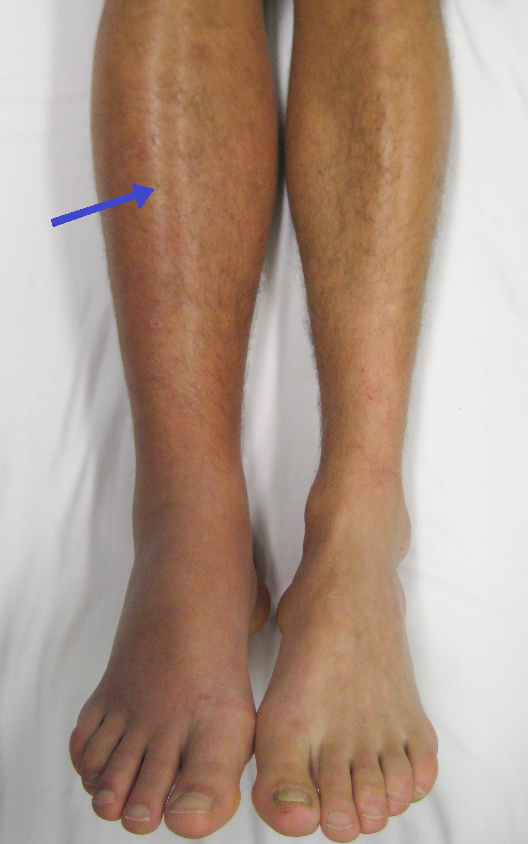
Pulmonary embolism (PE) is a blockage of an pulmonary artery, artery in the lungs by a substance that has moved from elsewhere in the body through the bloodstream (embolism). Symptoms of a PE may include dyspnea, shortness of breath, chest pain particularly upon breathing in, and coughing up blood. Symptoms of a deep vein thrombosis, blood clot in the leg may also be present, such as a erythema, red, warm, swollen, and painful leg. Signs of a PE include low blood oxygen saturation, oxygen levels, tachypnea, rapid breathing, tachycardia, rapid heart rate, and sometimes a mild fever. Severe cases can lead to Syncope (medicine), passing out, shock (circulatory), abnormally low blood pressure, obstructive shock, and cardiac arrest, sudden death. PE usually results from a blood clot in the leg that travels to the lung. The risk of blood clots is increased by advanced age, cancer, prolonged bed rest and immobilization, smoking, stroke, long-haul travel over 4 hours, certain genetics, g ... [...More Info...] [...Related Items...] OR: [Wikipedia] [Google] [Baidu] |
Bolus Tracking
Contrast CT, or contrast enhanced computed tomography (CECT), is X-ray computed tomography (CT) using radiocontrast. Radiocontrasts for X-ray CT are generally iodine-based types. This is useful to highlight structures such as blood vessels that otherwise would be difficult to delineate from their surroundings. Using contrast material can also help to obtain functional information about tissues. Often, images are taken both with and without radiocontrast. CT images are called ''precontrast'' or ''native-phase'' images before any radiocontrast has been administrated, and ''postcontrast'' after radiocontrast administration. Bolus tracking Bolus tracking is a technique to optimize timing of the imaging. A small bolus of radio-opaque contrast media is injected into a patient via a peripheral intravenous cannula. Depending on the vessel being imaged, the volume of contrast is tracked using a region of interest (abbreviated "R.O.I.") at a certain level and then followed by the CT ... [...More Info...] [...Related Items...] OR: [Wikipedia] [Google] [Baidu] |
Computed Tomography Angiography
Computed tomography angiography (also called CT angiography or CTA) is a computed tomography technique used for angiography—the visualization of arteries and veins—throughout the human body. Using contrast injected into the blood vessels, images are created to look for blockages, aneurysms (dilations of walls), dissections (tearing of walls), and stenosis (narrowing of vessel). CTA can be used to visualize the vessels of the heart, the aorta and other large blood vessels, the lungs, the kidneys, the head and neck, and the arms and legs. CTA can also be used to localise arterial or venous bleed of the gastrointestinal system. Medical uses CTA can be used to examine blood vessels in many key areas of the body including the brain, kidneys, pelvis, and the lungs. Coronary CT angiography Coronary CT angiography (CCTA) is the use of CT angiography to assess the arteries of the heart. The patient receives an intravenous injection of contrast and then the heart is scanned using a ... [...More Info...] [...Related Items...] OR: [Wikipedia] [Google] [Baidu] |
Ventilation/perfusion Scan
A ventilation/perfusion lung scan, also called a V/Q lung scan, or ventilation/perfusion scintigraphy, is a type of medical imaging using scintigraphy and medical isotopes to evaluate the circulation of air and blood within a patient's lungs, in order to determine the ventilation/perfusion ratio. The ventilation part of the test looks at the ability of air to reach all parts of the lungs, while the perfusion part evaluates how well blood circulates within the lungs. As Q in physiology is the letter used to describe bloodflow the term V/Q scan emerged. Uses This test is most commonly done in order to check for the presence of a blood clot or abnormal blood flow inside the lungs (such as a pulmonary embolism (PE) although computed tomography with radiocontrast is now more commonly used for this purpose. The V/Q scan may be used in some circumstances where radiocontrast would be inappropriate, as in Allergy to contrast agent or kidney failure. A V/Q lung scan may be performed in th ... [...More Info...] [...Related Items...] OR: [Wikipedia] [Google] [Baidu] |
D-dimer
D-dimer (or D dimer) is a fibrin degradation product (or FDP), a small protein fragment present in the blood after a blood clot is degraded by fibrinolysis. It is so named because it contains two D fragments of the fibrin protein joined by a cross-link, hence forming a protein dimer. D-dimer concentration may be determined by a blood test to help diagnose thrombosis. Since its introduction in the 1990s, it has become an important test performed in people with suspected thrombotic disorders, such as venous thromboembolism. While a negative result practically rules out thrombosis, a positive result can indicate thrombosis, but does not exclude other potential causes. Its main use, therefore, is to exclude thromboembolic disease where the probability is low. D-dimer levels are used as a predictive biomarker for the blood disorder, disseminated intravascular coagulation and in the coagulation disorders associated with COVID-19 infection. A four-fold increase in the protein is an ind ... [...More Info...] [...Related Items...] OR: [Wikipedia] [Google] [Baidu] |
Contrast Medium
A contrast agent (or contrast medium) is a substance used to increase the contrast of structures or fluids within the body in medical imaging. Contrast agents absorb or alter external electromagnetism or ultrasound, which is different from radiopharmaceuticals, which emit radiation themselves. In x-rays, contrast agents enhance the radiodensity in a target tissue or structure. In MRIs, contrast agents shorten (or in some instances increase) the relaxation times of nuclei within body tissues in order to alter the contrast in the image. Contrast agents are commonly used to improve the visibility of blood vessels and the gastrointestinal tract. Several types of contrast agent are in use in medical imaging and they can roughly be classified based on the imaging modalities where they are used. Most common contrast agents work based on X-ray attenuation and magnetic resonance signal enhancement. Radiocontrast media For radiography, which is based on X-rays, iodine and barium are the ... [...More Info...] [...Related Items...] OR: [Wikipedia] [Google] [Baidu] |
Kidney Failure
Kidney failure, also known as end-stage kidney disease, is a medical condition in which the kidneys can no longer adequately filter waste products from the blood, functioning at less than 15% of normal levels. Kidney failure is classified as either acute kidney failure, which develops rapidly and may resolve; and chronic kidney failure, which develops slowly and can often be irreversible. Symptoms may include leg swelling, feeling tired, vomiting, loss of appetite, and confusion. Complications of acute and chronic failure include uremia, high blood potassium, and volume overload. Complications of chronic failure also include heart disease, high blood pressure, and anemia. Causes of acute kidney failure include low blood pressure, blockage of the urinary tract, certain medications, muscle breakdown, and hemolytic uremic syndrome. Causes of chronic kidney failure include diabetes, high blood pressure, nephrotic syndrome, and polycystic kidney disease. Diagnosis of acute failure ... [...More Info...] [...Related Items...] OR: [Wikipedia] [Google] [Baidu] |
Multidetector Computed Tomography
A computed tomography scan (CT scan; formerly called computed axial tomography scan or CAT scan) is a medical imaging technique used to obtain detailed internal images of the body. The personnel that perform CT scans are called radiographers or radiology technologists. CT scanners use a rotating X-ray tube and a row of detectors placed in a gantry to measure X-ray attenuations by different tissues inside the body. The multiple X-ray measurements taken from different angles are then processed on a computer using tomographic reconstruction algorithms to produce tomographic (cross-sectional) images (virtual "slices") of a body. CT scans can be used in patients with metallic implants or pacemakers, for whom magnetic resonance imaging (MRI) is contraindicated. Since its development in the 1970s, CT scanning has proven to be a versatile imaging technique. While CT is most prominently used in medical diagnosis, it can also be used to form images of non-living objects. The 1979 Nob ... [...More Info...] [...Related Items...] OR: [Wikipedia] [Google] [Baidu] |
Intravenous Therapy
Intravenous therapy (abbreviated as IV therapy) is a medical technique that administers fluids, medications and nutrients directly into a person's vein. The intravenous route of administration is commonly used for rehydration or to provide nutrients for those who cannot, or will not—due to reduced mental states or otherwise—consume food or water by mouth. It may also be used to administer medications or other medical therapy such as blood products or electrolytes to correct electrolyte imbalances. Attempts at providing intravenous therapy have been recorded as early as the 1400s, but the practice did not become widespread until the 1900s after the development of techniques for safe, effective use. The intravenous route is the fastest way to deliver medications and fluid replacement throughout the body as they are introduced directly into the circulatory system and thus quickly distributed. For this reason, the intravenous route of administration is also used for the consump ... [...More Info...] [...Related Items...] OR: [Wikipedia] [Google] [Baidu] |
Contrast-induced Nephropathy
Contrast-induced nephropathy (CIN) is a purported form of kidney damage in which there has been recent exposure to medical imaging contrast material without another clear cause for the acute kidney injury. Despite extensive speculation, the actual occurrence of contrast-induced nephropathy has not been demonstrated in the literature. Analysis of observational studies has shown that radiocontrast use in CT scanning is not causally related to changes in kidney function. Terminology Given the increasing doubts about the contribution of radiocontrast to acute kidney injury, the American College of Radiology has proposed the name contrast-associated acute kidney injury (CA-AKI) (formerly referred to as post-contrast acute kidney injury; PC-AKI) does not imply a causal role, with the name contrast-induced acute kidney injury (CI-AKI) (formerly referred to as contrast-induced nephropathy; CIN) reserved for the rare cases where radiocontrast is likely to be causally related. Risk fact ... [...More Info...] [...Related Items...] OR: [Wikipedia] [Google] [Baidu] |
Iodinated Contrast
Iodinated contrast is a form of intravenous radiocontrast agent containing iodine, which enhances the visibility of vascular structures and organs during radiographic procedures. Some pathologies, such as cancer, have particularly improved visibility with iodinated contrast. The radiodensity of iodinated contrast is 25–30 Hounsfield units (HU) per milligram of iodine per milliliter at a tube voltage of 100–120 kVp. Types Iodine-based contrast media are usually classified as ionic or nonionic. Both types are used most commonly in radiology due to their relatively harmless interaction with the body and its solubility. Contrast media are primarily used to visualize vessels and changes in tissues on radiography and CT (computerized tomography). Contrast media can also be used for tests of the urinary tract, uterus and fallopian tubes. It may cause the patient to feel as if they have had urinary incontinence. It also puts a metallic taste in the mouth of the patient. The iodine ... [...More Info...] [...Related Items...] OR: [Wikipedia] [Google] [Baidu] |
Deep Vein Thrombosis
Deep vein thrombosis (DVT) is a type of venous thrombosis involving the formation of a blood clot in a deep vein, most commonly in the legs or pelvis. A minority of DVTs occur in the arms. Symptoms can include pain, swelling, redness, and enlarged veins in the affected area, but some DVTs have no symptoms. The most common life-threatening concern with DVT is the potential for a clot to embolize (detach from the veins), travel as an embolus through the right side of the heart, and become lodged in a pulmonary artery that supplies blood to the lungs. This is called a pulmonary embolism (PE). DVT and PE comprise the cardiovascular disease of venous thromboembolism (VTE). About two-thirds of VTE manifests as DVT only, with one-third manifesting as PE with or without DVT. The most frequent long-term DVT complication is post-thrombotic syndrome, which can cause pain, swelling, a sensation of heaviness, itching, and in severe cases, ulcers. Recurrent VTE occurs in about 30% of those i ... [...More Info...] [...Related Items...] OR: [Wikipedia] [Google] [Baidu] |


