|
C5a Receptor
The C5a receptor also known as complement component 5a receptor 1 (C5AR1) or CD88 (Cluster of Differentiation 88) is a G protein-coupled receptor for C5a. It functions as a complement receptor. C5a receptor modulates inflammatory responses, obesity, development and cancers. Cells The C5a receptor is expressed on: *Granulocytes *Monocytes *Dendritic cells *Hepatoma-derived cell line HepG2 *Astrocytes *Microglia Agonist and antagonists Potent and selective agonist and antagonists for C5aR have been developed. See also *Complement component 5a C5a is a protein fragment released from cleavage of complement component C5 by protease C5-convertase into C5a and C5b fragments. C5b is important in late events of the complement cascade, an orderly series of reactions which coordinates several ... for binding mechanism References Further reading * * * * * * * * * * * * * * * * * * * * External links * * * * {{Complement system ... [...More Info...] [...Related Items...] OR: [Wikipedia] [Google] [Baidu] |
G Protein-coupled Receptor
G protein-coupled receptors (GPCRs), also known as seven-(pass)-transmembrane domain receptors, 7TM receptors, heptahelical receptors, serpentine receptors, and G protein-linked receptors (GPLR), form a large group of evolutionarily-related proteins that are cell surface receptors that detect molecules outside the cell and activate cellular responses. Coupling with G proteins, they are called seven-transmembrane receptors because they pass through the cell membrane seven times. Text was copied from this source, which is available under Attribution 2.5 Generic (CC BY 2.5) license. Ligands can bind either to extracellular N-terminus and loops (e.g. glutamate receptors) or to the binding site within transmembrane helices (Rhodopsin-like family). They are all activated by agonists although a spontaneous auto-activation of an empty receptor can also be observed. G protein-coupled receptors are found only in eukaryotes, including yeast, choanoflagellates, and ... [...More Info...] [...Related Items...] OR: [Wikipedia] [Google] [Baidu] |
Complement Component 5a
C5a is a protein fragment released from cleavage of complement component C5 by protease C5-convertase into C5a and C5b fragments. C5b is important in late events of the complement cascade, an orderly series of reactions which coordinates several basic defense mechanisms, including formation of the membrane attack complex (MAC), one of the most basic weapons of the innate immune system, formed as an automatic response to intrusions from foreign particles and microbial invaders. It essentially pokes microscopic pinholes in these foreign objects, causing loss of water and sometimes death. C5a, the other cleavage product of C5, acts as a highly inflammatory peptide, encouraging complement activation, formation of the MAC, attraction of innate immune cells, and histamine release involved in allergic responses. The origin of C5 is in the hepatocyte, but its synthesis can also be found in macrophages, where it may cause local increase of C5a. C5a is a chemotactic agent and an anaphylatoxi ... [...More Info...] [...Related Items...] OR: [Wikipedia] [Google] [Baidu] |
Complement Receptor
A complement receptor is a membrane-bound receptor belonging to the complement system, which is part of the innate immune system. Complement receptors bind effector protein fragments that are produced in response to antigen-antibody complexes or damage-associated molecules. Complement receptor activation contributes to the regulation of inflammation, leukocyte extravasation, and phagocytosis; it also contributes to the adaptive immune response. Different complement receptors can participate in either the classical complement pathway, the alternative complement pathway, or both. Expression and function White blood cells, particularly monocytes and macrophages, express complement receptors on their surface. All four complement receptors can bind to fragments of complement component 3 or complement component 4 coated on pathogen surface, but the receptors trigger different downstream activities. Complement receptor (CR) 1, 3, and 4 function as opsonins which stimulate phagoc ... [...More Info...] [...Related Items...] OR: [Wikipedia] [Google] [Baidu] |
Granulocytes
Granulocytes are cells in the innate immune system characterized by the presence of specific granules in their cytoplasm. Such granules distinguish them from the various agranulocytes. All myeloblastic granulocytes are polymorphonuclear. They have varying shapes (morphology) of the nucleus (segmented, irregular; often lobed into three segments); and are referred to as polymorphonuclear leukocytes (PMN, PML, or PMNL). In common terms, ''polymorphonuclear granulocyte'' refers specifically to "neutrophil granulocytes", the most abundant of the granulocytes; the other types (eosinophils, basophils, and mast cells) have varying morphology. Granulocytes are produced via granulopoiesis in the bone marrow. Types There are four types of granulocytes (full name polymorphonuclear granulocytes): * Basophils * Eosinophils * Neutrophils * Mast cells Except for the mast cells, their names are derived from their staining characteristics; for example, the most abundant granulocyte is the neutrop ... [...More Info...] [...Related Items...] OR: [Wikipedia] [Google] [Baidu] |
Monocytes
Monocytes are a type of leukocyte or white blood cell. They are the largest type of leukocyte in blood and can differentiate into macrophages and conventional dendritic cells. As a part of the vertebrate innate immune system monocytes also influence adaptive immune responses and exert tissue repair functions. There are at least three subclasses of monocytes in human blood based on their phenotypic receptors. Structure Monocytes are amoeboid in appearance, and have nongranulated cytoplasm. Thus they are classified as agranulocytes, although they might occasionally display some azurophil granules and/or vacuoles. With a diameter of 15–22 μm, monocytes are the largest cell type in peripheral blood. Monocytes are mononuclear cells and the ellipsoidal nucleus is often lobulated/indented, causing a bean-shaped or kidney-shaped appearance. Monocytes compose 2% to 10% of all leukocytes in the human body. Development Monocytes are produced by the bone marrow from precursors cal ... [...More Info...] [...Related Items...] OR: [Wikipedia] [Google] [Baidu] |
Dendritic Cells
Dendritic cells (DCs) are antigen-presenting cells (also known as ''accessory cells'') of the mammalian immune system. Their main function is to process antigen material and present it on the cell surface to the T cells of the immune system. They act as messengers between the innate and the adaptive immune systems. Dendritic cells are present in those tissues that are in contact with the external environment, such as the skin (where there is a specialized dendritic cell type called the Langerhans cell) and the inner lining of the nose, lungs, stomach and intestines. They can also be found in an immature state in the blood. Once activated, they migrate to the lymph nodes where they interact with T cells and B cells to initiate and shape the adaptive immune response. At certain development stages they grow branched projections, the ''dendrites'' that give the cell its name (δένδρον or déndron being Greek for 'tree'). While similar in appearance, these are structures disti ... [...More Info...] [...Related Items...] OR: [Wikipedia] [Google] [Baidu] |
HepG2
Hep G2 (or HepG2) is a human liver cancer cell line. Hep G2 is an immortal cell line which was derived in 1975 from the liver tissue of a 15-year-old Caucasian male from Argentina with a well-differentiated hepatocellular carcinoma. These cells are epithelial in morphology, have a modal chromosome number of 55, and are not tumorigenic in nude mice. The cells secrete a variety of major plasma proteins, e.g., albumin, and the acute-phase proteins fibrinogen, alpha 2-macroglobulin, alpha 1-antitrypsin, transferrin and plasminogen. They have been grown successfully in large-scale cultivation systems. Hepatitis B virus surface antigens have not been detected. Hep G2 will respond to stimulation with human growth hormone. Hep G2 cells are a suitable ''in vitro'' model system for the study of polarized human hepatocytes. Another well-characterized polarized hepatocyte cell line is the rat hepatoma-derived hybrid cell line WIF-B. With the proper culture conditions, He ... [...More Info...] [...Related Items...] OR: [Wikipedia] [Google] [Baidu] |
Astrocytes
Astrocytes (from Ancient Greek , , "star" + , , "cavity", "cell"), also known collectively as astroglia, are characteristic star-shaped glial cells in the brain and spinal cord. They perform many functions, including biochemical control of endothelial cells that form the blood–brain barrier, provision of nutrients to the nervous tissue, maintenance of extracellular ion balance, regulation of cerebral blood flow, and a role in the repair and scarring process of the brain and spinal cord following infection and traumatic injuries. The proportion of astrocytes in the brain is not well defined; depending on the counting technique used, studies have found that the astrocyte proportion varies by region and ranges from 20% to 40% of all glia. Another study reports that astrocytes are the most numerous cell type in the brain. Astrocytes are the major source of cholesterol in the central nervous system. Apolipoprotein E transports cholesterol from astrocytes to neurons and other glial ... [...More Info...] [...Related Items...] OR: [Wikipedia] [Google] [Baidu] |
Microglia
Microglia are a type of neuroglia (glial cell) located throughout the brain and spinal cord. Microglia account for about 7% of cells found within the brain. As the resident macrophage cells, they act as the first and main form of active immune defense in the central nervous system (CNS). Microglia (and other neuroglia including astrocytes) are distributed in large non-overlapping regions throughout the CNS. Microglia are key cells in overall brain maintenance—they are constantly scavenging the CNS for plaques, damaged or unnecessary neurons and synapses, and infectious agents. Since these processes must be efficient to prevent potentially fatal damage, microglia are extremely sensitive to even small pathological changes in the CNS. This sensitivity is achieved in part by the presence of unique potassium channels that respond to even small changes in extracellular potassium. Recent evidence shows that microglia are also key players in the sustainment of normal brain functions und ... [...More Info...] [...Related Items...] OR: [Wikipedia] [Google] [Baidu] |
Agonist
An agonist is a chemical that activates a receptor to produce a biological response. Receptors are cellular proteins whose activation causes the cell to modify what it is currently doing. In contrast, an antagonist blocks the action of the agonist, while an inverse agonist causes an action opposite to that of the agonist. Etymology From the Greek αγωνιστής (agōnistēs), contestant; champion; rival < αγων (agōn), contest, combat; exertion, struggle < αγω (agō), I lead, lead towards, conduct; drive Types of agonists can be activated by either endogenous agonists (such as |
Receptor Antagonist
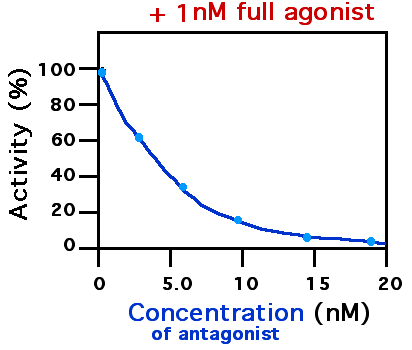
A receptor antagonist is a type of receptor ligand or drug that blocks or dampens a biological response by binding to and blocking a receptor rather than activating it like an agonist. Antagonist drugs interfere in the natural operation of receptor proteins.Pharmacology Guide: In vitro pharmacology: concentration-response curves " '' GlaxoWellcome.'' Retrieved on December 6, 2007. They are sometimes called blockers; examples include alpha blockers, |

.png)


