|
Uterus
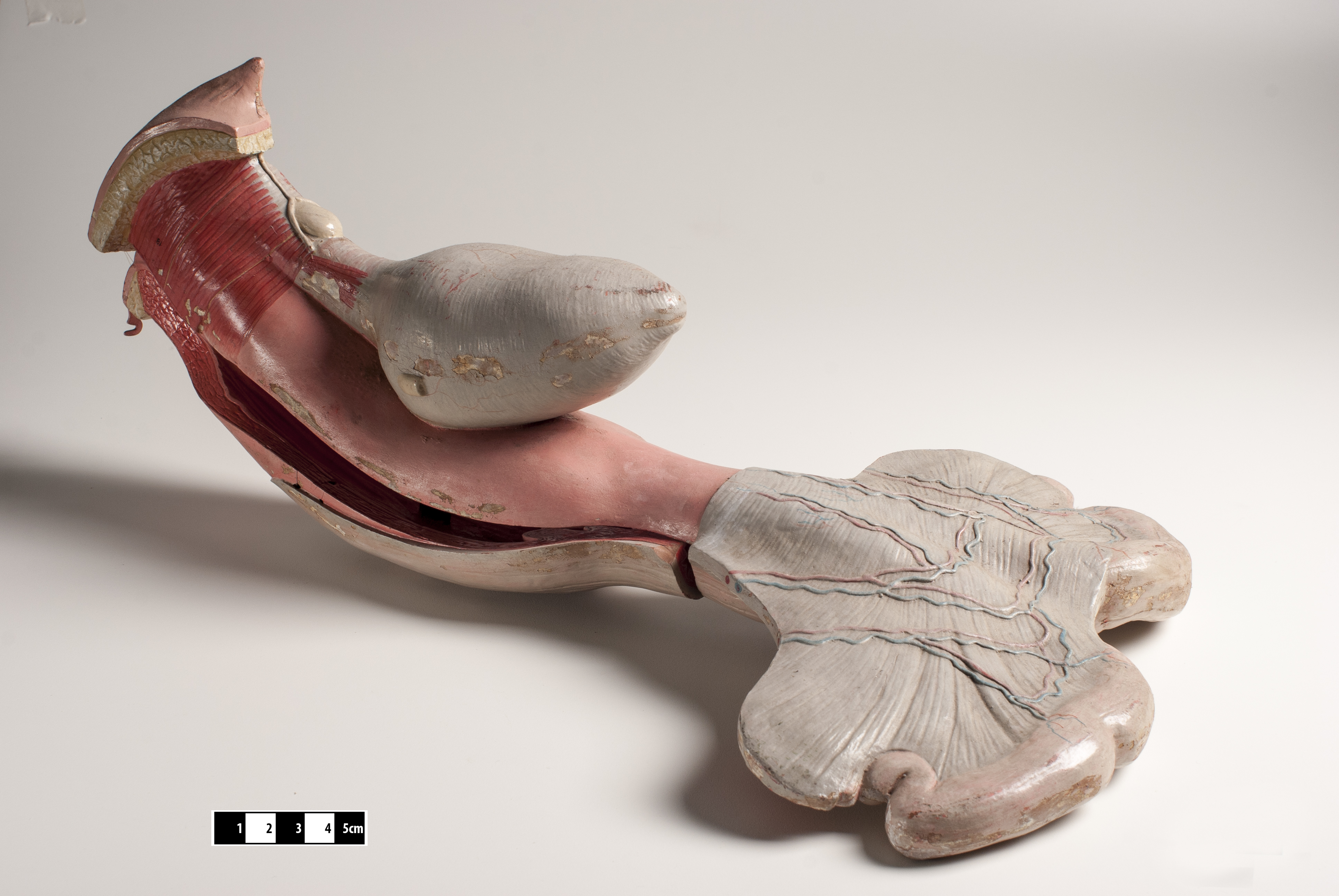
The uterus (from Latin ''uterus'', : uteri or uteruses) or womb () is the hollow organ, organ in the reproductive system of most female mammals, including humans, that accommodates the embryonic development, embryonic and prenatal development, fetal development of one or more Fertilized egg, fertilized eggs until birth. The uterus is a hormone-responsive sex organ that contains uterine gland, glands in its endometrium, lining that secrete uterine milk for embryonic nourishment. (The term ''uterus'' is also applied to analogous structures in some non-mammalian animals.) In humans, the lower end of the uterus is a narrow part known as the Uterine isthmus, isthmus that connects to the cervix, the anterior gateway leading to the vagina. The upper end, the body of the uterus, is connected to the fallopian tubes at the uterine horns; the rounded part, the fundus, is above the openings to the fallopian tubes. The connection of the uterine cavity with a fallopian tube is called the utero ... [...More Info...] [...Related Items...] OR: [Wikipedia] [Google] [Baidu] |
Cervix
The cervix (: cervices) or cervix uteri is a dynamic fibromuscular sexual organ of the female reproductive system that connects the vagina with the uterine cavity. The human female cervix has been documented anatomically since at least the time of Hippocrates, over 2,000 years ago. The cervix is approximately 4 cm long with a diameter of approximately 3 cm and tends to be described as a cylindrical shape, although the front and back walls of the cervix are contiguous. The size of the cervix changes throughout a woman's life cycle. For example, women in the fertile years of their reproductive cycle tend to have larger cervixes than postmenopausal women; likewise, women who have produced offspring have a larger cervix than those who have not. In relation to the vagina, the part of the cervix that opens to the uterus is called the ''internal os'' and the opening of the cervix in the vagina is called the ''external os''. Between them is a conduit commonly called the cervic ... [...More Info...] [...Related Items...] OR: [Wikipedia] [Google] [Baidu] |
Implantation (embryology)
Implantation, also known as nidation, is the stage in the mammalian embryonic development in which the blastocyst hatches, attaches, adheres, and penetrates into the endometrium of the female's uterus. Implantation is the first stage of gestation, and, when successful, the female is considered to be pregnant. An implanted embryo is detected by the presence of increased levels of human chorionic gonadotropin (hCG) in a pregnancy test. The implanted embryo will receive oxygen and nutrients in order to grow. For implantation to take place the uterus must become receptive. Uterine receptivity involves much cross-talk between the embryo and the uterus, initiating changes to the endometrium. This stage gives a synchrony that opens a window of implantation that enables successful implantation of a viable embryo. The endocannabinoid system plays a vital role in this synchrony in the uterus, influencing uterine receptivity, and embryo implantation. The embryo expresses cannabinoid r ... [...More Info...] [...Related Items...] OR: [Wikipedia] [Google] [Baidu] |
Fallopian Tubes
The fallopian tubes, also known as uterine tubes, oviducts or salpinges (: salpinx), are paired tubular sex organs in the human female body that stretch from the ovaries to the uterus. The fallopian tubes are part of the female reproductive system. In other vertebrates, they are only called oviducts. Each tube is a muscular hollow organ that is on average between in length, with an external diameter of . It has four described parts: the intramural part, isthmus, ampulla, and infundibulum with associated fimbriae. Each tube has two openings: a proximal opening nearest to the uterus, and a distal opening nearest to the ovary. The fallopian tubes are held in place by the mesosalpinx, a part of the broad ligament mesentery that wraps around the tubes. Another part of the broad ligament, the mesovarium suspends the ovaries in place. An egg cell is transported from an ovary to a fallopian tube where it may be fertilized in the ampulla of the tube. The fallopian tubes are line ... [...More Info...] [...Related Items...] OR: [Wikipedia] [Google] [Baidu] |
Endometrium
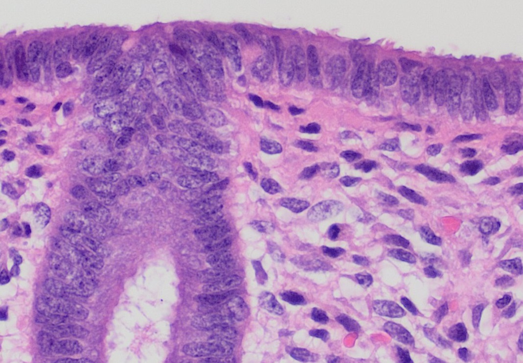
The endometrium is the inner epithelium, epithelial layer, along with its mucous membrane, of the mammalian uterus. It has a basal layer and a functional layer: the basal layer contains stem cells which regenerate the functional layer. The functional layer thickens and then is shed during menstruation in humans and some other mammals, including other apes, Old World monkeys, some species of bat, the elephant shrew and the Cairo spiny mouse. In most other mammals, the endometrium is reabsorbed in the estrous cycle. During pregnancy, the glands and blood vessels in the endometrium further increase in size and number. Vascular spaces fuse and become interconnected, forming the placenta, which supplies oxygen and nutrition to the embryo and fetus.Blue Histology - Female Reproductive System ... [...More Info...] [...Related Items...] OR: [Wikipedia] [Google] [Baidu] |
Uterine Artery
The uterine artery is an artery that supplies blood to the uterus in females. Structure The uterine artery usually arises from the anterior division of the internal iliac artery. It travels to the uterus, crossing the ureter anteriorly, to the uterus by traveling in the cardinal ligament. It travels through the parametrium of the inferior broad ligament of the uterus. It commonly anastomoses (connects with) the ovarian artery. The uterine artery is the major blood supply to the uterus and enlarges significantly during pregnancy. Branches and organs supplied * round ligament of the uterus * ovary ("ovarian branches") * uterus ( arcuate vessels) * vagina ( Vaginal branches of uterine artery) * uterine tube ("tubal branch") Anatomical variants Uterine artery can arise from the first branch of inferior gluteal artery. It can also arise as the 2nd or 3rd branch from the inferior gluteal artery. On the other hand, uterine artery can be first branch from internal iliac artery bef ... [...More Info...] [...Related Items...] OR: [Wikipedia] [Google] [Baidu] |
Reproductive System
The reproductive system of an organism, also known as the genital system, is the biological system made up of all the anatomical organs involved in sexual reproduction. Many non-living substances such as fluids, hormones, and pheromones are also important accessories to the reproductive system. Unlike most organ systems, the sexes of differentiated species often have significant differences. These differences allow for a combination of genetic material between two individuals, which allows for the possibility of greater genetic fitness of the offspring.Reproductive System 2001 Body Guide powered by Adam Animals In mammals, the major organs of the reproductive system include the external gen ...[...More Info...] [...Related Items...] OR: [Wikipedia] [Google] [Baidu] |
Vagina
In mammals and other animals, the vagina (: vaginas or vaginae) is the elastic, muscular sex organ, reproductive organ of the female genital tract. In humans, it extends from the vulval vestibule to the cervix (neck of the uterus). The #Vaginal opening and hymen, vaginal introitus is normally partly covered by a thin layer of mucous membrane, mucosal tissue called the hymen. The vagina allows for Copulation (zoology), copulation and birth. It also channels Menstruation (mammal), menstrual flow, which occurs in humans and closely related primates as part of the menstrual cycle. To accommodate smoother penetration of the vagina during sexual intercourse or other sexual activity, vaginal moisture increases during sexual arousal in human females and other female mammals. This increase in moisture provides vaginal lubrication, which reduces friction. The texture of the vaginal walls creates friction for the penis during sexual intercourse and stimulates it toward ejaculation, en ... [...More Info...] [...Related Items...] OR: [Wikipedia] [Google] [Baidu] |
Reproductive System
The reproductive system of an organism, also known as the genital system, is the biological system made up of all the anatomical organs involved in sexual reproduction. Many non-living substances such as fluids, hormones, and pheromones are also important accessories to the reproductive system. Unlike most organ systems, the sexes of differentiated species often have significant differences. These differences allow for a combination of genetic material between two individuals, which allows for the possibility of greater genetic fitness of the offspring.Reproductive System 2001 Body Guide powered by Adam Animals In mammals, the major organs of the reproductive system include the external gen ...[...More Info...] [...Related Items...] OR: [Wikipedia] [Google] [Baidu] |
Paramesonephric Duct
The paramesonephric ducts (or Müllerian ducts) are paired ducts of the embryo in the reproductive system of humans and other mammals that run down the lateral sides of the genital ridge and terminate at the sinus tubercle in the primitive urogenital sinus. They form in both sexes during 6th week of fetal development. In the female, go on to form the fallopian tubes/oviducts, uterus, cervix, and the upper one-third of the vagina. In males fetuses, they are normally made to regress by anti-Müllerian hormone which begins to be secreted by the testes during 8th week of fetal development. Each maramesonephric duct is situated just lateral to the mesonephric ducts, mesonephric ducts (Wolffian duct) of the same side. Development The female reproductive system is composed of two embryological segments: the urogenital sinus and the paramesonephric ducts. The two are conjoined at the sinus tubercle. Paramesonephric ducts are present on the embryo of both sexes. Only in females do they d ... [...More Info...] [...Related Items...] OR: [Wikipedia] [Google] [Baidu] |
Uterine Horns
The uterine horns (cornua of uterus) are the points in the upper uterus where the fallopian tubes or oviducts exit to meet the ovaries. They are one of the points of attachment for the round ligament of uterus (the other being the mons pubis). They also provide attachment to the ovarian ligament, which is located below the fallopian tube at the back, while the round ligament of uterus is located below the tube at the front. The uterine horns are far more prominent in other animals (such as cows and cats) than they are in humans. In the cat, implantation of the embryo occurs in one of the two uterine horns, not the body of the uterus itself. Occasionally, if a fallopian tube does not connect, the uterine horn will fill with blood each month, and a minor one-day surgery will be performed to remove it. Often, people who are born with this have trouble getting pregnant as both ovaries are functional and either may ovulate. The spare egg An egg is an organic vessel grown by ... [...More Info...] [...Related Items...] OR: [Wikipedia] [Google] [Baidu] |
Uterine Milk
Uterine glands or endometrial glands are tubular glands, lined by a simple columnar epithelium, found in the functional layer of the endometrium that lines the uterus. Their appearance varies during the menstrual cycle. During the proliferative phase, uterine glands appear long due to estrogen secretion by the ovaries. During the secretory phase, the uterine glands become very coiled with wide lumens and produce a glycogen-rich secretion known as ''histotroph'' or ''uterine milk''. This change corresponds with an increase in blood flow to spiral arteries due to increased progesterone secretion from the corpus luteum. During the pre-menstrual phase, progesterone secretion decreases as the corpus luteum degenerates, which results in decreased blood flow to the spiral arteries. The functional layer of the uterus containing the glands becomes necrotic, and eventually sloughs off during the menstrual phase of the cycle. They are of small size in the unimpregnated uterus, but shortly af ... [...More Info...] [...Related Items...] OR: [Wikipedia] [Google] [Baidu] |
Uterine Veins
The uterine vein is a vein of the uterus. It is found in the cardinal ligament. It drains into the internal iliac vein. It follows a similar course to the uterine artery. It helps to drain blood from the uterus, and removes waste from blood in the placenta during pregnancy. Structure The uterine vein is found in the cardinal ligament of the uterus. It travels through the broad ligament of the uterus to the lateral abdominal wall. It drains into the internal iliac vein. The uterine vein forms a venous plexus around the cervix. It follows a similar course to the uterine artery. Lymphatic vessels are associated with it. It also anastomoses with the ovarian vein. It may anastomose with the vaginal venous plexus. Function The uterine vein helps to drain blood from the uterus. This is also important for the removal of waste from blood in the placenta during pregnancy. Clinical significance Placenta measurement Measurements of the partial pressure of O2 in the uterine vein ... [...More Info...] [...Related Items...] OR: [Wikipedia] [Google] [Baidu] |


