|
Toes
Toes are the digits of the foot of a tetrapod. Animal species such as cats that walk on their toes are described as being ''digitigrade''. Humans, and other animals that walk on the soles of their feet, are described as being ''plantigrade''; ''unguligrade'' animals are those that walk on hooves at the tips of their toes. Structure There are normally five toes present on each human foot. Each toe consists of three phalanx bones, the proximal, middle, and distal, with the exception of the big toe (). For a minority of people, the little toe also is missing a middle bone. The hallux only contains two phalanx bones, the proximal and distal. The joints between each phalanx are the interphalangeal joints. The proximal phalanx bone of each toe articulates with the metatarsal bone of the foot at the metatarsophalangeal joint. Each toe is surrounded by skin, and present on all five toes is a toenail. The toes are, from medial to lateral: * the first toe, also known as the ha ... [...More Info...] [...Related Items...] OR: [Wikipedia] [Google] [Baidu] |
Digit (anatomy)
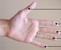
A digit is one of several most distal parts of a limb, such as fingers or toes, present in many vertebrates. Names Some languages have different names for hand and foot digits (English: respectively "finger" and " toe", German: "Finger" and "Zeh", French: "doigt" and "orteil"). In other languages, e.g. Arabic, Russian, Polish, Spanish, Portuguese, Italian, Czech, Tagalog, Turkish, Bulgarian, and Persian, there are no specific one-word names for fingers and toes; these are called "digit of the hand" or "digit of the foot" instead. In Japanese, yubi (指) can mean either, depending on context. Human digits Humans normally have five digits on each extremity. Each digit is formed by several bones called phalanges, surrounded by soft tissue. Human fingers normally have a nail at the distal phalanx. The phenomenon of polydactyly occurs when extra digits are present; fewer digits than normal are also possible, for instance in ectrodactyly. Whether such a mutation can ... [...More Info...] [...Related Items...] OR: [Wikipedia] [Google] [Baidu] |
Metatarsophalangeal Joints
The metatarsophalangeal joints (MTP joints) are the joints between the metatarsal bones of the foot and the proximal bones (proximal phalanges) of the toes. They are analogous to the knuckles of the hand, and are consequently known as toe knuckles in common speech. They are condyloid joints, meaning that an elliptical or rounded surface (of the metatarsal bones) comes close to a shallow cavity (of the proximal phalanges). The region of skin directly below the joints forms the ball of the foot. The ligaments are the plantar and two collateral. Movements The movements permitted in the metatarsophalangeal joints are flexion, extension, abduction, adduction and circumduction. File:The feet of C. H. Unthan, the armless fiddler Wellcome L0034227.jpg, Left: toes adducted (pulled towards the center) and spread (abducted); right, both feet clenched (plantar flexed) File:Footgym rings1.jpg, The upper foot is clenching (plantarflexing) at the MTP joints and at the joints of the to ... [...More Info...] [...Related Items...] OR: [Wikipedia] [Google] [Baidu] |
Toenail
A nail is a protective plate characteristically found at the tip of the digits (fingers and toes) of all primates, corresponding to the claws in other tetrapod animals. Fingernails and toenails are made of a tough rigid protein called alpha-keratin, a polymer also found in the claws, hooves, and horns of vertebrates. Structure The nail consists of the nail plate, the nail matrix and the nail bed below it, and the grooves surrounding it. Parts of the nail The nail matrix is the active tissue (or germinal matrix) that generates cells. The cells harden as they move outward from the nail root to the nail plate. The nail matrix is also known as the ''matrix unguis'', keratogenous membrane, or onychostroma. It is the part of the nail bed that is beneath the nail and contains nerves, lymph, and blood vessels. The matrix produces cells that become the nail plate. The width and thickness of the nail plate is determined by the size, length, and thickness of the matrix, while ... [...More Info...] [...Related Items...] OR: [Wikipedia] [Google] [Baidu] |
Metti (cropped)
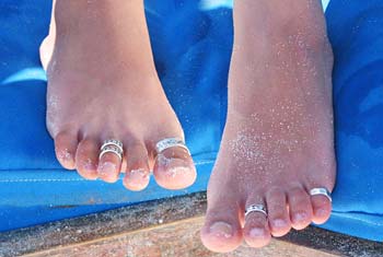
A toe ring is a ring made out of metals and non-metals worn on any of the toes. The second toe of either foot is where they are worn most commonly. This is because proportionately it is the longest toe and thus the easiest toe to put a ring on and stay without being connected to anything else. In most western countries they are a relatively new fashion accessory, and typically have no symbolic meaning. They are usually worn with barefoot sandals, anklets, bare feet or flip flops. Like finger rings, toe rings come in many shapes and forms, from intricately designed flowers embedded with jewels to simple bands. Fitted toe rings are rings that are of one size, whereas adjustable toe rings have a gap at the bottom so they can be easily made to fit snugly. Toe rings in India The wearing of toe rings has been practised in India since ancient times. In the Ramayana, there is a mention of Sita, on being abducted by Ravana, throwing her toe ring down so that lord Rama could find ... [...More Info...] [...Related Items...] OR: [Wikipedia] [Google] [Baidu] |
Medial (anatomy)
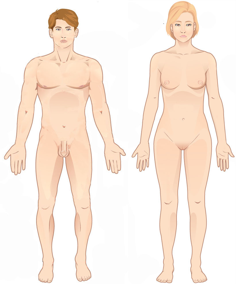
Standard anatomical terms of location are used to describe unambiguously the anatomy of humans and other animals. The terms, typically derived from Latin or Greek roots, describe something in its standard anatomical position. This position provides a definition of what is at the front ("anterior"), behind ("posterior") and so on. As part of defining and describing terms, the body is described through the use of anatomical planes and axes. The meaning of terms that are used can change depending on whether a vertebrate is a biped or a quadruped, due to the difference in the neuraxis, or if an invertebrate is a non-bilaterian. A non-bilaterian has no anterior or posterior surface for example but can still have a descriptor used such as proximal or distal in relation to a body part that is nearest to, or furthest from its middle. International organisations have determined vocabularies that are often used as standards for subdisciplines of anatomy. For example, '' Terminolog ... [...More Info...] [...Related Items...] OR: [Wikipedia] [Google] [Baidu] |
Anatomical Terms Of Movement
Motion, the process of movement, is described using specific anatomical terms. Motion includes movement of organs, joints, limbs, and specific sections of the body. The terminology used describes this motion according to its direction relative to the anatomical position of the body parts involved. Anatomists and others use a unified set of terms to describe most of the movements, although other, more specialized terms are necessary for describing unique movements such as those of the hands, feet, and eyes. In general, motion is classified according to the anatomical plane it occurs in. ''Flexion'' and ''extension'' are examples of ''angular'' motions, in which two axes of a joint are brought closer together or moved further apart. ''Rotational'' motion may occur at other joints, for example the shoulder, and are described as ''internal'' or ''external''. Other terms, such as ''elevation'' and ''depression'', describe movement above or below the horizontal plane. Many anatomical ... [...More Info...] [...Related Items...] OR: [Wikipedia] [Google] [Baidu] |
Tendon
A tendon or sinew is a tough band of fibrous connective tissue, dense fibrous connective tissue that connects skeletal muscle, muscle to bone. It sends the mechanical forces of muscle contraction to the skeletal system, while withstanding tension (physics), tension. Tendons, like ligaments, are made of collagen. The difference is that ligaments connect bone to bone, while tendons connect muscle to bone. There are about 4,000 tendons in the adult human body. Structure A tendon is made of dense regular connective tissue, whose main cellular components are special fibroblasts called tendon cells (tenocytes). Tendon cells synthesize the tendon's extracellular matrix, which abounds with densely-packed collagen fibers. The collagen fibers run parallel to each other and are grouped into fascicles. Each fascicle is bound by an endotendineum, which is a delicate loose connective tissue containing thin collagen fibrils and elastic fibers. A set of fascicles is bound by an epitenon, whi ... [...More Info...] [...Related Items...] OR: [Wikipedia] [Google] [Baidu] |
Flexor Digitorum Brevis
The flexor digitorum brevis or flexor digitorum communis brevishttps://www.anatomyatlases.org/AnatomicVariants/MuscularSystem/Text/F/14Flexor.shtml is a muscle which lies in the middle of the sole of the foot, immediately above the central part of the plantar aponeurosis, with which it is firmly united. Its deep surface is separated from the lateral plantar vessels and nerves by a thin layer of fascia. Structure It arises by a narrow tendon, from the medial process of the tuberosity of the calcaneus, from the central part of the plantar aponeurosis, and from the intermuscular septa between it and the adjacent muscles. It passes forward, and divides into four tendons, one for each of the four lesser toes. Opposite the bases of the first phalanges, each tendon divides into two slips, to allow of the passage of the corresponding tendon of the flexor digitorum longus; the two portions of the tendon then unite and form a grooved channel for the reception of the accompanying long F ... [...More Info...] [...Related Items...] OR: [Wikipedia] [Google] [Baidu] |
Phalanges Of The Foot
The phalanges (: phalanx ) are digit (anatomy), digital bones in the hands and foot, feet of most vertebrates. In primates, the Thumb, thumbs and Hallux, big toes have two phalanges while the other Digit (anatomy), digits have three phalanges. The phalanges are classed as long bones. Structure The phalanges are the bones that make up the fingers of the hand and the toes of the foot. There are 56 phalanges in the human body, with fourteen on each hand and foot. Three phalanges are present on each finger and toe, with the exception of the thumb and hallux, big toe, which possess only two. The middle and far phalanges of the fifth toes are often fused together (symphalangism). The phalanges of the hand are commonly known as the finger bones. The phalanges of the foot differ from the hand in that they are often shorter and more compressed, especially in the proximal phalanges, those closest to the torso. A phalanx is named according to whether it is Anatomical terms of locatio ... [...More Info...] [...Related Items...] OR: [Wikipedia] [Google] [Baidu] |
Extensor Digitorum Brevis
The extensor digitorum brevis muscle (sometimes EDB) is a muscle on the upper surface of the foot that helps extend digits 2 through 4. Structure The muscle originates from the forepart of the upper and lateral surface of the calcaneus (in front of the groove for the peroneus brevis tendon), from the interosseous talocalcaneal ligament and the stem of the inferior extensor retinaculum. The fibres pass obliquely forwards and medially across the dorsum of the foot and end in four tendons. The medial part of the muscle, also known as extensor hallucis brevis, ends in a tendon which crosses the dorsalis pedis artery and inserts into the dorsal surface of the base of the proximal phalanx of the great toe. The other three tendons insert into the lateral sides of the tendons of extensor digitorum longus for the second, third and fourth toes. Nerve supply Nerve supply: lateral terminal branch of Deep Peroneal Nerve (deep fibular nerve) (proximal sciatic branches L4-L5, but most clinica ... [...More Info...] [...Related Items...] OR: [Wikipedia] [Google] [Baidu] |
Flexor Hallucis Longus Muscle
The flexor hallucis longus muscle (FHL) attaches to the plantar surface of phalanx of the great toe and is responsible for flexing that toe. The FHL is one of the three deep muscles of the posterior compartment of the leg, the others being the flexor digitorum longus and the tibialis posterior. The tibialis posterior is the most powerful of these deep muscles. All three muscles are innervated by the tibial nerve which comprises half of the sciatic nerve. Structure The flexor hallucis longus is situated on the fibular side of the leg. It arises from the inferior two-thirds of the posterior surface of the body of the fibula, with the exception of 2.5 cm at its lowest part; from the lower part of the interosseous membrane; from an intermuscular septum between it and the peroneus muscles, laterally, and from the fascia covering the tibialis posterior, medially. The fibers pass obliquely downward and backward, where it passes through the tarsal tunnel on the medial side ... [...More Info...] [...Related Items...] OR: [Wikipedia] [Google] [Baidu] |
Posterior (anatomy)
Standard anatomical terms of location are used to describe unambiguously the anatomy of humans and other animals. The terms, typically derived from Latin or Greek language, Greek roots, describe something in its standard anatomical position. This position provides a definition of what is at the front ("anterior"), behind ("posterior") and so on. As part of defining and describing terms, the body is described through the use of anatomical planes and anatomical axes, axes. The meaning of terms that are used can change depending on whether a vertebrate is a biped or a quadruped, due to the difference in the neuraxis, or if an invertebrate is a non-bilaterian. A non-bilaterian has no anterior or posterior surface for example but can still have a descriptor used such as proximal or distal in relation to a body part that is nearest to, or furthest from its middle. International organisations have determined vocabularies that are often used as standards for subdisciplines of anatomy. ... [...More Info...] [...Related Items...] OR: [Wikipedia] [Google] [Baidu] |


