|
Cortical Magnification
In neuroscience, cortical magnification describes how many neurons in an area of the visual cortex are 'responsible' for processing a stimulus of a given size, as a function of visual field location. In the center of the visual field, corresponding to the center of the fovea of the retina, a very large number of neurons process information from a small region of the visual field. If the same stimulus is seen in the periphery of the visual field (i.e. away from the center), it would be processed by a much smaller number of neurons. The reduction of the number of neurons per visual field area from foveal to peripheral representations is achieved in several steps along the visual pathway, starting already in the retina. For quantitative purposes, the cortical magnification factor is normally expressed in millimeters of cortical surface per degree of visual angle. When expressed in this way, the values of cortical magnification factor vary by a factor of approximately 30 – 90 ... [...More Info...] [...Related Items...] OR: [Wikipedia] [Google] [Baidu] |
Neuroscience
Neuroscience is the scientific study of the nervous system (the brain, spinal cord, and peripheral nervous system), its functions, and its disorders. It is a multidisciplinary science that combines physiology, anatomy, molecular biology, developmental biology, cytology, psychology, physics, computer science, chemistry, medicine, statistics, and mathematical modeling to understand the fundamental and emergent properties of neurons, glia and neural circuits. The understanding of the biological basis of learning, memory, behavior, perception, and consciousness has been described by Eric Kandel as the "epic challenge" of the biological sciences. The scope of neuroscience has broadened over time to include different approaches used to study the nervous system at different scales. The techniques used by neuroscientists have expanded enormously, from molecular and cellular studies of individual neurons to imaging of sensory, motor and cognitive tasks in the brain. Hist ... [...More Info...] [...Related Items...] OR: [Wikipedia] [Google] [Baidu] |
Neurons
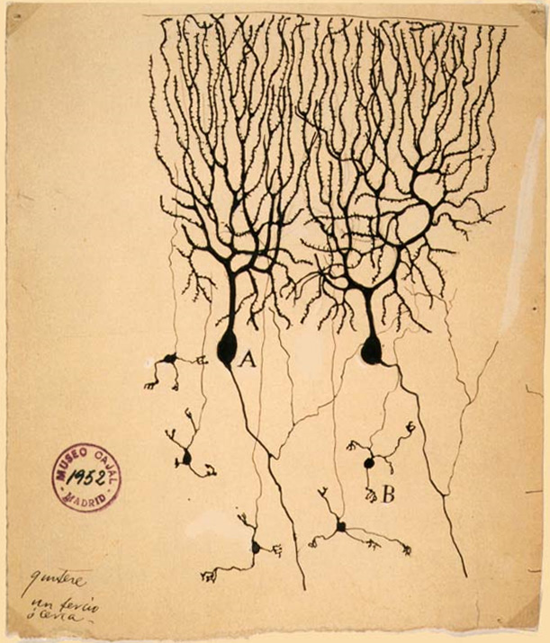
A neuron (American English), neurone (British English), or nerve cell, is an membrane potential#Cell excitability, excitable cell (biology), cell that fires electric signals called action potentials across a neural network (biology), neural network in the nervous system. They are located in the nervous system and help to receive and conduct impulses. Neurons communicate with other cells via synapses, which are specialized connections that commonly use minute amounts of chemical neurotransmitters to pass the electric signal from the presynaptic neuron to the target cell through the synaptic gap. Neurons are the main components of nervous tissue in all Animalia, animals except sponges and placozoans. Plants and fungi do not have nerve cells. Molecular evidence suggests that the ability to generate electric signals first appeared in evolution some 700 to 800 million years ago, during the Tonian period. Predecessors of neurons were the peptidergic secretory cells. They eventually ga ... [...More Info...] [...Related Items...] OR: [Wikipedia] [Google] [Baidu] |
Cortical Area
The cerebral cortex, also known as the cerebral mantle, is the outer layer of neural tissue of the cerebrum of the brain in humans and other mammals. It is the largest site of Neuron, neural integration in the central nervous system, and plays a key role in attention, perception, awareness, thought, memory, language, and consciousness. The six-layered neocortex makes up approximately 90% of the Cortex (anatomy), cortex, with the allocortex making up the remainder. The cortex is divided into left and right parts by the longitudinal fissure, which separates the two cerebral hemispheres that are joined beneath the cortex by the corpus callosum and other commissural fibers. In most mammals, apart from small mammals that have small brains, the cerebral cortex is folded, providing a greater surface area in the confined volume of the neurocranium, cranium. Apart from minimising brain and cranial volume, gyrification, cortical folding is crucial for the Neural circuit, brain circuitry ... [...More Info...] [...Related Items...] OR: [Wikipedia] [Google] [Baidu] |
Visual Cortex
The visual cortex of the brain is the area of the cerebral cortex that processes visual information. It is located in the occipital lobe. Sensory input originating from the eyes travels through the lateral geniculate nucleus in the thalamus and then reaches the visual cortex. The area of the visual cortex that receives the sensory input from the lateral geniculate nucleus is the primary visual cortex, also known as visual area 1 ( V1), Brodmann area 17, or the striate cortex. The extrastriate areas consist of visual areas 2, 3, 4, and 5 (also known as V2, V3, V4, and V5, or Brodmann area 18 and all Brodmann area 19). Both hemispheres of the brain include a visual cortex; the visual cortex in the left hemisphere receives signals from the right visual field, and the visual cortex in the right hemisphere receives signals from the left visual field. Introduction The primary visual cortex (V1) is located in and around the calcarine fissure in the occipital lobe. Each h ... [...More Info...] [...Related Items...] OR: [Wikipedia] [Google] [Baidu] |
Stimulus (physiology)
In physiology, a stimulus is a change in a living thing's internal or external environment. This change can be detected by an organism or organ using sensitivity, and leads to a physiological reaction. Sensory receptors can receive stimuli from outside the body, as in touch receptors found in the skin or light receptors in the eye, as well as from inside the body, as in chemoreceptors and mechanoreceptors. When a stimulus is detected by a sensory receptor, it can elicit a reflex via stimulus transduction. An internal stimulus is often the first component of a homeostatic control system. External stimuli are capable of producing systemic responses throughout the body, as in the fight-or-flight response. In order for a stimulus to be detected with high probability, its level of strength must exceed the absolute threshold; if a signal does reach threshold, the information is transmitted to the central nervous system (CNS), where it is integrated and a decision on how to ... [...More Info...] [...Related Items...] OR: [Wikipedia] [Google] [Baidu] |
Visual Field
The visual field is "that portion of space in which objects are visible at the same moment during steady fixation of the gaze in one direction"; in ophthalmology and neurology the emphasis is mostly on the structure inside the visual field and it is then considered “the field of functional capacity obtained and recorded by means of perimetry”.Strasburger, Hans; Pöppel, Ernst (2002). Visual Field. In G. Adelman & B.H. Smith (Eds): ''Encyclopedia of Neuroscience''; 3rd edition, on CD-ROM. Elsevier Science B.V., Amsterdam, New York. However, the visual field can also be understood as a predominantly ''perceptual'' concept and its definition then becomes that of the "spatial array of visual sensations available to observation in introspectionist psychological experiments" (for example in van Doorn et al., 2013). The corresponding concept for optical instruments and image sensors is the field of view (FOV). In humans and animals, the FOV refers to the area visible when eye mov ... [...More Info...] [...Related Items...] OR: [Wikipedia] [Google] [Baidu] |
Retinotopy
Retinotopy () is the mapping of visual input from the retina to neurons, particularly those neurons within the visual stream. For clarity, 'retinotopy' can be replaced with 'retinal mapping', and 'retinotopic' with 'retinally mapped'. Visual field maps (retinotopic maps) are found in many amphibian and mammalian species, though the specific size, number, and spatial arrangement of these maps can differ considerably. Sensory topographies can be found throughout the brain and are critical to the understanding of one's external environment. Moreover, the study of sensory topographies and retinotopy in particular has furthered our understanding of how neurons encode and organize sensory signals. Retinal mapping of the visual field is maintained through various points of the visual pathway including but not limited to the retina, the dorsal lateral geniculate nucleus, the optic tectum, the primary visual cortex (V1), and higher visual areas (V2-V4). Retinotopic maps in cortical ar ... [...More Info...] [...Related Items...] OR: [Wikipedia] [Google] [Baidu] |
Fovea Centralis
The ''fovea centralis'' is a small, central pit composed of closely packed Cone cell, cones in the eye. It is located in the center of the ''macula lutea'' of the retina. The ''fovea'' is responsible for sharp central visual perception, vision (also called foveal vision), which is necessary in humans for activities for which visual detail is of primary importance, such as reading (activity), reading and driving. The fovea is surrounded by the ''parafovea'' belt and the ''perifovea'' outer region. The ''parafovea'' is the intermediate belt, where the Retinal ganglion cell, ganglion cell layer is composed of more than five layers of cells, as well as the highest density of cones; the ''perifovea'' is the outermost region where the ganglion cell layer contains two to four layers of cells, and is where visual acuity is below the optimum. The ''perifovea'' contains an even more diminished density of cones, having 12 per 100 micrometres versus 50 per 100 micrometres in the most centra ... [...More Info...] [...Related Items...] OR: [Wikipedia] [Google] [Baidu] |
Retina
The retina (; or retinas) is the innermost, photosensitivity, light-sensitive layer of tissue (biology), tissue of the eye of most vertebrates and some Mollusca, molluscs. The optics of the eye create a focus (optics), focused two-dimensional image of the visual world on the retina, which then processes that image within the retina and sends nerve impulses along the optic nerve to the visual cortex to create visual perception. The retina serves a function which is in many ways analogous to that of the photographic film, film or image sensor in a camera. The neural retina consists of several layers of neurons interconnected by Chemical synapse, synapses and is supported by an outer layer of pigmented epithelial cells. The primary light-sensing cells in the retina are the photoreceptor cells, which are of two types: rod cell, rods and cone cell, cones. Rods function mainly in dim light and provide monochromatic vision. Cones function in well-lit conditions and are responsible fo ... [...More Info...] [...Related Items...] OR: [Wikipedia] [Google] [Baidu] |
Peripheral Vision
Peripheral vision, or ''indirect vision'', is vision as it occurs outside the point of fixation, i.e. away from the center of gaze or, when viewed at large angles, in (or out of) the "corner of one's eye". The vast majority of the area in the visual field is included in the notion of peripheral vision. "Far peripheral" vision refers to the area at the edges of the visual field, "mid-peripheral" vision refers to medium eccentricities, and "near-peripheral", sometimes referred to as "para-central" vision, exists adjacent to the center of gaze. Boundaries Inner boundaries The inner boundaries of peripheral vision can be defined in any of several ways depending on the context. In everyday language the term "peripheral vision" is often used to refer to what in technical usage would be called "far peripheral vision." This is vision outside of the range of stereoscopic vision. It can be conceived as bounded at the center by a circle 60° in radius or 120° in diameter, centered aro ... [...More Info...] [...Related Items...] OR: [Wikipedia] [Google] [Baidu] |
Visual System
The visual system is the physiological basis of visual perception (the ability to perception, detect and process light). The system detects, phototransduction, transduces and interprets information concerning light within the visible range to construct an imaging, image and build a mental model of the surrounding environment. The visual system is associated with the eye and functionally divided into the optics, optical system (including cornea and crystalline lens, lens) and the nervous system, neural system (including the retina and visual cortex). The visual system performs a number of complex tasks based on the ''image forming'' functionality of the eye, including the formation of monocular images, the neural mechanisms underlying stereopsis and assessment of distances to (depth perception) and between objects, motion perception, pattern recognition, accurate motor coordination under visual guidance, and colour vision. Together, these facilitate higher order tasks, such as ... [...More Info...] [...Related Items...] OR: [Wikipedia] [Google] [Baidu] |
Visual Acuity
Visual acuity (VA) commonly refers to the clarity of visual perception, vision, but technically rates an animal's ability to recognize small details with precision. Visual acuity depends on optical and neural factors. Optical factors of the eye influence the sharpness of an image on its retina. Neural factors include the health and functioning of the retina, of the neural pathways to the brain, and of the interpretative faculty of the brain. The most commonly referred-to visual acuity is ''distance acuity'' or ''far acuity'' (e.g., "20/20 vision"), which describes someone's ability to recognize small details at a far distance. This ability is compromised in people with myopia, also known as short-sightedness or near-sightedness. Another visual acuity is ''Near visual acuity, near acuity'', which describes someone's ability to recognize small details at a near distance. This ability is compromised in people with hyperopia, also known as long-sightedness or far-sightedness. A com ... [...More Info...] [...Related Items...] OR: [Wikipedia] [Google] [Baidu] |


