|
Coracoclavicular Ligament
The coracoclavicular ligament is a ligament of the shoulder. It connects the clavicle to the coracoid process of the scapula. Structure The coracoclavicular ligament connects the clavicle to the coracoid process of the scapula. It it is not part of the acromioclavicular joint articulation, but is usually described with it, since it keeps the clavicle in contact with the acromion. It consists of two fasciculi, the trapezoid ligament in front, and the conoid ligament behind. These ligaments are in relation, in front, with the subclavius muscle and the deltoid muscle; behind, with the trapezius. Variation The insertions of the coracoclavicular ligament can occur in slightly different places in different people. It may contain three fascicles rather than two. Function The coracoclavicular ligament is a strong stabilizer of the acromioclavicular joint. It is also important in the transmission of weight of the upper limb to the axial skeleton. There is very little movement at the ... [...More Info...] [...Related Items...] OR: [Wikipedia] [Google] [Baidu] |
Shoulder
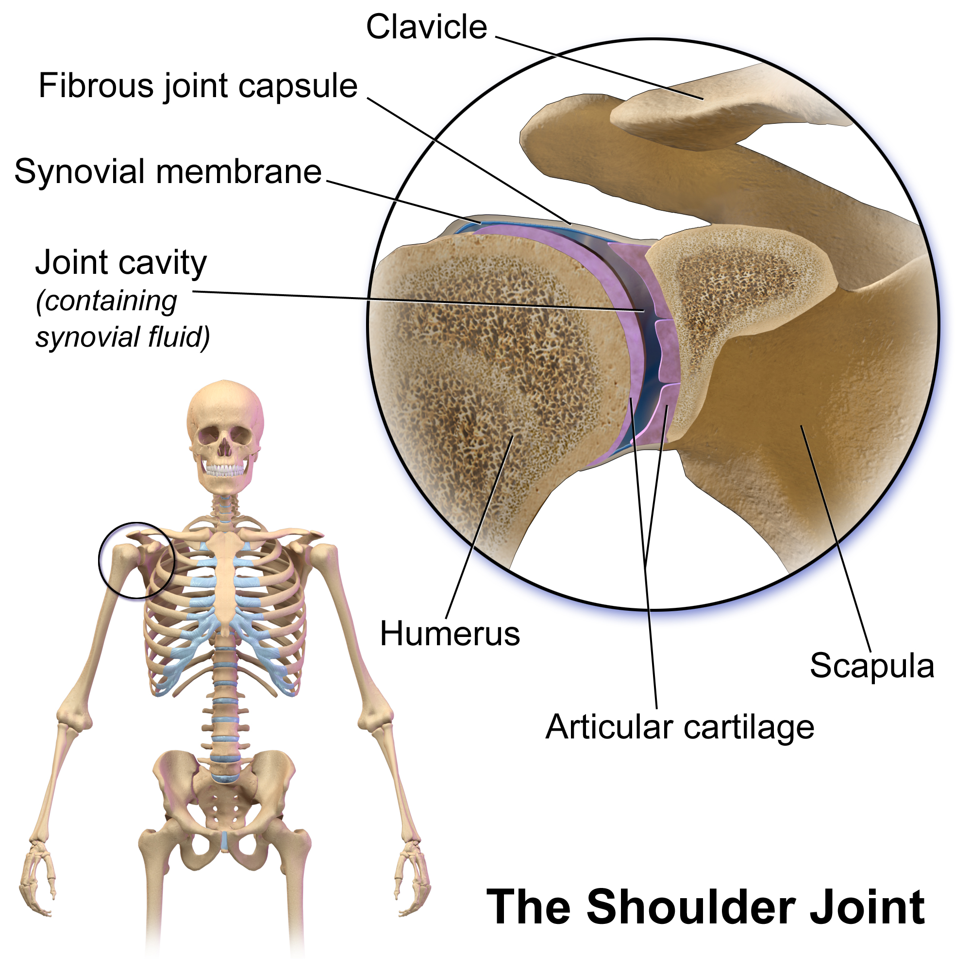
The human shoulder is made up of three bones: the clavicle (collarbone), the scapula (shoulder blade), and the humerus (upper arm bone) as well as associated muscles, ligaments and tendons. The articulations between the bones of the shoulder make up the shoulder joints. The shoulder joint, also known as the glenohumeral joint, is the major joint of the shoulder, but can more broadly include the acromioclavicular joint. In human anatomy, the shoulder joint comprises the part of the body where the humerus attaches to the scapula, and the head sits in the glenoid cavity. The shoulder is the group of structures in the region of the joint. The shoulder joint is the main joint of the shoulder. It is a ball and socket joint that allows the arm to rotate in a circular fashion or to hinge out and up away from the body. The joint capsule is a soft tissue envelope that encircles the glenohumeral joint and attaches to the scapula, humerus, and head of the biceps. It is lined by a thin, ... [...More Info...] [...Related Items...] OR: [Wikipedia] [Google] [Baidu] |
Deltoid Muscle
The deltoid muscle is the muscle forming the rounded contour of the human shoulder. It is also known as the 'common shoulder muscle', particularly in other animals such as the domestic cat. Anatomically, the deltoid muscle appears to be made up of three distinct sets of muscle fibers, namely the # anterior or clavicular part (pars clavicularis) # posterior or scapular part (pars scapularis) # intermediate or acromial part (pars acromialis) However, electromyography suggests that it consists of at least seven groups that can be independently coordinated by the nervous system. It was previously called the deltoideus (plural ''deltoidei'') and the name is still used by some anatomists. It is called so because it is in the shape of the Greek capital letter delta (Δ). Deltoid is also further shortened in slang as "delt". A study of 30 shoulders revealed an average mass of in humans, ranging from to . Structure Previous studies showed that the insertions of the tendons of the delto ... [...More Info...] [...Related Items...] OR: [Wikipedia] [Google] [Baidu] |
Orthopedic Surgery
Orthopedic surgery or orthopedics ( alternatively spelt orthopaedics), is the branch of surgery concerned with conditions involving the musculoskeletal system. Orthopedic surgeons use both surgical and nonsurgical means to treat musculoskeletal trauma, spine diseases, sports injuries, degenerative diseases, infections, tumors, and congenital disorders. Etymology Nicholas Andry coined the word in French as ', derived from the Ancient Greek words ὀρθός ''orthos'' ("correct", "straight") and παιδίον ''paidion'' ("child"), and published ''Orthopedie'' (translated as ''Orthopædia: Or the Art of Correcting and Preventing Deformities in Children'') in 1741. The word was assimilated into English as ''orthopædics''; the ligature ''æ'' was common in that era for ''ae'' in Greek- and Latin-based words. As the name implies, the discipline was initially developed with attention to children, but the correction of spinal and bone deformities in all stages of life eventually ... [...More Info...] [...Related Items...] OR: [Wikipedia] [Google] [Baidu] |
Joint Dislocation
A joint dislocation, also called luxation, occurs when there is an abnormal separation in the joint, where two or more bones meet.Dislocations. Lucile Packard Children’s Hospital at Stanford. Retrieved 3 March 2013 A partial dislocation is referred to as a subluxation. Dislocations are often caused by sudden trauma on the joint like an impact or fall. A joint dislocation can cause damage to the surrounding ligaments, tendons, muscles, and nerves. Dislocations can occur in any major joint (shoulder, knees, etc.) or minor joint (toes, fingers, etc.). The most common joint dislocation is a shoulder dislocation. Treatment for joint dislocation is usually by closed reduction, that is, skilled manipulation to return the bones to their normal position. Reduction should only be performed by trained medical professionals, because it can cause injury to soft tissue and/or the nerves and vascular structures around the dislocation. Symptoms and signs The following symptoms are common with ... [...More Info...] [...Related Items...] OR: [Wikipedia] [Google] [Baidu] |
Axial Skeleton
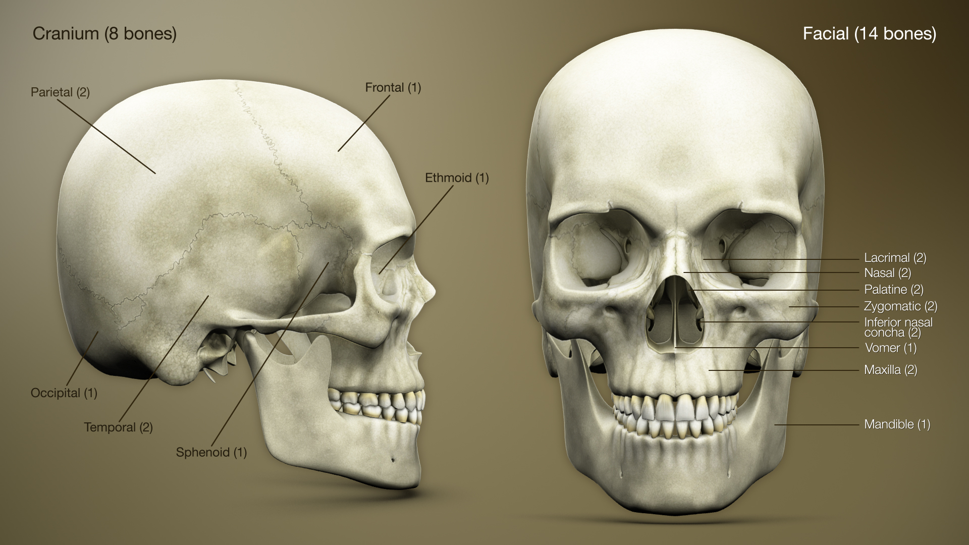
The axial skeleton is the part of the skeleton that consists of the bones of the head and trunk (anatomy), trunk of a vertebrate. In the human skeleton, it consists of 80 bones and is composed of six parts; the human skull, skull (22 bones), also the ossicles of the middle ear, the hyoid bone, the rib cage, sternum and the vertebral column. The axial skeleton together with the appendicular skeleton form the complete skeleton. Another definition of axial skeleton is the bones including the vertebrae, sacrum, coccyx, skull, ribs, and sternum. Structure Flat bones house the brain and other vital organs. This article mainly deals with the axial skeletons of humans; however, it is important to understand the evolutionary lineage of the axial skeleton. The human axial skeleton consists of 81 different bones. It is the medial core of the body and connects the pelvis to the body, where the appendix skeleton attaches. As the skeleton grows older the bones get weaker with the exception ... [...More Info...] [...Related Items...] OR: [Wikipedia] [Google] [Baidu] |
Upper Limb
The upper limbs or upper extremities are the forelimbs of an upright-postured tetrapod vertebrate, extending from the scapulae and clavicles down to and including the digits, including all the musculatures and ligaments involved with the shoulder, elbow, wrist and knuckle joints. In humans, each upper limb is divided into the arm, forearm and hand, and is primarily used for climbing, lifting and manipulating objects. Definition In formal usage, the term "arm" only refers to the structures from the shoulder to the elbow, explicitly excluding the forearm, and thus "upper limb" and "arm" are not synonymous. However, in casual usage, the terms are often used interchangeably. The term "upper arm" is redundant in anatomy, but in informal usage is used to distinguish between the two terms. Structure In the human body the muscles of the upper limb can be classified by origin, topography, function, or innervation. While a grouping by innervation reveals embryological and phylogenet ... [...More Info...] [...Related Items...] OR: [Wikipedia] [Google] [Baidu] |
Journal Of Shoulder And Elbow Surgery
The ''Journal of Shoulder and Elbow Surgery'' is a peer-reviewed medical journal covering orthopedic surgery related to the shoulder, elbow, and upper extremities. It is the official journal of multiple shoulder and elbow societies, including the American Shoulder and Elbow Surgeons, the European Society for Surgery of Shoulder and Elbow, the Japan Shoulder Society, the Shoulder and Elbow Society of Australia, the South American Shoulder and Elbow Society, the South African Shoulder and Elbow Surgeons, the Asian Shoulder Association, the Korean Shoulder and Elbow Society, the International Congress of Shoulder and Elbow Surgery, and the American Society of Shoulder and Elbow Therapists. History The journal was established in January 1991, with Mosby, now an imprint of Elsevier, as the publisher. The founding editor-in-chief was Robert Cofield (Mayo Clinic). In 1997, Robert Neviaser ( George Washington University Medical Center) took over as editor-in-chief. In 2008, Bil ... [...More Info...] [...Related Items...] OR: [Wikipedia] [Google] [Baidu] |
Trapezius
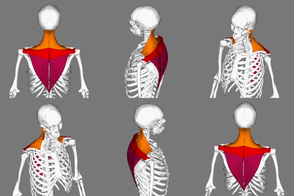
The trapezius is a large paired trapezoid-shaped surface muscle that extends longitudinally from the occipital bone to the lower thoracic vertebrae of the spine and laterally to the spine of the scapula. It moves the scapula and supports the arm. The trapezius has three functional parts: an upper (descending) part which supports the weight of the arm; a middle region (transverse), which retracts the scapula; and a lower (ascending) part which medially rotates and depresses the scapula. Name and history The trapezius muscle resembles a trapezium, also known as a trapezoid, or diamond-shaped quadrilateral. The word "spinotrapezius" refers to the human trapezius, although it is not commonly used in modern texts. In other mammals, it refers to a portion of the analogous muscle. Similarly, the term "tri-axle back plate" was historically used to describe the trapezius muscle. Structure The ''superior'' or ''upper'' (or descending) fibers of the trapezius originate from the sp ... [...More Info...] [...Related Items...] OR: [Wikipedia] [Google] [Baidu] |
Subclavius Muscle
The subclavius is a small triangular muscle, placed between the clavicle and the first rib. Along with the pectoralis major and pectoralis minor muscles, the subclavius muscle makes up the anterior axioappendicular muscles, also known as anterior wall of the axilla. Structure It arises by a short, thick tendon from the first rib and its cartilage at their junction, in front of the costoclavicular ligament. The fleshy fibers proceed obliquely superolaterally, to be inserted into the groove on the under surface of the clavicle. Innervation The nerve to subclavius (or subclavian nerve) innervates the muscle. This arises from the junction of the fifth and sixth cervical nerves, from the superior/upper trunk of the brachial plexus. Variation Insertion into coracoid process instead of clavicle or into both clavicle and coracoid process. Sternoscapular fasciculus to the upper border of scapula. Sternoclavicularis from manubrium to clavicle between pectoralis major and coracoclavicular ... [...More Info...] [...Related Items...] OR: [Wikipedia] [Google] [Baidu] |
Scapula
The scapula (plural scapulae or scapulas), also known as the shoulder blade, is the bone that connects the humerus (upper arm bone) with the clavicle (collar bone). Like their connected bones, the scapulae are paired, with each scapula on either side of the body being roughly a mirror image of the other. The name derives from the Classical Latin word for trowel or small shovel, which it was thought to resemble. In compound terms, the prefix omo- is used for the shoulder blade in medical terminology. This prefix is derived from ὦμος (ōmos), the Ancient Greek word for shoulder, and is cognate with the Latin , which in Latin signifies either the shoulder or the upper arm bone. The scapula forms the back of the shoulder girdle. In humans, it is a flat bone, roughly triangular in shape, placed on a posterolateral aspect of the thoracic cage. Structure The scapula is a thick, flat bone lying on the thoracic wall that provides an attachment for three groups of muscles: intrin ... [...More Info...] [...Related Items...] OR: [Wikipedia] [Google] [Baidu] |
Conoid Ligament
The conoid ligament is the posterior and medial fasciculus of the coracoclavicular ligament. It is formed by a dense band of fibers, conical in form, with its base directed upward. It is attached by its apex to a rough impression at the base of the coracoid process on the scapula, medial to the trapezoid ligament; above, by its expanded base, to the conoid tubercle on the under surface of the clavicle, and to a line proceeding medialward from it for 1.25 cm. These ligaments are in relation, in front, with the subclavius and deltoid muscles; behind, with the trapezius The trapezius is a large paired trapezoid-shaped surface muscle that extends longitudinally from the occipital bone to the lower thoracic vertebrae of the spine and laterally to the spine of the scapula. It moves the scapula and supports th .... References External links * Ligaments of the upper limb {{ligament-stub ... [...More Info...] [...Related Items...] OR: [Wikipedia] [Google] [Baidu] |
Trapezoid Ligament
The trapezoid ligament is a ligament connecting the coracoid process of the scapula (the shoulder blade) to the trapezoid line of the clavicle (collarbone). It is an anterior and lateral fasciculus, and is broad, thin, and quadrilateral. Its anterior border is free; its posterior border is joined with the conoid ligament The conoid ligament is the posterior and medial fasciculus of the coracoclavicular ligament. It is formed by a dense band of fibers, conical in form, with its base directed upward. It is attached by its apex to a rough impression at the base of th ..., the two forming, by their junction, an angle projecting backward. References External links * Ligaments of the upper limb {{ligament-stub ... [...More Info...] [...Related Items...] OR: [Wikipedia] [Google] [Baidu] |


