|
Condylar Canal
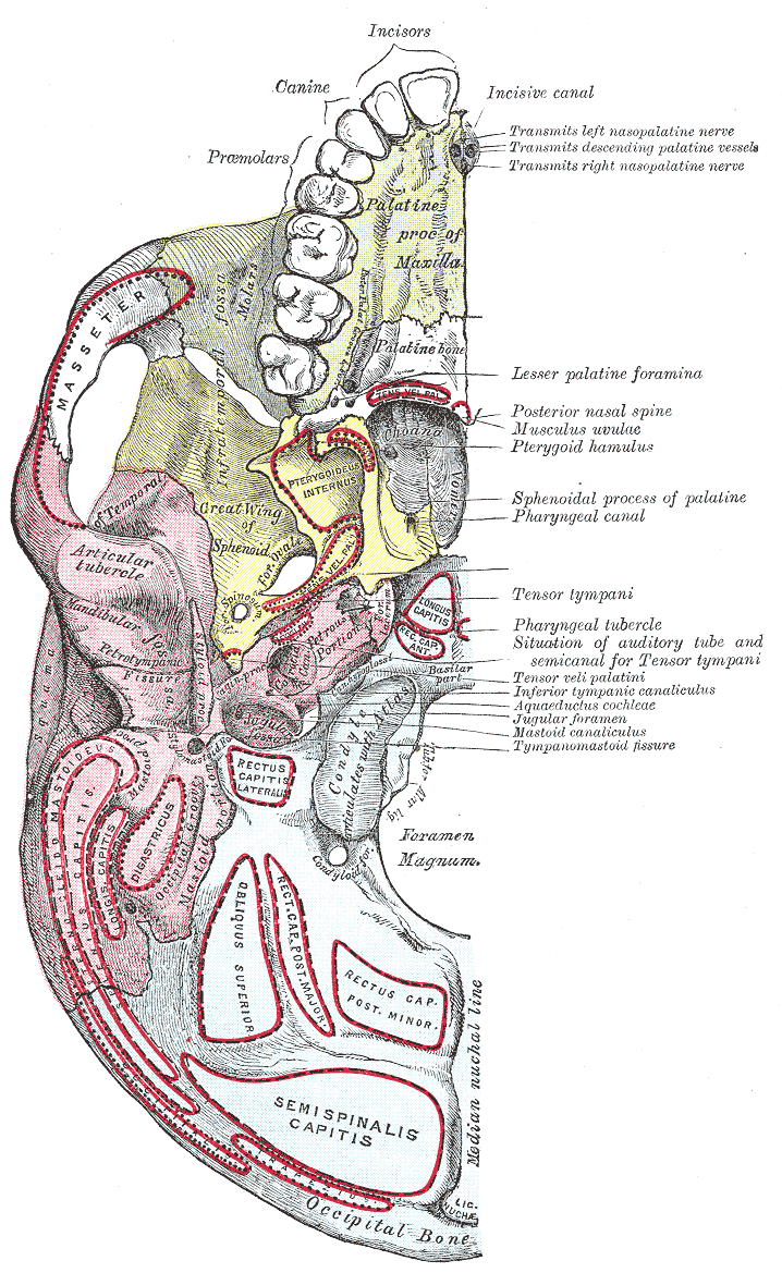
The condylar canal (or condyloid canal) is a canal in the condyloid fossa of the lateral parts of occipital bone behind the occipital condyle. Resection of the rectus capitis posterior major and minor muscles reveals the bony recess leading to the condylar canal, which is situated posterior and lateral to the occipital condyle. It is immediately superior to the extradural vertebral artery, which makes a loop above the posterior C1 ring to enter the foramen magnum. The anteriomedial wall of the condylar canal thickens to join the foramen magnum rim and connect to the occipital condyle. Through the condylar canal, the occipital emissary vein connects to the venous system including the suboccipital venous plexus, occipital sinus and sigmoid sinus The sigmoid sinuses (sigma- or s-shaped hollow curve), also known as the , are venous sinuses within the skull that receive blood from posterior dural venous sinus veins. Structure The sigmoid sinus is a dural venous sinus situated ... [...More Info...] [...Related Items...] OR: [Wikipedia] [Google] [Baidu] |
Occipital Bone
The occipital bone () is a neurocranium, cranial dermal bone and the main bone of the occiput (back and lower part of the skull). It is trapezoidal in shape and curved on itself like a shallow dish. The occipital bone overlies the occipital lobes of the cerebrum. At the base of skull in the occipital bone, there is a large oval opening called the foramen magnum, which allows the passage of the spinal cord. Like the other cranial bones, it is classed as a flat bone. Due to its many attachments and features, the occipital bone is described in terms of separate parts. From its front to the back is the basilar part of occipital bone, basilar part, also called the basioccipital, at the sides of the foramen magnum are the lateral parts of occipital bone, lateral parts, also called the exoccipitals, and the back is named as the squamous part of occipital bone, squamous part. The basilar part is a thick, somewhat quadrilateral piece in front of the foramen magnum and directed towards the ... [...More Info...] [...Related Items...] OR: [Wikipedia] [Google] [Baidu] |
Base Of The Skull
The base of skull, also known as the cranial base or the cranial floor, is the most inferior area of the skull. It is composed of the endocranium and the lower parts of the calvaria. Structure Structures found at the base of the skull are for example: Bones There are five bones that make up the base of the skull: *Ethmoid bone * Sphenoid bone * Occipital bone *Frontal bone *Temporal bone Sinuses *Occipital sinus * Superior sagittal sinus *Superior petrosal sinus Foramina of the skull * Foramen cecum *Optic foramen *Foramen lacerum *Foramen rotundum * Foramen magnum * Foramen ovale *Jugular foramen *Internal auditory meatus *Mastoid foramen *Sphenoidal emissary foramen *Foramen spinosum Sutures *Frontoethmoidal suture *Sphenofrontal suture *Sphenopetrosal suture *Sphenoethmoidal suture * Petrosquamous suture *Sphenosquamosal suture Other *Sphenoidal lingula *Subarcuate fossa *Dorsum sellae *Jugular process *Petro-occipital fissure *Condylar canal * Jugular tubercle * ... [...More Info...] [...Related Items...] OR: [Wikipedia] [Google] [Baidu] |
Occipital Bone
The occipital bone () is a neurocranium, cranial dermal bone and the main bone of the occiput (back and lower part of the skull). It is trapezoidal in shape and curved on itself like a shallow dish. The occipital bone overlies the occipital lobes of the cerebrum. At the base of skull in the occipital bone, there is a large oval opening called the foramen magnum, which allows the passage of the spinal cord. Like the other cranial bones, it is classed as a flat bone. Due to its many attachments and features, the occipital bone is described in terms of separate parts. From its front to the back is the basilar part of occipital bone, basilar part, also called the basioccipital, at the sides of the foramen magnum are the lateral parts of occipital bone, lateral parts, also called the exoccipitals, and the back is named as the squamous part of occipital bone, squamous part. The basilar part is a thick, somewhat quadrilateral piece in front of the foramen magnum and directed towards the ... [...More Info...] [...Related Items...] OR: [Wikipedia] [Google] [Baidu] |
Condyloid Fossa
Behind either condyle of the lateral parts of occipital bone is a depression, the condyloid fossa (or condylar fossa), which receives the posterior margin of the superior facet of the atlas when the head is bent backward; the floor of this fossa is sometimes perforated by the condyloid canal, through which an emissary vein passes from the transverse sinus. Additional images File:Condyloid fossa - animation02.gif, Human skull seen from below. Position of condyloid fossa shown in red. File:Condyloid fossa - animation04.gif, Skull and cervical vertebra. Position of condyloid fossa shown in red. File:Cervical XRayFlexionExtension.jpg, X-ray of cervical spine (neck) in flexion and extension (bending backwards) See also * Occipital condyle * Atlas An atlas is a collection of maps; it is typically a bundle of maps of Earth or of a region of Earth. Atlases have traditionally been bound into book form, but today many atlases are in multimedia formats. In addition to presenting ... [...More Info...] [...Related Items...] OR: [Wikipedia] [Google] [Baidu] |
Lateral Parts Of Occipital Bone
The lateral parts of the occipital bone (also called the exoccipitals) are situated at the sides of the foramen magnum; on their under surfaces are the condyles for articulation with the superior facets of the atlas. Description The condyles are oval or reniform (kidney-shaped) in shape, and their anterior extremities, directed forward and medialward, are closer together than their posterior, and encroach on the basilar portion of the bone; the posterior extremities extend back to the level of the middle of the foramen magnum. The articular surfaces of the condyles are convex from before backward and from side to side, and look downward and lateralward. To their margins are attached the capsules of the atlantoöccipital articulations, and on the medial side of each is a rough impression or tubercle for the alar ligament. At the base of either condyle the bone is tunnelled by a short canal, the hypoglossal canal (anterior condyloid foramen). This begins on the cranial surface o ... [...More Info...] [...Related Items...] OR: [Wikipedia] [Google] [Baidu] |
Occipital Condyle
The occipital condyles are undersurface protuberances of the occipital bone in vertebrates, which function in articulation with the superior facets of the atlas vertebra. The condyles are oval or reniform (kidney-shaped) in shape, and their anterior extremities, directed forward and medialward, are closer together than their posterior, and encroach on the basilar portion of the bone; the posterior extremities extend back to the level of the middle of the foramen magnum. The articular surfaces of the condyles are convex from before backward and from side to side, and look downward and lateralward. To their margins are attached the capsules of the atlanto-occipital joints, and on the medial side of each is a rough impression or tubercle for the alar ligament. At the base of either condyle the bone is tunnelled by a short canal, the hypoglossal canal. Clinical significance Fracture of an occipital condyle may occur in isolation, or as part of a more extended basilar skull fracture ... [...More Info...] [...Related Items...] OR: [Wikipedia] [Google] [Baidu] |
Vertebral Artery
The vertebral arteries are major arteries An artery (plural arteries) () is a blood vessel in humans and most animals that takes blood away from the heart to one or more parts of the body (tissues, lungs, brain etc.). Most arteries carry oxygenated blood; the two exceptions are the pu ... of the neck. Typically, the vertebral arteries originate from the subclavian arteries. Each vessel courses superiorly along each side of the neck, merging within the skull to form the single, midline basilar artery. As the supplying component of the ''vertebrobasilar vascular system'', the vertebral arteries supply blood to the upper spinal cord, brainstem, cerebellum, and Cerebral circulation#Posterior cerebral circulation, posterior part of brain. Structure The vertebral arteries usually arise from the posterosuperior aspect of the central subclavian arteries on each side of the body, then enter deep to the transverse process at the level of the 6th cervical vertebrae (C6), or occasio ... [...More Info...] [...Related Items...] OR: [Wikipedia] [Google] [Baidu] |
Foramen Magnum
The foramen magnum ( la, great hole) is a large, oval-shaped opening in the occipital bone of the skull. It is one of the several oval or circular openings (foramina) in the base of the skull. The spinal cord, an extension of the medulla oblongata, passes through the foramen magnum as it exits the cranial cavity. Apart from the transmission of the medulla oblongata and its membranes, the foramen magnum transmits the vertebral arteries, the anterior and posterior spinal arteries, the tectorial membranes and alar ligaments. It also transmits the accessory nerve into the skull. The foramen magnum is a very important feature in bipedal mammals. One of the attributes of a biped's foramen magnum is a forward shift of the anterior border of the cerebellar tentorium; this is caused by the shortening of the cranial base. Studies on the foramen magnum position have shown a connection to the functional influences of both posture and locomotion. The forward shift of the foramen magnum i ... [...More Info...] [...Related Items...] OR: [Wikipedia] [Google] [Baidu] |
Occipital Emissary Vein
The occipital emissary vein is a small emissary vein which passes through the condylar canal The condylar canal (or condyloid canal) is a canal in the condyloid fossa of the lateral parts of occipital bone behind the occipital condyle. Resection of the rectus capitis posterior major and minor muscles reveals the bony recess leading to t .... References Veins of the head and neck {{circulatory-stub ... [...More Info...] [...Related Items...] OR: [Wikipedia] [Google] [Baidu] |
Suboccipital Venous Plexus
The suboccipital venous plexus drains deoxygenated blood from the back of the head. It communicates with the external vertebral venous plexuses. The external vertebral venous plexuses travel inferiorly from this suboccipital region to drain into the brachiocephalic vein. The occipital vein joins in the formation of the plexus deep to the musculature of the back and from here drains into the external jugular vein. The plexus surrounds segments of the vertebral artery The vertebral arteries are major arteries An artery (plural arteries) () is a blood vessel in humans and most animals that takes blood away from the heart to one or more parts of the body (tissues, lungs, brain etc.). Most arteries carry o .... Veins of the head and neck {{circulatory-stub ... [...More Info...] [...Related Items...] OR: [Wikipedia] [Google] [Baidu] |
Occipital Sinus
The occipital sinus is the smallest of the dural venous sinuses. It is usually unpaired, and is sometimes altogether absent. It is situated in the attached margin of the falx cerebelli. It commences near the foramen magnum, and ends by draining into the confluence of sinuses. Occipital sinuses were discovered by Guichard Joseph Duverney. Anatomy The occipital sinus is present in around 65% of individuals. It is usually single, but occasionally paired. It is situated in the attached margin of the falx cerebelli. Course The occipital sinus commences around the margin of the foramen magnum by several small venous channels (one of which joins the terminal part of the sigmoid sinus). It terminates by draining into the confluence of the sinuses. Communications The occipital sinus communicates with the marginal sinus Marginal may refer to: * ''Marginal'' (album), the third album of the Belgian rock band Dead Man Ray, released in 2001 * ''Marginal'' (manga) * '' El Marginal'' ... [...More Info...] [...Related Items...] OR: [Wikipedia] [Google] [Baidu] |
Sigmoid Sinus
The sigmoid sinuses (sigma- or s-shaped hollow curve), also known as the , are venous sinuses within the skull that receive blood from posterior dural venous sinus veins. Structure The sigmoid sinus is a dural venous sinus situated within the dura mater. The sigmoid sinus receives blood from the transverse sinuses, which track the posterior wall of the cranial cavity, travels inferiorly along the parietal bone, temporal bone and occipital bone, and converges with the inferior petrosal sinuses to form the internal jugular vein. Each sigmoid sinus begins beneath the temporal bone and follows a tortuous course to the jugular foramen, at which point the sinus becomes continuous with the internal jugular vein. Function The sigmoid sinus receives blood from the transverse sinuses, which receive blood from the posterior aspect of the skull. Along its course, the sigmoid sinus also receives blood from the cerebral veins, cerebellar veins, diploic veins, and emissary veins. See als ... [...More Info...] [...Related Items...] OR: [Wikipedia] [Google] [Baidu] |


