|
Computed Tomography Of The Abdomen And Pelvis
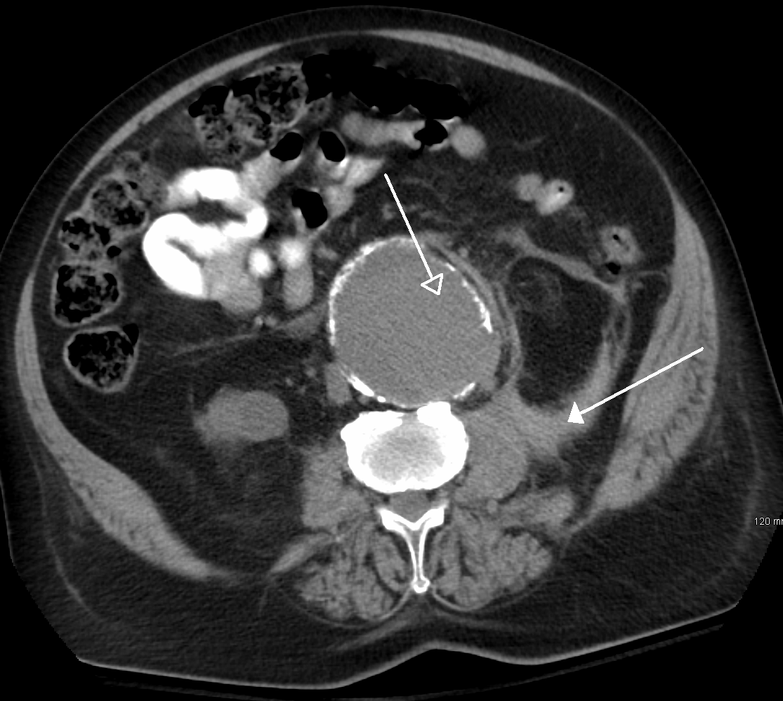
Computed tomography of the abdomen and pelvis is an application of computed tomography (CT) and is a sensitive method for diagnosis of Human abdomen, abdominal diseases. It is used frequently to determine stage of cancer and to follow progress. It is also a useful test to investigate acute abdominal pain (especially of the lower quadrants, whereas ultrasound is the preferred first line investigation for right upper quadrant pain). Kidney stone, Renal stones, appendicitis, pancreatitis, diverticulitis, aortic aneurysm, abdominal aortic aneurysm, and bowel obstruction are conditions that are readily diagnosed and assessed with CT. CT is also the first line for detecting solid organ injury after trauma. Advantages Multidetector CT (MDCT) can clearly delineate anatomic structures in the abdomen, which is critical in the diagnosis of internal diaphragmatic and other nonpalpable or unsuspected hernias. MDCT also offers clear detail of the abdominal wall allowing wall hernias to be i ... [...More Info...] [...Related Items...] OR: [Wikipedia] [Google] [Baidu] [Amazon] |
CT Scan
A computed tomography scan (CT scan), formerly called computed axial tomography scan (CAT scan), is a medical imaging technique used to obtain detailed internal images of the body. The personnel that perform CT scans are called radiographers or radiology technologists. CT scanners use a rotating X-ray tube and a row of detectors placed in a gantry (medical), gantry to measure X-ray Attenuation#Radiography, attenuations by different tissues inside the body. The multiple X-ray measurements taken from different angles are then processed on a computer using tomographic reconstruction algorithms to produce Tomography, tomographic (cross-sectional) images (virtual "slices") of a body. CT scans can be used in patients with metallic implants or pacemakers, for whom magnetic resonance imaging (MRI) is Contraindication, contraindicated. Since its development in the 1970s, CT scanning has proven to be a versatile imaging technique. While CT is most prominently used in medical diagnosis, i ... [...More Info...] [...Related Items...] OR: [Wikipedia] [Google] [Baidu] [Amazon] |
Contrast CT
Contrast CT, or contrast-enhanced computed tomography (CECT), is CT scan, X-ray computed tomography (CT) using radiocontrast. Radiocontrasts for X-ray CT are generally Iodinated contrast, iodine-based types. This is useful to highlight structures such as blood vessels that otherwise would be difficult to delineate from their surroundings. Using contrast material can also help to obtain functional information about tissues. Often, images are taken both with and without radiocontrast. CT images are called ''precontrast'' or ''native-phase'' images before any radiocontrast has been administered, and ''postcontrast'' after radiocontrast administration. Bolus tracking Bolus tracking is a technique to optimize timing of the imaging. A small Bolus (medicine), bolus of radio-opaque contrast media is Injection (medicine), injected into a patient via a peripheral intravenous cannula. Depending on the vessel being imaged, the volume of contrast is tracked using a region of interest (abbr ... [...More Info...] [...Related Items...] OR: [Wikipedia] [Google] [Baidu] [Amazon] |
Diagnostic Radiology
Medical imaging is the technique and process of imaging the interior of a body for clinical analysis and medical intervention, as well as visual representation of the function of some organs or tissues (physiology). Medical imaging seeks to reveal internal structures hidden by the skin and bones, as well as to diagnose and treat disease. Medical imaging also establishes a database of normal anatomy and physiology to make it possible to identify abnormalities. Although imaging of removed organ (anatomy), organs and Tissue (biology), tissues can be performed for medical reasons, such procedures are usually considered part of pathology instead of medical imaging. Measurement and recording techniques that are not primarily designed to produce images, such as electroencephalography (EEG), magnetoencephalography (MEG), electrocardiography (ECG), and others, represent other technologies that produce data susceptible to representation as a parameter graph versus time or maps that contain ... [...More Info...] [...Related Items...] OR: [Wikipedia] [Google] [Baidu] [Amazon] |
Abdominal CT With Scan Range And Field Of View, Without Annotations
The abdomen (colloquially called the gut, belly, tummy, midriff, tucky, or stomach) is the front part of the torso between the thorax (chest) and pelvis in humans and in other vertebrates. The area occupied by the abdomen is called the abdominal cavity. In arthropods, it is the posterior tagma of the body; it follows the thorax or cephalothorax. In humans, the abdomen stretches from the thorax at the thoracic diaphragm to the pelvis at the pelvic brim. The pelvic brim stretches from the lumbosacral joint (the intervertebral disc between L5 and S1) to the pubic symphysis and is the edge of the pelvic inlet. The space above this inlet and under the thoracic diaphragm is termed the abdominal cavity. The boundary of the abdominal cavity is the abdominal wall in the front and the peritoneal surface at the rear. In vertebrates, the abdomen is a large body cavity enclosed by the abdominal muscles, at the front and to the sides, and by part of the vertebral column at the back. Low ... [...More Info...] [...Related Items...] OR: [Wikipedia] [Google] [Baidu] [Amazon] |
CT Urogram
A computed tomography urography (CT urography or CT urogram) is a computed tomography scan that examines the urinary tract after contrast dye is injected into a vein. In a CT urogram, the contrast agent is through a cannula into a vein, allowed to be cleared by the kidneys and excreted through the urinary tract as part of the urine. Uses Plain CT urography (without contrast) is used to evaluate stone diseases, calcifications within kidneys, density of renal masses and presence of any bleeding before contrast is given. Then iodinated contrast is given intravenously to evaluate kidney tumours (if any) and urinary tract. CT scan is then taken at 30 to 70 seconds (when the contrast reaches the renal cortex and renal medulla The renal medulla (Latin: ''medulla renis'' 'marrow of the kidney') is the innermost part of the kidney. The renal medulla is split up into a number of sections, known as the renal pyramids. Blood enters into the kidney via the renal artery, which ...) to ... [...More Info...] [...Related Items...] OR: [Wikipedia] [Google] [Baidu] [Amazon] |
Renal Parenchymal Phase CT Of Transitional Cell Carcinoma
In humans, the kidneys are two reddish-brown bean-shaped blood-filtering organs that are a multilobar, multipapillary form of mammalian kidneys, usually without signs of external lobulation. They are located on the left and right in the retroperitoneal space, and in adult humans are about in length. They receive blood from the paired renal arteries; blood exits into the paired renal veins. Each kidney is attached to a ureter, a tube that carries excreted urine to the bladder. The kidney participates in the control of the volume of various body fluids, fluid osmolality, acid-base balance, various electrolyte concentrations, and removal of toxins. Filtration occurs in the glomerulus: one-fifth of the blood volume that enters the kidneys is filtered. Examples of substances reabsorbed are solute-free water, sodium, bicarbonate, glucose, and amino acids. Examples of substances secreted are hydrogen, ammonium, potassium and uric acid. The nephron is the structural and functional un ... [...More Info...] [...Related Items...] OR: [Wikipedia] [Google] [Baidu] [Amazon] |
Transient Hepatic Attenuation Differences
Transient hepatic attenuation differences (THAD) are areas of Contrast medium, enhancement during the arterial phase of contrast CT of the liver. THAD is thought to be a physiological phenomenon resulting from regional variation in the blood supply by the portal vein and/or the hepatic artery. THAD may in some cases be associated with liver tumors such as a hepatocellular carcinoma. References Hepatology Radiology Medical imaging {{med-imaging-stub ... [...More Info...] [...Related Items...] OR: [Wikipedia] [Google] [Baidu] [Amazon] |
Late Arterial And Portal Venous Phase CT Of Focal Nodular Hyperplasia
Late or LATE may refer to: Everyday usage * Tardy, or late, not being on time * Late (or the late) may refer to a person who is dead Music * ''Late'' (The 77s album), 2000 * Late (Alvin Batiste album), 1993 * Late!, a pseudonym used by Dave Grohl on his ''Pocketwatch'' album * Late (rapper), an underground rapper from Wolverhampton * "Late", a song by Kanye West from ''Late Registration'' Other uses * Late (Tonga), an uninhabited volcanic island southwest of Vavau in the kingdom of Tonga * "Late" (''The Handmaid's Tale''), a television episode * LaTe, Oy Laivateollisuus Ab, a defunct shipbuilding company * Limbic-predominant age-related TDP-43 encephalopathy, a proposed form of dementia * Local-authority trading enterprise, a New Zealand business law * Local average treatment effect, a concept in econometrics * Late, a synonym for ''cooler'' in stellar classification See also * * * ''Lates'', a genus of fish in the lates perch family * Later (other) Later may refer ... [...More Info...] [...Related Items...] OR: [Wikipedia] [Google] [Baidu] [Amazon] |
Arterial And Portal Venous Phase CT Of Cholangiocarcinoma
An artery () is a blood vessel in humans and most other animals that takes oxygenated blood away from the heart in the systemic circulation to one or more parts of the body. Exceptions that carry deoxygenated blood are the pulmonary arteries in the pulmonary circulation that carry blood to the lungs for oxygenation, and the umbilical arteries in the fetal circulation that carry deoxygenated blood to the placenta. It consists of a multi-layered artery wall wrapped into a tube-shaped channel. Arteries contrast with veins, which carry deoxygenated blood back towards the heart; or in the pulmonary and fetal circulations carry oxygenated blood to the lungs and fetus respectively. Structure The anatomy of arteries can be separated into gross anatomy, at the macroscopic level, and microanatomy, which must be studied with a microscope. The arterial system of the human body is divided into systemic arteries, carrying blood from the heart to the whole body, and pulmonary arteries, c ... [...More Info...] [...Related Items...] OR: [Wikipedia] [Google] [Baidu] [Amazon] |
Abdominal CT Angiography
The abdomen (colloquially called the gut, belly, tummy, midriff, tucky, or stomach) is the front part of the torso between the thorax (chest) and pelvis in humans and in other vertebrates. The area occupied by the abdomen is called the abdominal cavity. In arthropods, it is the posterior tagma of the body; it follows the thorax or cephalothorax. In humans, the abdomen stretches from the thorax at the thoracic diaphragm to the pelvis at the pelvic brim. The pelvic brim stretches from the lumbosacral joint (the intervertebral disc between L5 and S1) to the pubic symphysis and is the edge of the pelvic inlet. The space above this inlet and under the thoracic diaphragm is termed the abdominal cavity. The boundary of the abdominal cavity is the abdominal wall in the front and the peritoneal surface at the rear. In vertebrates, the abdomen is a large body cavity enclosed by the abdominal muscles, at the front and to the sides, and by part of the vertebral column at the back. Lowe ... [...More Info...] [...Related Items...] OR: [Wikipedia] [Google] [Baidu] [Amazon] |


