|
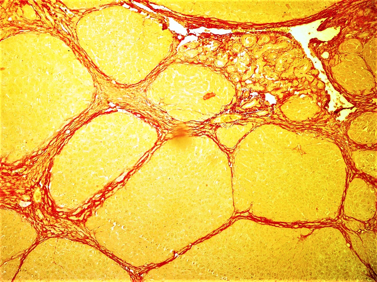
Chronic Sclerosing Sialadenitis
Chronic sclerosing sialadenitis is a chronic (long-lasting) inflammatory condition affecting the salivary gland. Relatively rare in occurrence, this condition is benign, but presents as hard, indurated and enlarged masses that are clinically indistinguishable from salivary gland neoplasms or tumors. It is now regarded as a manifestation of IgG4-related disease. Involvement of the submandibular glands is also known as Küttner's tumor, named after Hermann Küttner (1870–1932), a German Oral and Maxillofacial Surgeon, who reported four cases of submandibular gland lesions for the first time in 1896. Presentation The inflammatory lesions in Küttner's tumor may occur on one side (unilateral) or both sides (bilateral), predominantly involving the submandibular gland, but is also known to occur in other major and minor salivary glands, including the parotid gland. Overall, salivary gland tumors are relatively rare, with approximately 2.5–3 cases per 100,000 people per year s ... [...More Info...] [...Related Items...] OR: [Wikipedia] [Google] [Baidu] |
Salivary Gland
The salivary glands in mammals are exocrine glands that produce saliva through a system of ducts. Humans have three paired major salivary glands (parotid, submandibular, and sublingual), as well as hundreds of minor salivary glands. Salivary glands can be classified as serous, mucous, or seromucous (mixed). In serous secretions, the main type of protein secreted is alpha-amylase, an enzyme that breaks down starch into maltose and glucose, whereas in mucous secretions, the main protein secreted is mucin, which acts as a lubricant. In humans, 1200 to 1500 ml of saliva are produced every day. The secretion of saliva (salivation) is mediated by parasympathetic stimulation; acetylcholine is the active neurotransmitter and binds to muscarinic receptors in the glands, leading to increased salivation. The fourth pair of salivary glands, the tubarial glands discovered in 2020, are named for their location, being positioned in front and over the torus tubarius. However, this find ... [...More Info...] [...Related Items...] OR: [Wikipedia] [Google] [Baidu] |
Sclerosis (medicine)
Sclerosis (from Greek σκληρός ''sklērós'', "hard") is the stiffening of a tissue or anatomical feature, usually caused by a replacement of the normal organ-specific tissue with connective tissue. The structure may be said to have undergone sclerotic changes or display sclerotic lesions, which refers to the process of sclerosis. Common medical conditions whose pathology involves sclerosis include: * Amyotrophic lateral sclerosis—also known as Lou Gehrig's disease or motor neurone disease—a progressive, incurable, usually fatal disease of motor neurons. * Atherosclerosis, a deposit of fatty materials, such as cholesterol, in the arteries which causes hardening. * Focal segmental glomerulosclerosis is a disease that attacks the kidney's filtering system (glomeruli) causing serious scarring and thus a cause of nephrotic syndrome in children and adolescents, as well as an important cause of kidney failure in adults. * Hippocampal sclerosis, a brain damage often seen in ... [...More Info...] [...Related Items...] OR: [Wikipedia] [Google] [Baidu] |
Sialadenitis
Sialadenitis (sialoadenitis) is inflammation of salivary glands, usually the major ones, the most common being the parotid gland, followed by submandibular and sublingual glands. It should not be confused with sialadenosis (sialosis) which is a non-inflammatory enlargement of the major salivary glands. Sialadenitis can be further classed as acute or chronic. Acute sialadenitis is an acute inflammation of a salivary gland which may present itself as a red, painful swelling that is tender to touch. Chronic sialadenitis is typically less painful but presents as recurrent swellings, usually after meals, without redness. Causes of sialadenitis are varied, including bacterial (most commonly ''Staphylococcus aureus''), viral and autoimmune conditions. Types Acute ;Predisposing factors * sialolithiasis * decreased flow (dehydration, post-operative, drugs) * poor oral hygiene * exacerbation of low grade chronic sialoadenitis ;Clinical features * painful swelling * reddened skin * edema ... [...More Info...] [...Related Items...] OR: [Wikipedia] [Google] [Baidu] |
Magnetic Resonance Imaging
Magnetic resonance imaging (MRI) is a medical imaging technique used in radiology to form pictures of the anatomy and the physiological processes of the body. MRI scanners use strong magnetic fields, magnetic field gradients, and radio waves to generate images of the organs in the body. MRI does not involve X-rays or the use of ionizing radiation, which distinguishes it from CT and PET scans. MRI is a medical application of nuclear magnetic resonance (NMR) which can also be used for imaging in other NMR applications, such as NMR spectroscopy. MRI is widely used in hospitals and clinics for medical diagnosis, staging and follow-up of disease. Compared to CT, MRI provides better contrast in images of soft-tissues, e.g. in the brain or abdomen. However, it may be perceived as less comfortable by patients, due to the usually longer and louder measurements with the subject in a long, confining tube, though "Open" MRI designs mostly relieve this. Additionally, implants and oth ... [...More Info...] [...Related Items...] OR: [Wikipedia] [Google] [Baidu] |
Fine-needle Aspiration
Fine-needle aspiration (FNA) is a diagnostic procedure used to investigate lumps or masses. In this technique, a thin (23–25 gauge (0.52 to 0.64 mm outer diameter)), hollow needle is inserted into the mass for sampling of cells that, after being stained, are examined under a microscope (biopsy). The sampling and biopsy considered together are called fine-needle aspiration biopsy (FNAB) or fine-needle aspiration cytology (FNAC) (the latter to emphasize that any aspiration biopsy involves cytopathology, not histopathology). Fine-needle aspiration biopsies are very safe minor surgical procedures. Often, a major surgical (excisional or open) biopsy can be avoided by performing a needle aspiration biopsy instead, eliminating the need for hospitalization. In 1981, the first fine-needle aspiration biopsy in the United States was done at Maimonides Medical Center. Today, this procedure is widely used in the diagnosis of cancer and inflammatory conditions. Aspiration is safer and ... [...More Info...] [...Related Items...] OR: [Wikipedia] [Google] [Baidu] |
Ultrasonography
Ultrasound is sound waves with frequencies higher than the upper audible limit of human hearing. Ultrasound is not different from "normal" (audible) sound in its physical properties, except that humans cannot hear it. This limit varies from person to person and is approximately 20 kilohertz (20,000 hertz) in healthy young adults. Ultrasound devices operate with frequencies from 20 kHz up to several gigahertz. Ultrasound is used in many different fields. Ultrasonic devices are used to detect objects and measure distances. Ultrasound imaging or sonography is often used in medicine. In the nondestructive testing of products and structures, ultrasound is used to detect invisible flaws. Industrially, ultrasound is used for cleaning, mixing, and accelerating chemical processes. Animals such as bats and porpoises use ultrasound for locating prey and obstacles. History Acoustics, the science of sound, starts as far back as Pythagoras in the 6th century BC, who wrote on ... [...More Info...] [...Related Items...] OR: [Wikipedia] [Google] [Baidu] |
Immunoglobulin G
Immunoglobulin G (Ig G) is a type of antibody. Representing approximately 75% of serum antibodies in humans, IgG is the most common type of antibody found in blood circulation. IgG molecules are created and released by plasma B cells. Each IgG antibody has two paratopes. Function Antibodies are major components of humoral immunity. IgG is the main type of antibody found in blood and extracellular fluid, allowing it to control infection of body tissues. By binding many kinds of pathogens such as viruses, bacteria, and fungi, IgG protects the body from infection. It does this through several mechanisms: * IgG-mediated binding of pathogens causes their immobilization and binding together via agglutination; IgG coating of pathogen surfaces (known as opsonization) allows their recognition and ingestion by phagocytic immune cells leading to the elimination of the pathogen itself; * IgG activates all the classical pathway of the complement system, a cascade of immune protein pr ... [...More Info...] [...Related Items...] OR: [Wikipedia] [Google] [Baidu] |
Sialolithiasis
Sialolithiasis (also termed salivary calculi, or salivary stones) is a crystallopathy where a calcified mass or ''sialolith'' forms within a salivary gland, usually in the duct of the submandibular gland (also termed "Wharton's duct"). Less commonly the parotid gland or rarely the sublingual gland or a minor salivary gland may develop salivary stones. The usual symptoms are pain and swelling of the affected salivary gland, both of which get worse when salivary flow is stimulated, e.g. with the sight, thought, smell or taste of food, or with hunger or chewing. This is often termed "mealtime syndrome". Inflammation or infection of the gland may develop as a result. Sialolithiasis may also develop because of the presence of existing chronic infection of the glands, dehydration (e.g. use of phenothiazines), Sjögren's syndrome and/or increased local levels of calcium, but in many instances the cause is idiopathic (unknown). The condition is usually managed by removing the stone, and ... [...More Info...] [...Related Items...] OR: [Wikipedia] [Google] [Baidu] |
Fibrosis
Fibrosis, also known as fibrotic scarring, is a pathological wound healing in which connective tissue replaces normal parenchymal tissue to the extent that it goes unchecked, leading to considerable tissue remodelling and the formation of permanent scar tissue. Repeated injuries, chronic inflammation and repair are susceptible to fibrosis where an accidental excessive accumulation of extracellular matrix components, such as the collagen is produced by fibroblasts, leading to the formation of a permanent fibrotic scar. In response to injury, this is called scarring, and if fibrosis arises from a single cell line, this is called a fibroma. Physiologically, fibrosis acts to deposit connective tissue, which can interfere with or totally inhibit the normal architecture and function of the underlying organ or tissue. Fibrosis can be used to describe the pathological state of excess deposition of fibrous tissue, as well as the process of connective tissue deposition in healing. Define ... [...More Info...] [...Related Items...] OR: [Wikipedia] [Google] [Baidu] |
IgG4-related Disease
IgG4-related disease (IgG4-RD), formerly known as IgG4-related systemic disease, is a chronic inflammatory condition characterized by tissue infiltration with lymphocytes and IgG4-secreting plasma cells, various degrees of fibrosis (scarring) and a usually prompt response to oral steroids. In approximately 51–70% of people with this disease, ''serum'' IgG4 concentrations are elevated during an acute phase. It is a relapsing-remitting disease associated with a tendency to mass forming, tissue-destructive lesions in multiple sites, with a characteristic histopathological appearance in whichever site is involved. Inflammation and the deposition of connective tissue in affected anatomical sites can lead to organ dysfunction, organ failure, or even death if not treated. Early detection is important to avoid organ damage and potentially serious complications. Treatment is recommended in all symptomatic cases of IgG4-RD and also in asymptomatic IgG4-RD involving certain anatomical site ... [...More Info...] [...Related Items...] OR: [Wikipedia] [Google] [Baidu] |
Atrophy
Atrophy is the partial or complete wasting away of a part of the body. Causes of atrophy include mutations (which can destroy the gene to build up the organ), poor nourishment, poor circulation, loss of hormonal support, loss of nerve supply to the target organ, excessive amount of apoptosis of cells, and disuse or lack of exercise or disease intrinsic to the tissue itself. In medical practice, hormonal and nerve inputs that maintain an organ or body part are said to have ''trophic'' effects. A diminished muscular trophic condition is designated as ''atrophy''. Atrophy is reduction in size of cell, organ or tissue, after attaining its normal mature growth. In contrast, hypoplasia is the reduction in the cellular numbers of an organ, or tissue that has not attained normal maturity. Atrophy is the general physiological process of reabsorption and breakdown of tissues, involving apoptosis. When it occurs as a result of disease or loss of trophic support because of other diseases ... [...More Info...] [...Related Items...] OR: [Wikipedia] [Google] [Baidu] |
Plasma Cell
Plasma cells, also called plasma B cells or effector B cells, are white blood cells that originate in the lymphoid organs as B lymphocytes and secrete large quantities of proteins called antibodies in response to being presented specific substances called antigens. These antibodies are transported from the plasma cells by the blood plasma and the lymphatic system to the site of the target antigen (foreign substance), where they initiate its neutralization or destruction. B cells differentiate into plasma cells that produce antibody molecules closely modeled after the receptors of the precursor B cell. Structure Plasma cells are large lymphocytes with abundant cytoplasm and a characteristic appearance on light microscopy. They have basophilic cytoplasm and an eccentric nucleus with heterochromatin in a characteristic cartwheel or clock face arrangement. Their cytoplasm also contains a pale zone that on electron microscopy contains an extensive Golgi apparatus and centrioles ... [...More Info...] [...Related Items...] OR: [Wikipedia] [Google] [Baidu] |


