|
Cervical Vertebra 1
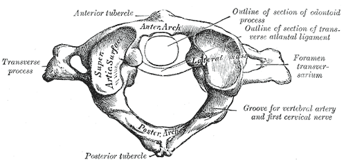
In anatomy, the atlas (C1) is the most superior (first) cervical vertebra of the spine and is located in the neck. It is named for Atlas of Greek mythology because, just as Atlas supported the globe, it supports the entire head. The atlas is the topmost vertebra and, with the axis (the vertebra below it), forms the joint connecting the skull and spine. The atlas and axis are specialized to allow a greater range of motion than normal vertebrae. They are responsible for the nodding and rotation movements of the head. The atlanto-occipital joint allows the head to nod up and down on the vertebral column. The dens acts as a pivot that allows the atlas and attached head to rotate on the axis, side to side. The atlas's chief peculiarity is that it has no body. It is ring-like and consists of an anterior and a posterior arch and two lateral masses. The atlas and axis are important neurologically because the brainstem extends down to the axis. Structure Anterior arch The anterio ... [...More Info...] [...Related Items...] OR: [Wikipedia] [Google] [Baidu] |
Anatomy
Anatomy () is the branch of biology concerned with the study of the structure of organisms and their parts. Anatomy is a branch of natural science that deals with the structural organization of living things. It is an old science, having its beginnings in prehistoric times. Anatomy is inherently tied to developmental biology, embryology, comparative anatomy, evolutionary biology, and phylogeny, as these are the processes by which anatomy is generated, both over immediate and long-term timescales. Anatomy and physiology, which study the structure and function of organisms and their parts respectively, make a natural pair of related disciplines, and are often studied together. Human anatomy is one of the essential basic sciences that are applied in medicine. The discipline of anatomy is divided into macroscopic and microscopic. Macroscopic anatomy, or gross anatomy, is the examination of an animal's body parts using unaided eyesight. Gross anatomy also includes the br ... [...More Info...] [...Related Items...] OR: [Wikipedia] [Google] [Baidu] |
Anterior Longitudinal Ligament
The anterior longitudinal ligament is a ligament that runs down the anterior surface of the spine. It traverses all of the vertebral bodies and intervertebral discs on their ventral side. It may be partially cut to treat certain abnormal curvatures in the vertebral column, such as kyphosis. Structure The anterior longitudinal ligament runs down the vertebral bodies and intervertebral discs of all of the vertebrae on their ventral side. The ligament is thick and slightly more narrow over the vertebral bodies and thinner but slightly wider over the intervertebral discs. This effect is much less pronounced than that seen in the posterior longitudinal ligament. It tends to be narrower and thicker around thoracic vertebrae, but wider and thinner around cervical vertebrae and lumbar vertebrae. The anterior longitudinal ligament has three layers: superficial, intermediate and deep. The superficial layer traverses 3 – 4 vertebrae, the intermediate layer covers 2 – 3 and the deep ... [...More Info...] [...Related Items...] OR: [Wikipedia] [Google] [Baidu] |
Vertebral Artery
The vertebral arteries are major arteries of the neck. Typically, the vertebral arteries originate from the subclavian arteries. Each vessel courses superiorly along each side of the neck, merging within the skull to form the single, midline basilar artery. As the supplying component of the ''vertebrobasilar vascular system'', the vertebral arteries supply blood to the upper spinal cord, brainstem, cerebellum, and posterior part of brain. Structure The vertebral arteries usually arise from the posterosuperior aspect of the central subclavian arteries on each side of the body, then enter deep to the transverse process at the level of the 6th cervical vertebrae (C6), or occasionally (in 7.5% of cases) at the level of C7. They then proceed superiorly, in the transverse foramen of each cervical vertebra. Once they have passed through the transverse foramen of C1 (also known as the atlas), the vertebral arteries travel across the posterior arch of C1 and through the suboccip ... [...More Info...] [...Related Items...] OR: [Wikipedia] [Google] [Baidu] |
Arcuate Foramen
In human anatomy, arcuate foramen, also known as ponticulus posticus (Latin for "little posterior bridge") or Kimmerle's anomaly, refers to a bony bridge on the atlas (C1 vertebra) that covers the groove for the vertebral artery. It is a common anatomical variation and estimated to occur in approximately 3-15% of the population.Full Text It occurs in females more commonly than males. The ponticulus posticus is created through ossification of the posterior atlantooccipital ligament. Pathology The presence of arcuate foramen is associated with |
Anatomic Variant
An anatomical variation, anatomical variant, or anatomical variability is a presentation of body structure with morphological features different from those that are typically described in the majority of individuals. Anatomical variations are categorized into three types including morphometric (size or shape), consistency (present or absent), and spatial (proximal/distal or right/left). Variations are seen as normal in the sense that they are found consistently among different individuals, are mostly without symptoms, and are termed anatomical variations rather than abnormalities. Anatomical variations are mainly caused by genetics and may vary considerably between different populations. The rate of variation considerably differs between single organs, particularly in muscles. Knowledge of anatomical variations is important in order to distinguish them from pathological conditions. A very early paper published in 1898, presented anatomic variations to have a wide range and signi ... [...More Info...] [...Related Items...] OR: [Wikipedia] [Google] [Baidu] |
Sulcus Arteriae Vertebralis
{{disambiguation ...
''Sulcus'' (plural ''sulci'') may refer to: * Gingival sulcus, the space between a tooth and surrounding tissue * Sulcus (morphology), a groove, crevice or furrow in medicine, botany, and zoology * Sulcus (neuroanatomy), a crevice on the surface of the brain * Sulcus (geology), a long parallel groove on a planet or a moon * Coronal sulcus, the groove under the corona of the Glans penis * In botany, sulci in seeds or pollen grains are colpi See also * Sulci, an ancient town in southwest Sardinia notable for the Battle of Sulci in 258 BC * Sulcalization, a term in phonetics and phonology * Gyrification Gyrification is the process of forming the characteristic folds of the cerebral cortex. The peak of such a fold is called a '' gyrus'' (pl. ''gyri''), and its trough is called a '' sulcus'' (pl. ''sulci''). The neurons of the cerebral cortex r ... [...More Info...] [...Related Items...] OR: [Wikipedia] [Google] [Baidu] |
Posterior Atlantooccipital Membrane
The posterior atlantooccipital membrane (posterior atlantooccipital ligament) is a broad but thin membrane. It is connected above to the posterior margin of the foramen magnum and below to the upper border of the posterior arch of the atlas. On each side of this membrane there is a defect above the groove for the vertebral artery which serves as an opening for the entrance of the artery. The suboccipital nerve also passes through this defect. The free border of the membrane arches over the artery and nerve and is sometimes ossified. The membrane is deep to the Recti capitis posteriores minores and Obliqui capitis superiores and is superficial to the dura mater of the vertebral canal to which it is closely associated. In 2015, Scali et al. revisited the anatomy of the posterior atlantooccipital membrane via plastination. Their findings revealed that the PAO membrane superiorly consisted of periosteum of the occiput, whereas inferiorly it formed part of the dura at the cerebro ... [...More Info...] [...Related Items...] OR: [Wikipedia] [Google] [Baidu] |
Ligamentum Nuchae
The nuchal ligament is a ligament at the back of the neck that is continuous with the supraspinous ligament. Structure The nuchal ligament extends from the external occipital protuberance on the skull and median nuchal line to the spinous process of the seventh cervical vertebra in the lower part of the neck. From the anterior border of the nuchal ligament, a fibrous lamina is given off. This is attached to the posterior tubercle of the atlas, and to the spinous processes of the cervical vertebrae, and forms a septum between the muscles on either side of the neck. The trapezius and splenius capitis muscle attach to the nuchal ligament. Function It is a tendon-like structure that has developed independently in humans and other animals well adapted for running. In some four-legged animals, particularly ungulates, the nuchal ligament serves to sustain the weight of the head. Clinical significance In Chiari malformation treatment, decompression and duraplasty with a harvested n ... [...More Info...] [...Related Items...] OR: [Wikipedia] [Google] [Baidu] |
Recti Capitis Posteriores Minores
The rectus capitis posterior minor (or rectus capitis posticus minor, both being Latin for ''lesser posterior straight muscle of the head'') arises by a narrow pointed tendon from the tubercle on the posterior arch of the atlas, and, widening as it ascends, is inserted into the medial part of the inferior nuchal line of the occipital bone and the surface between it and the foramen magnum, and also takes some attachment to the spinal dura mater. The synergists are the rectus capitis posterior major and the obliquus capitis superior. Connective tissue bridges were noted at the atlanto-occipital joint between the rectus capitis posterior minor (RCPm) muscle and the dorsal spinal dura. Similar connective tissue connections of the rectus capitis posterior major have been reported recently as well. The perpendicular arrangement of these fibers appears to restrict dural movement toward the spinal cord. The ligamentum nuchae was found to be continuous with the posterior cervical spi ... [...More Info...] [...Related Items...] OR: [Wikipedia] [Google] [Baidu] |
Rudiment (biology)
Vestigiality is the retention, during the process of evolution, of genetically determined structures or attributes that have lost some or all of the ancestral function in a given species. Assessment of the vestigiality must generally rely on comparison with homologous features in related species. The emergence of vestigiality occurs by normal evolutionary processes, typically by loss of function of a feature that is no longer subject to positive selection pressures when it loses its value in a changing environment. The feature may be selected against more urgently when its function becomes definitively harmful, but if the lack of the feature provides no advantage, and its presence provides no disadvantage, the feature may not be phased out by natural selection and persist across species. Examples of vestigial structures (also called degenerate, atrophied, or rudimentary organs) are the loss of functional wings in island-dwelling birds; the human vomeronasal organ; and the h ... [...More Info...] [...Related Items...] OR: [Wikipedia] [Google] [Baidu] |
Occipital Bone
The occipital bone () is a cranial dermal bone and the main bone of the occiput (back and lower part of the skull). It is trapezoidal in shape and curved on itself like a shallow dish. The occipital bone overlies the occipital lobes of the cerebrum. At the base of skull in the occipital bone, there is a large oval opening called the foramen magnum, which allows the passage of the spinal cord. Like the other cranial bones, it is classed as a flat bone. Due to its many attachments and features, the occipital bone is described in terms of separate parts. From its front to the back is the basilar part, also called the basioccipital, at the sides of the foramen magnum are the lateral parts, also called the exoccipitals, and the back is named as the squamous part. The basilar part is a thick, somewhat quadrilateral piece in front of the foramen magnum and directed towards the pharynx. The squamous part is the curved, expanded plate behind the foramen magnum and is the largest ... [...More Info...] [...Related Items...] OR: [Wikipedia] [Google] [Baidu] |


