|
Cecum
The cecum or caecum is a pouch within the peritoneum that is considered to be the beginning of the large intestine. It is typically located on the right side of the body (the same side of the body as the appendix (anatomy), appendix, to which it is joined). The word cecum (, plural ceca ) stems from the Latin ''wikt:caecus, caecus'' meaning blindness, blind. It receives chyme from the ileum, and connects to the ascending colon of the large intestine. It is separated from the ileum by the ileocecal valve (ICV) or Bauhin's valve. It is also separated from the Large intestine#Structure, colon by the cecocolic junction. While the cecum is usually intraperitoneal, the ascending colon is Retroperitoneal space, retroperitoneal. In herbivores, the cecum stores food material where bacteria are able to break down the cellulose. In humans, the cecum is involved in absorption of salts and electrolytes and lubricates the solid waste that passes into the large intestine. Structure Develo ... [...More Info...] [...Related Items...] OR: [Wikipedia] [Google] [Baidu] |
Appendiceal Carcinoid Tumor
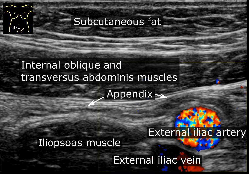
The appendix (or vermiform appendix; also cecal [or caecal] appendix; vermix; or vermiform process) is a finger-like, blind-ended tube connected to the cecum, from which it prenatal development, develops in the embryo. The cecum is a pouch-like structure of the large intestine, located at the junction of the small intestine, small and the large intestines. The term "vermiform" comes from Latin and means "worm-shaped". The appendix was once considered a Human vestigiality, vestigial organ, but this view has changed since the early 2000s. Research suggests that the appendix may serve an important purpose. In particular, it may serve as a reservoir for beneficial Gut flora, gut bacteria. Structure The human appendix averages in length but can range from . The diameter of the appendix is , and more than is considered a thickened or inflamed appendix. The longest appendix ever removed was long. The appendix is usually located in the lower right quadrant (anatomy), quadrant of th ... [...More Info...] [...Related Items...] OR: [Wikipedia] [Google] [Baidu] |
Appendix (anatomy)
The appendix (or vermiform appendix; also cecal r caecalappendix; vermix; or vermiform process) is a finger-like, blind-ended tube connected to the cecum, from which it develops in the embryo. The cecum is a pouch-like structure of the large intestine, located at the junction of the small and the large intestines. The term "vermiform" comes from Latin and means "worm-shaped". The appendix was once considered a vestigial organ, but this view has changed since the early 2000s. Research suggests that the appendix may serve an important purpose. In particular, it may serve as a reservoir for beneficial gut bacteria. Structure The human appendix averages in length but can range from . The diameter of the appendix is , and more than is considered a thickened or inflamed appendix. The longest appendix ever removed was long. The appendix is usually located in the lower right quadrant of the abdomen, near the right hip bone. The base of the appendix is located beneath the ileoce ... [...More Info...] [...Related Items...] OR: [Wikipedia] [Google] [Baidu] |
Large Intestine
The large intestine, also known as the large bowel, is the last part of the gastrointestinal tract and of the digestive system in tetrapods. Water is absorbed here and the remaining waste material is stored in the rectum as feces before being removed by defecation. The colon is the longest portion of the large intestine, and the terms are often used interchangeably but most sources define the large intestine as the combination of the cecum, colon, rectum, and anal canal. Some other sources exclude the anal canal. In humans, the large intestine begins in the right iliac region of the pelvis, just at or below the waist, where it is joined to the end of the small intestine at the cecum, via the ileocecal valve. It then continues as the colon ascending the abdomen, across the width of the abdominal cavity as the transverse colon, and then descending to the rectum and its endpoint at the anal canal. Overall, in humans, the large intestine is about long, which is about one-fi ... [...More Info...] [...Related Items...] OR: [Wikipedia] [Google] [Baidu] |
Ileocecal Valve
The ileocecal valve (ileal papilla, ileocaecal valve, Tulp's valve, Tulpius valve, Bauhin's valve, ileocecal eminence, valve of Varolius or colic valve) is a sphincter muscle valve that separates the small intestine and the large intestine. Its critical function is to limit the reflux of colonic contents into the ileum. Approximately two liters of fluid enters the colon daily through the ileocecal valve. Microanatomy The histology of the ileocecal valve shows an abrupt change from a villous mucosa pattern of the ileum to a more colonic mucosa. A thickening of the muscularis mucosa, which is the smooth muscle tissue found beneath the mucosal layer of the digestive tract. A thickening of the muscularis externa is also noted. There is also a variable amount of lymphatic tissue found at the valve. The ileocecal valve has a papillose structure. Clinical significance Colonoscopy During colonoscopy, the ileocecal valve is used, along with the appendiceal orifice, in the identifi ... [...More Info...] [...Related Items...] OR: [Wikipedia] [Google] [Baidu] |
Ileum
The ileum () is the final section of the small intestine in most higher vertebrates, including mammals, reptiles, and birds. In fish, the divisions of the small intestine are not as clear and the terms posterior intestine or distal intestine may be used instead of ileum. Its main function is to absorb vitamin B12, bile salts, and whatever products of digestion that were not absorbed by the jejunum. The ileum follows the duodenum and jejunum and is separated from the cecum by the ileocecal valve (ICV). In humans, the ileum is about 2–4 m long, and the pH is usually between 7 and 8 (neutral or slightly basic). ''Ileum ''is derived from the Greek word ''eilein'', meaning "to twist up tightly". Structure The ileum is the third and final part of the small intestine. It follows the jejunum and ends at the ileocecal junction, where the terminal ileum communicates with the cecum of the large intestine through the ileocecal valve. The ileum, along with the jejunum, is suspended ... [...More Info...] [...Related Items...] OR: [Wikipedia] [Google] [Baidu] |
Human Gastrointestinal Tract
The gastrointestinal tract (GI tract, digestive tract, alimentary canal) is the tract or passageway of the digestive system that leads from the mouth to the anus. The GI tract contains all the major organs of the digestive system, in humans and other animals, including the esophagus, stomach, and intestines. Food taken in through the mouth is digested to extract nutrients and absorb energy, and the waste expelled at the anus as feces. ''Gastrointestinal'' is an adjective meaning of or pertaining to the stomach and intestines. Most animals have a "through-gut" or complete digestive tract. Exceptions are more primitive ones: sponges have small pores ( ostia) throughout their body for digestion and a larger dorsal pore (osculum) for excretion, comb jellies have both a ventral mouth and dorsal anal pores, while cnidarians and acoels have a single pore for both digestion and excretion. The human gastrointestinal tract consists of the esophagus, stomach, and intestines, and is ... [...More Info...] [...Related Items...] OR: [Wikipedia] [Google] [Baidu] |
Gastrointestinal Tract
The gastrointestinal tract (GI tract, digestive tract, alimentary canal) is the tract or passageway of the digestive system that leads from the mouth to the anus. The GI tract contains all the major organ (biology), organs of the digestive system, in humans and other animals, including the esophagus, stomach, and intestines. Food taken in through the mouth is digestion, digested to extract nutrients and absorb energy, and the waste expelled at the anus as feces. ''Gastrointestinal'' is an adjective meaning of or pertaining to the stomach and intestines. Nephrozoa, Most animals have a "through-gut" or complete digestive tract. Exceptions are more primitive ones: sponges have small pores (ostium (sponges), ostia) throughout their body for digestion and a larger dorsal pore (osculum) for excretion, comb jellies have both a ventral mouth and dorsal anal pores, while cnidarians and acoels have a single pore for both digestion and excretion. The human gastrointestinal tract consists o ... [...More Info...] [...Related Items...] OR: [Wikipedia] [Google] [Baidu] |
Neutropenic Enterocolitis
Neutropenic enterocolitis is inflammation of the cecum (part of the large intestine) that may be associated with infection. It is particularly associated with neutropenia, a low level of neutrophil granulocytes (the most common form of white blood cells) in the blood. Signs and symptoms Signs and symptoms of typhlitis may include diarrhea, a distended abdomen, fever, chills, nausea, vomiting, and abdominal pain or tenderness. Cause The condition is usually caused by Gram-positive enteric commensal bacteria of the gut (gut flora). '' Clostridium difficile'' is a species of Gram-positive bacteria that commonly causes severe diarrhea and other intestinal diseases when competing bacteria are wiped out by antibiotics, causing pseudomembranous colitis, whereas Clostridium septicum is responsible for most cases of neutropenic enterocolitis. Typhlitis most commonly occurs in immunocompromised patients, such as those undergoing chemotherapy, patients with AIDS, kidney transplant patie ... [...More Info...] [...Related Items...] OR: [Wikipedia] [Google] [Baidu] |
Carcinoid Tumor
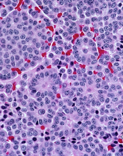
A carcinoid (also carcinoid tumor) is a slow-growing type of neuroendocrine tumor originating in the cells of the neuroendocrine system. In some cases, metastasis may occur. Carcinoid tumors of the midgut (jejunum, ileum, appendix, and cecum) are associated with carcinoid syndrome. Carcinoid tumors are the most common malignant tumor of the appendix, but they are most commonly associated with the small intestine, and they can also be found in the rectum and stomach. They are known to grow in the liver, but this finding is usually a manifestation of metastatic disease from a primary carcinoid occurring elsewhere in the body. They have a very slow growth rate compared to most malignant tumors. The median age at diagnosis for all patients with neuroendocrine tumors is 63 years. Signs and symptoms While most carcinoids are asymptomatic through the natural life and are discovered only upon surgery for unrelated reasons (so-called ''coincidental carcinoids''), all carcinoids are co ... [...More Info...] [...Related Items...] OR: [Wikipedia] [Google] [Baidu] |
Small Intestine
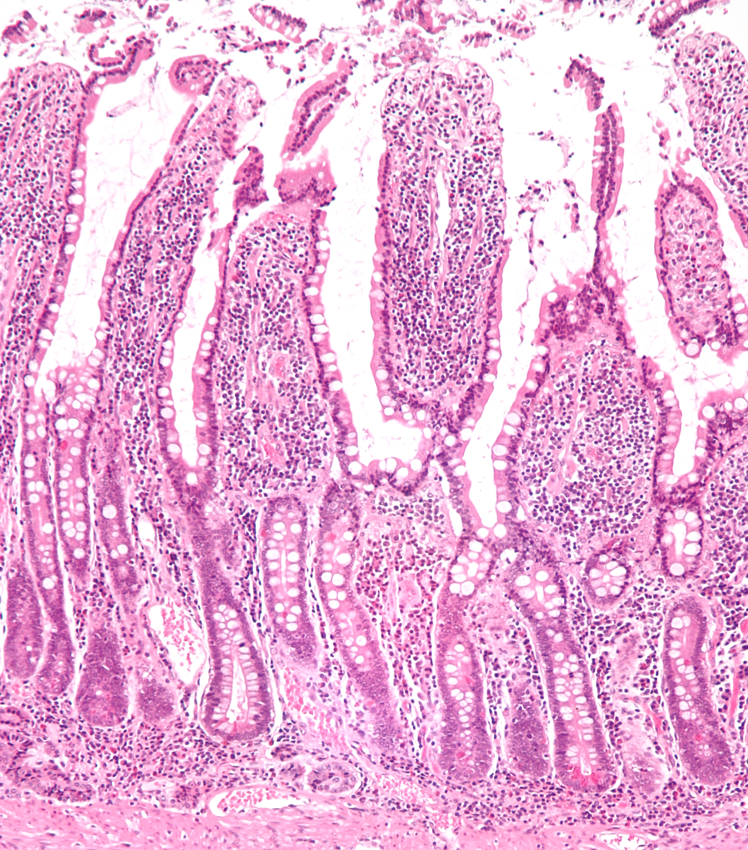
The small intestine or small bowel is an organ in the gastrointestinal tract where most of the absorption of nutrients from food takes place. It lies between the stomach and large intestine, and receives bile and pancreatic juice through the pancreatic duct to aid in digestion. The small intestine is about long and folds many times to fit in the abdomen. Although it is longer than the large intestine, it is called the small intestine because it is narrower in diameter. The small intestine has three distinct regions – the duodenum, jejunum, and ileum. The duodenum, the shortest, is where preparation for absorption through small finger-like protrusions called villi begins. The jejunum is specialized for the absorption through its lining by enterocytes: small nutrient particles which have been previously digested by enzymes in the duodenum. The main function of the ileum is to absorb vitamin B12, bile salts, and whatever products of digestion that were not absorbed by the ... [...More Info...] [...Related Items...] OR: [Wikipedia] [Google] [Baidu] |
Retroperitoneal Space
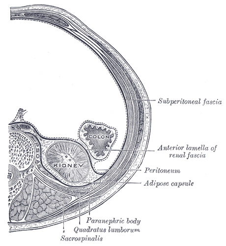
The retroperitoneal space (retroperitoneum) is the anatomical space (sometimes a potential space) behind (''retro'') the peritoneum. It has no specific delineating anatomical structures. Organs are retroperitoneal if they have peritoneum on their anterior side only. Structures that are not suspended by mesentery in the abdominal cavity and that lie between the parietal peritoneum and abdominal wall are classified as retroperitoneal. This is different from organs that are not retroperitoneal, which have peritoneum on their posterior side and are suspended by mesentery in the abdominal cavity. The retroperitoneum can be further subdivided into the following: *Perirenal (or perinephric) space *Anterior pararenal (or paranephric) space *Posterior pararenal (or paranephric) space Retroperitoneal structures Structures that lie behind the peritoneum are termed "retroperitoneal". Organs that were once suspended within the abdominal cavity by mesentery but migrated posterior to the p ... [...More Info...] [...Related Items...] OR: [Wikipedia] [Google] [Baidu] |


