|
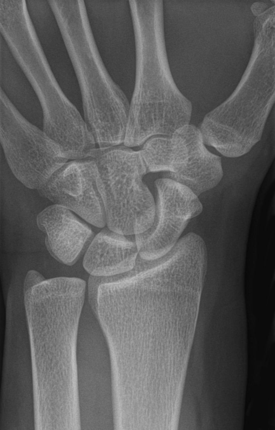
Carpal Coalition
Carpal coalition is the abnormal fusion of two or more carpal bones when they fail to segment during intrauterine development. First described by Eduard Sandifort in 1779, carpal coalitions are often an isolated issue which connect two carpal bones in the same row of the wrist. These issues are congenital and occur at various rates throughout the population. Signs and symptoms Patients with carpal coalition often offer no clinical significance and patients rarely have any associated issues. Though infrequent, some patients may complain of pain. Causes Carpal coalition result from an incomplete separation of a common embryological carpal precursor in utero, during the fifth to eighth weeks. Subtypes The lunate and triquetral bones are the most common carpal bones to fuse together, resulting in a lunotriquetral coalition in 1% of people. 60% of patients with a lunotriquetral coalition will have it bilaterally. Among isolated incidents the capitate and hamate bones are the next mos ... [...More Info...] [...Related Items...] OR: [Wikipedia] [Google] [Baidu] |
Congenital
A birth defect, also known as a congenital disorder, is an abnormal condition that is present at birth regardless of its cause. Birth defects may result in disabilities that may be physical, intellectual, or developmental. The disabilities can range from mild to severe. Birth defects are divided into two main types: structural disorders in which problems are seen with the shape of a body part and functional disorders in which problems exist with how a body part works. Functional disorders include metabolic and degenerative disorders. Some birth defects include both structural and functional disorders. Birth defects may result from genetic or chromosomal disorders, exposure to certain medications or chemicals, or certain infections during pregnancy. Risk factors include folate deficiency, drinking alcohol or smoking during pregnancy, poorly controlled diabetes, and a mother over the age of 35 years old. Many are believed to involve multiple factors. Birth defects may be vi ... [...More Info...] [...Related Items...] OR: [Wikipedia] [Google] [Baidu] |
Scaphoid
The scaphoid bone is one of the carpal bones of the wrist. It is situated between the hand and forearm on the thumb side of the wrist (also called the lateral or radial side). It forms the radial border of the carpal tunnel. The scaphoid bone is the largest bone of the proximal row of wrist bones, its long axis being from above downward, lateralward, and forward. It is approximately the size and shape of a medium cashew. Structure The scaphoid is situated between the proximal and distal rows of carpal bones. It is located on the radial side of the wrist, and articulates with the radius, lunate, trapezoid, trapezium, and capitate. Over 80% of the bone is covered in articular cartilage. Bone The palmar surface of the scaphoid is concave, and forming a distal tubercle, giving attachment to the transverse carpal ligament. The proximal surface is triangular, smooth and convex. The lateral surface is narrow and gives attachment to the radial collateral ligament. The medial su ... [...More Info...] [...Related Items...] OR: [Wikipedia] [Google] [Baidu] |
Tarsal Coalition
Tarsal coalition is an abnormal connecting bridge of tissue between two normally-separate tarsal bones. The term 'coalition' means a coming together of two or more entities to merge into one mass. The tissue connecting the bones, often referred to as a "bar", may be composed of fibrous or osseous tissue. The two most common types of tarsal coalitions are calcaneo-navicular (calcaneonavicular bar) and talo- calcaneal (talocalcaneal bar), comprising 90% of all tarsal coalitions. There are other bone coalition combinations possible, but they are very rare.Tarsal coalition and painful flatfoot, K.A. Vincent, Shriners Hospital for Children, Portland, Oregon and Department of Orthopedics, Oregon Health Sciences University, Portland, OR 97201-3905, USA Symptoms tend to occur in the same location, regardless of the location of coalition: on the lateral foot, just anterior and below the lateral malleolus. This area is called the sinus tarsi. Symptoms The bones of children are very ma ... [...More Info...] [...Related Items...] OR: [Wikipedia] [Google] [Baidu] |
Turner Syndrome
Turner syndrome (TS), also known as 45,X, or 45,X0, is a genetic condition in which a female is partially or completely missing an X chromosome. Signs and symptoms vary among those affected. Often, a short and webbed neck, low-set ears, low hairline at the back of the neck, short stature, and swollen hands and feet are seen at birth. Typically, those affected do not develop menstrual periods, or breasts without hormone treatment and are unable to have children without reproductive technology. Heart defects, diabetes, and low thyroid hormone occur in the disorder more frequently than average. Most people with Turner syndrome have normal intelligence; however, many have problems with spatial visualization that may be needed in order to learn mathematics. Vision and hearing problems also occur more often than average. Turner syndrome is not usually inherited; rather, it occurs during formation of the reproductive cells in a parent or in early cell division during development. ... [...More Info...] [...Related Items...] OR: [Wikipedia] [Google] [Baidu] |
Holt–Oram Syndrome
Holt–Oram syndrome (also called atrio-digital syndrome, atriodigital dysplasia, cardiac-limb syndrome, heart-hand syndrome type 1, HOS, ventriculo-radial syndrome) is an autosomal dominant disorder that affects bones in the arms and hands (the upper limbs) and often causes heart problems. The syndrome may include an absent radial bone in the forearm, an atrial septal defect in the heart, or heart block. It affects approximately 1 in 100,000 people. Presentation All people with Holt-Oram syndrome have, at least one, abnormal wrist bone, which can often only be detected by X-ray. Other bone abnormalities are associated with the syndrome. These vary widely in severity, and include a missing thumb, a thumb that looks like a finger, upper arm bones of unequal length or underdeveloped, partial or complete absence of bones in the forearm, and abnormalities in the collar bone or shoulder blade. Bone abnormalities may affect only one side of the body or both sides; if both sides ar ... [...More Info...] [...Related Items...] OR: [Wikipedia] [Google] [Baidu] |
Quantitative Trait Locus
A quantitative trait locus (QTL) is a locus (section of DNA) that correlates with variation of a quantitative trait in the phenotype of a population of organisms. QTLs are mapped by identifying which molecular markers (such as SNPs or AFLPs) correlate with an observed trait. This is often an early step in identifying the actual genes that cause the trait variation. Definition A quantitative trait locus (QTL) is a region of DNA which is associated with a particular phenotypic trait, which varies in degree and which can be attributed to polygenic effects, i.e., the product of two or more genes, and their environment. . These QTLs are often found on different chromosomes. The number of QTLs which explain variation in the phenotypic trait indicates the genetic architecture of a trait. It may indicate that plant height is controlled by many genes of small effect, or by a few genes of large effect. Typically, QTLs underlie continuous traits (those traits which vary continuously, ... [...More Info...] [...Related Items...] OR: [Wikipedia] [Google] [Baidu] |
Arthrodesis
Arthrodesis, also known as artificial ankylosis or syndesis, is the artificial induction of joint ossification between two bones by surgery. This is done to relieve intractable pain in a joint which cannot be managed by pain medication, splints, or other normally indicated treatments. The typical causes of such pain are fractures which disrupt the joint, severe sprains, and arthritis. It is most commonly performed on joints in the spine, hand, ankle, and foot. Historically, knee and hip arthrodeses were also performed as pain-relieving procedures, but with the great successes achieved in hip and knee arthroplasty, arthrodesis of these large joints has fallen out of favour as a primary procedure, and now is only used as a procedure of last resort in some failed arthroplasties. Method Arthrodesis can be done in several ways: * A bone graft can be created between the two bones using a bone from elsewhere in the person's body (autograft) or using donor bone (allograft) from a ... [...More Info...] [...Related Items...] OR: [Wikipedia] [Google] [Baidu] |
Trapezoid Bone
The trapezoid bone (lesser multangular bone) is a carpal bone in tetrapods, including humans. It is the smallest bone in the distal row of carpal bones that give structure to the palm of the hand. It may be known by its wedge-shaped form, the broad end of the wedge constituting the dorsal, the narrow end the palmar surface; and by its having four articular facets touching each other, and separated by sharp edges. It is homologous with the "second distal carpal" of reptiles and amphibians. Structure The trapezoid is a four-sided carpal bone found within the hand. The trapezoid is found within the distal row of carpal bones. Surfaces The '' superior surface'', quadrilateral, smooth, and slightly concave, articulates with the scaphoid. The '' inferior surface'' articulates with the proximal end of the second metacarpal bone; it is convex from side to side, concave from before backward and subdivided by an elevated ridge into two unequal facets. The ''dorsal'' and '' palmar surfa ... [...More Info...] [...Related Items...] OR: [Wikipedia] [Google] [Baidu] |
Eduard Sandifort
Eduard Sandifort (November 14, 1742 – February 12, 1814) was a Dutch physician and anatomist. He received his medical doctorate degree (Ph.D.) from Leiden University in 1763, and worked as a general practitioner in The Hague. He was fluent in Dutch, German, Swedish, and Italian. He became a professor of anatomy and surgery in 1771 at Leiden University. His most important writings are ''Observationes Anatomico-pathologicæ'' (1778), ''Excercitationes anatomicoacademicæ'' (1783–85), and the ''Museum Anatomicum Academiae Lugduno-Batavæ'' (1789–93), which was finished by his son, Gerard Sandifort (1779–1848). Sandifort translated Nils Rosén von Rosenstein's ''Underrättelser om barn-sjukdomar och deras botemedel'' (''The diseases of children, and their remedies'') to Dutch in 1768. Sandifort was elected in 1768 as a foreign member of the Swedish Royal Academy of Sciences in Stockholm. In 1779 he was the first to document a case of carpal coalition Carpal coalition is th ... [...More Info...] [...Related Items...] OR: [Wikipedia] [Google] [Baidu] |
Trapezium (bone)
The trapezium bone (greater multangular bone) is a carpal bone in the hand. It forms the radial border of the carpal tunnel. Structure The trapezium is distinguished by a deep groove on its anterior surface. It is situated at the radial side of the carpus, between the scaphoid and the first metacarpal bone (the metacarpal bone of the thumb). It is homologous with the first distal carpal of reptiles and amphibians. Surfaces The trapezium is an irregular-shaped carpal bone found within the hand. The trapezium is found within the distal row of carpal bones, and is directly adjacent to the metacarpal bone of the thumb. On its ulnar surface are found the trapezoid and scaphoid bones. The '' superior surface'' is directed upward and medialward; medially it is smooth, and articulates with the scaphoid; laterally it is rough and continuous with the lateral surface. The '' inferior surface'' is oval, concave from side to side, convex from before backward, so as to form a saddle-shaped ... [...More Info...] [...Related Items...] OR: [Wikipedia] [Google] [Baidu] |
Pisiform
The pisiform bone ( or ), also spelled pisiforme (from the Latin ''pisifomis'', pea-shaped), is a small knobbly, sesamoid bone that is found in the wrist. It forms the ulnar border of the carpal tunnel. Structure The pisiform is a sesamoid bone, with no covering membrane of periosteum. It is the last carpal bone to ossify. The pisiform bone is a small bone found in the proximal row of the wrist (carpus). It is situated where the ulna joins the wrist, within the tendon of the flexor carpi ulnaris muscle. It only has one side that acts as a joint, articulating with the triquetral bone. It is on a plane anterior to the other carpal bones and is spheroidal in form. The pisiform bone has four surfaces: # The ''dorsal surface'' is smooth and oval, and articulates with the triquetral: this facet approaches the superior, but not the inferior border of the bone. # The ''palmar surface'' is rounded and rough, and gives attachment to the transverse carpal ligament, the flexor carpi ulnaris ... [...More Info...] [...Related Items...] OR: [Wikipedia] [Google] [Baidu] |


