|
Calyx Of Held
The Calyx of Held is a particularly large synapse in the mammalian auditory central nervous system, so named after Hans Held who first described it in his 1893 article ''Die centrale Gehörleitung''Held, H. "Die centrale Gehörleitung" Arch. Anat. Physiol. Anat. Abt, 1893 because of its resemblance to the calyx of a flower. Globular bushy cells in the anteroventral cochlear nucleus (AVCN) send axons to the contralateral medial nucleus of the trapezoid body (MNTB), where they synapse via these calyces on MNTB principal cells. These principal cells then project to the ipsilateral lateral superior olive (LSO), where they inhibit postsynaptic neurons and provide a basis for interaural level detection (ILD), required for high frequency sound localization. This synapse has been described as the largest in the brain. The related endbulb of Held is also a large axon terminal smaller synapse (15-30 μm in diameter) found in other auditory brainstem structures, namely the cochlear ... [...More Info...] [...Related Items...] OR: [Wikipedia] [Google] [Baidu] |
Calyx Of Held V3
Calyx or calyce (plural "calyces"), from the Latin ''calix'' which itself comes from the Ancient Greek ''κάλυξ'' (''kálux'') meaning "husk" or "pod", may refer to: Biology * Calyx (anatomy), collective name for several cup-like structures in animal anatomy * Calyx (botany), the collective name for sepals of a flower * ''Calyce'' (beetle), a genus of beetles * ''Calyx'' (sponge), a genus of sea sponges * Calyx of Held, a large synapse in the auditory brainstem structure * '' Eubela calyx'', species of sea snail * Renal calyx, a chamber in the kidney that surrounds the apex of the renal pyramids Other uses * Calyx (fictional moon), a fictional moon in ''Colony Wars'' * ''Calyx'' (magazine), a literary journal * Calyx (musician), UK producer of drum and bass * Calyce (mythology), several figures in Greek mythology * CALYX, publisher * Calyx AI, a biopharmaceutical services company * Calyx Institute, a non-profit education and research organization * "Calyx", a textil ... [...More Info...] [...Related Items...] OR: [Wikipedia] [Google] [Baidu] |
Hyperpolarization (biology)
Hyperpolarization is a change in a cell's membrane potential that makes it more negative. It is the opposite of a depolarization. It inhibits action potentials by increasing the stimulus required to move the membrane potential to the action potential threshold. Hyperpolarization is often caused by efflux of K+ (a cation) through K+ channels, or influx of Cl– (an anion) through Cl– channels. On the other hand, influx of cations, e.g. Na+ through Na+ channels or Ca2+ through Ca2+ channels, inhibits hyperpolarization. If a cell has Na+ or Ca2+ currents at rest, then inhibition of those currents will also result in a hyperpolarization. This voltage-gated ion channel response is how the hyperpolarization state is achieved. In neurons, the cell enters a state of hyperpolarization immediately following the generation of an action potential. While hyperpolarized, the neuron is in a refractory period that lasts roughly 2 milliseconds, during which the neuron is unabl ... [...More Info...] [...Related Items...] OR: [Wikipedia] [Google] [Baidu] |
NG2 Glia
Oligodendrocyte progenitor cells (OPCs), also known as oligodendrocyte precursor cells, NG2-glia, O2A cells, or polydendrocytes, are a subtype of glia in the central nervous system named for their essential role as precursor cell, precursors to oligodendrocytes. They are typically identified by coexpression of PDGFRA and NG2 proteoglycan, NG2. OPCs play a critical role in developmental and adult myelinogenesis by giving rise to oligodendrocytes, which then ensheath axons and provide electrical insulation in the form of a myelin sheath, enabling faster action potential propagation and high fidelity transmission without a need for an increase in axonal diameter. The loss or lack of OPCs, and consequent lack of differentiated oligodendrocytes, is associated with a loss of myelination and subsequent impairment of neurological functions. In addition, OPCs express receptors for various neurotransmitters and undergo membrane depolarization when they receive synaptic inputs from neurons. ... [...More Info...] [...Related Items...] OR: [Wikipedia] [Google] [Baidu] |
Astrocyte
Astrocytes (from Ancient Greek , , "star" + , , "cavity", "cell"), also known collectively as astroglia, are characteristic star-shaped glial cells in the brain and spinal cord. They perform many functions, including biochemical control of endothelial cells that form the blood–brain barrier, provision of nutrients to the nervous tissue, maintenance of extracellular ion balance, regulation of cerebral blood flow, and a role in the repair and scarring process of the brain and spinal cord following infection and traumatic injuries. The proportion of astrocytes in the brain is not well defined; depending on the counting technique used, studies have found that the astrocyte proportion varies by region and ranges from 20% to 40% of all glia. Another study reports that astrocytes are the most numerous cell type in the brain. Astrocytes are the major source of cholesterol in the central nervous system. Apolipoprotein E transports cholesterol from astrocytes to neurons and other gli ... [...More Info...] [...Related Items...] OR: [Wikipedia] [Google] [Baidu] |
Neuroglia
Glia, also called glial cells (gliocytes) or neuroglia, are non-neuronal cells in the central nervous system (brain and spinal cord) and the peripheral nervous system that do not produce electrical impulses. They maintain homeostasis, form myelin in the peripheral nervous system, and provide support and protection for neurons. In the central nervous system, glial cells include oligodendrocytes, astrocytes, ependymal cells, and microglia, and in the peripheral nervous system they include Schwann cells and satellite cells. Function They have four main functions: *to surround neurons and hold them in place *to supply nutrients and oxygen to neurons *to insulate one neuron from another *to destroy pathogens and remove dead neurons. They also play a role in neurotransmission and synaptic connections, and in physiological processes such as breathing. While glia were thought to outnumber neurons by a ratio of 10:1, recent studies using newer methods and reappraisal of historical ... [...More Info...] [...Related Items...] OR: [Wikipedia] [Google] [Baidu] |
Calcium Buffering
Calcium buffering describes the processes which help stabilise the concentration of free calcium ions within cells, in a similar manner to how pH buffers maintain a stable concentration of hydrogen ions. The majority of calcium ions within the cell are bound to intracellular proteins, leaving a minority freely dissociated. When calcium is added to or removed from the cytoplasm by transport across the cell membrane or sarcoplasmic reticulum, calcium buffers minimise the effect on changes in cytoplasmic free calcium concentration by binding calcium to or releasing calcium from intracellular proteins. As a result, 99% of the calcium added to the cytosol of a cardiomyocyte during each cardiac cycle becomes bound to calcium buffers, creating a relatively small change in free calcium. The regulation of free calcium is of particular importance in excitable cells like cardiomyocytes and neurons. Within these cells, many intracellular proteins can act as calcium buffers. In cardiac muscl ... [...More Info...] [...Related Items...] OR: [Wikipedia] [Google] [Baidu] |
Depolarization
In biology, depolarization or hypopolarization is a change within a cell, during which the cell undergoes a shift in electric charge distribution, resulting in less negative charge inside the cell compared to the outside. Depolarization is essential to the function of many cells, communication between cells, and the overall physiology of an organism. Most cells in higher organisms maintain an internal environment that is negatively charged relative to the cell's exterior. This difference in charge is called the cell's membrane potential. In the process of depolarization, the negative internal charge of the cell temporarily becomes more positive (less negative). This shift from a negative to a more positive membrane potential occurs during several processes, including an action potential. During an action potential, the depolarization is so large that the potential difference across the cell membrane briefly reverses polarity, with the inside of the cell becoming positively char ... [...More Info...] [...Related Items...] OR: [Wikipedia] [Google] [Baidu] |
Mitochondrion
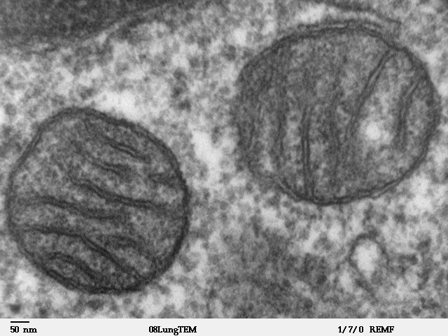
A mitochondrion (; ) is an organelle found in the cells of most Eukaryotes, such as animals, plants and fungi. Mitochondria have a double membrane structure and use aerobic respiration to generate adenosine triphosphate (ATP), which is used throughout the cell as a source of chemical energy. They were discovered by Albert von Kölliker in 1857 in the voluntary muscles of insects. The term ''mitochondrion'' was coined by Carl Benda in 1898. The mitochondrion is popularly nicknamed the "powerhouse of the cell", a phrase coined by Philip Siekevitz in a 1957 article of the same name. Some cells in some multicellular organisms lack mitochondria (for example, mature mammalian red blood cells). A large number of unicellular organisms, such as microsporidia, parabasalids and diplomonads, have reduced or transformed their mitochondria into other structures. One eukaryote, '' Monocercomonoides'', is known to have completely lost its mitochondria, and one multicellular organ ... [...More Info...] [...Related Items...] OR: [Wikipedia] [Google] [Baidu] |
Microtubules
Microtubules are polymers of tubulin that form part of the cytoskeleton and provide structure and shape to eukaryotic cells. Microtubules can be as long as 50 micrometres, as wide as 23 to 27 nm and have an inner diameter between 11 and 15 nm. They are formed by the polymerization of a dimer of two globular proteins, alpha and beta tubulin into protofilaments that can then associate laterally to form a hollow tube, the microtubule. The most common form of a microtubule consists of 13 protofilaments in the tubular arrangement. Microtubules play an important role in a number of cellular processes. They are involved in maintaining the structure of the cell and, together with microfilaments and intermediate filaments, they form the cytoskeleton. They also make up the internal structure of cilia and flagella. They provide platforms for intracellular transport and are involved in a variety of cellular processes, including the movement of secretory vesicles, or ... [...More Info...] [...Related Items...] OR: [Wikipedia] [Google] [Baidu] |
Neuropeptides
Neuropeptides are chemical messengers made up of small chains of amino acids that are synthesized and released by neurons. Neuropeptides typically bind to G protein-coupled receptors (GPCRs) to modulate neural activity and other tissues like the gut, muscles, and heart. There are over 100 known neuropeptides, representing the largest and most diverse class of signaling molecules in the nervous system. Neuropeptides are synthesized from large precursor proteins which are cleaved and post-translationally processed then packaged into dense core vesicles. Neuropeptides are often co-released with other neuropeptides and neurotransmitters in a single neuron, yielding a multitude of effects. Once released, neuropeptides can diffuse widely to affect a broad range of targets. Synthesis Neuropeptides are synthesized from large, inactive precursor proteins called prepropeptides. Prepropeptides contain sequences for a family of distinct peptides and often contain repeated copies of the ... [...More Info...] [...Related Items...] OR: [Wikipedia] [Google] [Baidu] |
Cochlea
The cochlea is the part of the inner ear involved in hearing. It is a spiral-shaped cavity in the bony labyrinth, in humans making 2.75 turns around its axis, the modiolus. A core component of the cochlea is the Organ of Corti, the sensory organ of hearing, which is distributed along the partition separating the fluid chambers in the coiled tapered tube of the cochlea. The name cochlea derives . Structure The cochlea (plural is cochleae) is a spiraled, hollow, conical chamber of bone, in which waves propagate from the base (near the middle ear and the oval window) to the apex (the top or center of the spiral). The spiral canal of the cochlea is a section of the bony labyrinth of the inner ear that is approximately 30 mm long and makes 2 turns about the modiolus. The cochlear structures include: * Three ''scalae'' or chambers: ** the vestibular duct or ''scala vestibuli'' (containing perilymph), which lies superior to the cochlear duct and abuts the oval window ** the ty ... [...More Info...] [...Related Items...] OR: [Wikipedia] [Google] [Baidu] |


