|
Cajal–Retzius Cell
Cajal–Retzius cells (CR cells) (also known as Horizontal cells of Cajal) are a heterogeneous population of morphologically and molecularly distinct reelin-producing cell types in the marginal zone/layer I of the developmental cerebral cortex and in the immature hippocampus of different species and at different times during embryogenesis and postnatal life. These cells were discovered by two scientists, Santiago Ramón y Cajal and Gustaf Retzius, at two different times and in different species. They are originated in the developing brain in multiple sites within the neocortex and hippocampus. From there, Cajal–Retzius (CR) cells migrate through the marginal zone, originating the layer I of the cortex. As these cells are involved in the correct organization of the developing brain, there are several studies implicating CR cells in neurodevelopmental disorders, especially schizophrenia, bipolar disorder, autism, lissencephaly and temporal lobe epilepsy. Development In 1971 it ... [...More Info...] [...Related Items...] OR: [Wikipedia] [Google] [Baidu] |
Santiago Ramón Y Cajal
Santiago Ramón y Cajal (; 1 May 1852 – 17 October 1934) was a Spanish neuroscientist, pathologist, and histologist specializing in neuroanatomy and the central nervous system. He and Camillo Golgi received the Nobel Prize in Physiology or Medicine in 1906. Ramón y Cajal was the first person of Spanish origin to win a scientific Nobel Prize. His original investigations of the microscopic structure of the brain made him a pioneer of modern neuroscience. Hundreds of his drawings illustrating the arborizations ("tree growing") of brain cells are still in use, since the mid-20th century, for educational and training purposes. Biography Santiago Ramón y Cajal was born on the 1st of May 1852 in the town of Petilla de Aragón, Navarre, Spain. As a child he was transferred many times from one school to another because of behavior that was declared poor, rebellious, and showing an anti-authoritarian attitude. An extreme example of his precociousness and rebelliousness at the age ... [...More Info...] [...Related Items...] OR: [Wikipedia] [Google] [Baidu] |
Patch Clamp
The patch clamp technique is a laboratory technique in electrophysiology used to study ionic currents in individual isolated living cells, tissue sections, or patches of cell membrane. The technique is especially useful in the study of excitable cells such as neurons, cardiomyocytes, muscle fibers, and pancreatic beta cells, and can also be applied to the study of bacterial ion channels in specially prepared giant spheroplasts. Patch clamping can be performed using the voltage clamp technique. In this case, the voltage across the cell membrane is controlled by the experimenter and the resulting currents are recorded. Alternatively, the current clamp technique can be used. In this case, the current passing across the membrane is controlled by the experimenter and the resulting changes in voltage are recorded, generally in the form of action potentials. Erwin Neher and Bert Sakmann developed the patch clamp in the late 1970s and early 1980s. This discovery made it possib ... [...More Info...] [...Related Items...] OR: [Wikipedia] [Google] [Baidu] |
Lissencephaly
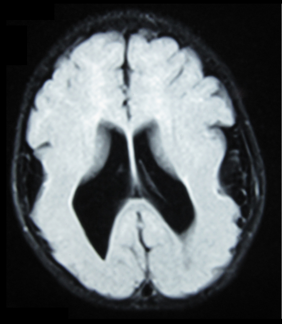
Lissencephaly (, meaning "smooth brain") is a set of rare brain disorders whereby the whole or parts of the surface of the brain appear smooth. It is caused by defective neuronal migration during the 12th to 24th weeks of gestation resulting in a lack of development of brain folds ( gyri) and grooves (sulci). It is a form of cephalic disorder. Terms such as ''agyria'' (no gyri) and '' pachygyria'' (broad gyri) are used to describe the appearance of the surface of the brain. Children with lissencephaly generally have significant developmental delays, but these vary greatly from child to child depending on the degree of brain malformation and seizure control. Life expectancy can be shortened, generally due to respiratory problems. Symptoms and signs Affected children display severe psychomotor impairment, failure to thrive, seizures, and muscle spasticity or hypotonia. Other symptoms of the disorder may include unusual facial appearance, difficulty swallowing, and anomalies of th ... [...More Info...] [...Related Items...] OR: [Wikipedia] [Google] [Baidu] |
Schizophrenia
Schizophrenia is a mental disorder characterized by continuous or relapsing episodes of psychosis. Major symptoms include hallucinations (typically hearing voices), delusions, and disorganized thinking. Other symptoms include social withdrawal, decreased emotional expression, and apathy. Symptoms typically develop gradually, begin during young adulthood, and in many cases never become resolved. There is no objective diagnostic test; diagnosis is based on observed behavior, a history that includes the person's reported experiences, and reports of others familiar with the person. To be diagnosed with schizophrenia, symptoms and functional impairment need to be present for six months (DSM-5) or one month ( ICD-11). Many people with schizophrenia have other mental disorders, especially substance use disorders, depressive disorders, anxiety disorders, and obsessive–compulsive disorder. About 0.3% to 0.7% of people are diagnosed with schizophrenia during their lifet ... [...More Info...] [...Related Items...] OR: [Wikipedia] [Google] [Baidu] |
Alzheimer's Disease
Alzheimer's disease (AD) is a neurodegeneration, neurodegenerative disease that usually starts slowly and progressively worsens. It is the cause of 60–70% of cases of dementia. The most common early symptom is difficulty in short-term memory, remembering recent events. As the disease advances, symptoms can include primary progressive aphasia, problems with language, Orientation (mental), disorientation (including easily getting lost), mood swings, loss of motivation, self-neglect, and challenging behaviour, behavioral issues. As a person's condition declines, they often withdraw from family and society. Gradually, bodily functions are lost, ultimately leading to death. Although the speed of progression can vary, the typical life expectancy following diagnosis is three to nine years. The cause of Alzheimer's disease is poorly understood. There are many environmental and genetic risk factors associated with its development. The strongest genetic risk factor is from an alle ... [...More Info...] [...Related Items...] OR: [Wikipedia] [Google] [Baidu] |
Reeler
A reeler is a mouse mutant, so named because of its characteristic "reeling" gait. This is caused by the profound underdevelopment of the mouse's cerebellum, a segment of the brain responsible for locomotion. The mutation is autosomal and recessive, and prevents the typical cerebellar folia from forming. Cortical neurons are generated normally but are abnormally placed, resulting in disorganization of cortical laminar layers in the central nervous system. The reason is the lack of Reelin, an extracellular matrix glycoprotein, which, during the corticogenesis, is secreted mainly by the Cajal-Retzius cells. In the reeler neocortex, cortical plate neurons are aligned in a practically inverted fashion (‘‘outside-in’’). In the ventricular zone of the cortex fewer neurons have been found to have radial glial processes. In the dentate gyrus of hippocampus, no characteristic radial glial scaffold is formed and no compact granule cell layer is established. Therefore, the reeler m ... [...More Info...] [...Related Items...] OR: [Wikipedia] [Google] [Baidu] |
Cerebral Cortex
The cerebral cortex, also known as the cerebral mantle, is the outer layer of neural tissue of the cerebrum of the brain in humans and other mammals. The cerebral cortex mostly consists of the six-layered neocortex, with just 10% consisting of allocortex. It is separated into two cortices, by the longitudinal fissure that divides the cerebrum into the left and right cerebral hemispheres. The two hemispheres are joined beneath the cortex by the corpus callosum. The cerebral cortex is the largest site of neural integration in the central nervous system. It plays a key role in attention, perception, awareness, thought, memory, language, and consciousness. The cerebral cortex is part of the brain responsible for cognition. In most mammals, apart from small mammals that have small brains, the cerebral cortex is folded, providing a greater surface area in the confined volume of the cranium. Apart from minimising brain and cranial volume, cortical folding is crucial for ... [...More Info...] [...Related Items...] OR: [Wikipedia] [Google] [Baidu] |
Cerebellum
The cerebellum (Latin for "little brain") is a major feature of the hindbrain of all vertebrates. Although usually smaller than the cerebrum, in some animals such as the mormyrid fishes it may be as large as or even larger. In humans, the cerebellum plays an important role in motor control. It may also be involved in some cognitive functions such as attention and language as well as emotional control such as regulating fear and pleasure responses, but its movement-related functions are the most solidly established. The human cerebellum does not initiate movement, but contributes to coordination, precision, and accurate timing: it receives input from sensory systems of the spinal cord and from other parts of the brain, and integrates these inputs to fine-tune motor activity. Cerebellar damage produces disorders in fine movement, equilibrium, posture, and motor learning in humans. Anatomically, the human cerebellum has the appearance of a separate structure attached to th ... [...More Info...] [...Related Items...] OR: [Wikipedia] [Google] [Baidu] |
Reeler
A reeler is a mouse mutant, so named because of its characteristic "reeling" gait. This is caused by the profound underdevelopment of the mouse's cerebellum, a segment of the brain responsible for locomotion. The mutation is autosomal and recessive, and prevents the typical cerebellar folia from forming. Cortical neurons are generated normally but are abnormally placed, resulting in disorganization of cortical laminar layers in the central nervous system. The reason is the lack of Reelin, an extracellular matrix glycoprotein, which, during the corticogenesis, is secreted mainly by the Cajal-Retzius cells. In the reeler neocortex, cortical plate neurons are aligned in a practically inverted fashion (‘‘outside-in’’). In the ventricular zone of the cortex fewer neurons have been found to have radial glial processes. In the dentate gyrus of hippocampus, no characteristic radial glial scaffold is formed and no compact granule cell layer is established. Therefore, the reeler m ... [...More Info...] [...Related Items...] OR: [Wikipedia] [Google] [Baidu] |
DAB1
The Disabled-1 (Dab1) gene encodes a key regulator of Reelin signaling. Reelin is a large glycoprotein secreted by neurons of the developing brain, particularly Cajal-Retzius cells. DAB1 functions downstream of Reln in a signaling pathway that controls cell positioning in the developing brain and during adult neurogenesis. It docks to the intracellular part of the Reelin very low density lipoprotein receptor ( VLDLR) and apoE receptor type 2 ( ApoER2) and becomes tyrosine-phosphorylated following binding of Reelin to cortical neurons. In mice, mutations of Dab1 and Reelin generate identical phenotypes. In humans, Reelin mutations are associated with brain malformations and mental retardation. In mice, Dab1 mutation results in the ''scrambler'' mouse phenotype. With a genomic length of 1.1 Mbp for a coding region of 5.5 kb, DAB1 provides a rare example of genomic complexity, which will impede the identification of human mutations. Gene function Cortical neurons form in special ... [...More Info...] [...Related Items...] OR: [Wikipedia] [Google] [Baidu] |
Low-density Lipoprotein Receptor-related Protein 8
Low-density lipoprotein receptor-related protein 8 (LRP8), also known as apolipoprotein E receptor 2 (ApoER2), is a protein that in humans is encoded by the ''LRP8'' gene. ApoER2 is a cell surface receptor that is part of the low-density lipoprotein receptor family. These receptors function in signal transduction and endocytosis of specific ligands. Through interactions with one of its ligands, reelin, ApoER2 plays an important role in embryonic neuronal migration and postnatal long-term potentiation. Another LDL family receptor, VLDLR, also interacts with reelin, and together these two receptors influence brain development and function. Decreased expression of ApoER2 is associated with certain neurological diseases. Structure ApoER2 is a protein made up of 870 amino acids. It is separated into a ligand binding domain of eight ligand binding regions, an EGF-like domain containing three cysteine-rich repeats, an O-linked glycosylation domain of 89 amino acids, a transmembrane do ... [...More Info...] [...Related Items...] OR: [Wikipedia] [Google] [Baidu] |
VLDL Receptor
The very-low-density-lipoprotein receptor (VLDLR) is a transmembrane lipoprotein receptor of the low-density-lipoprotein (LDL) receptor family. VLDLR shows considerable homology with the members of this lineage. Discovered in 1992 by T. Yamamoto, VLDLR is widely distributed throughout the tissues of the body, including the heart, skeletal muscle, adipose tissue, and the brain, but is absent from the liver. This receptor has an important role in cholesterol uptake, metabolism of apolipoprotein E-containing triacylglycerol-rich lipoproteins, and neuronal migration in the developing brain. In humans, VLDLR is encoded by the ''VLDLR'' gene. Mutations of this gene may lead to a variety of symptoms and diseases, which include type I lissencephaly, cerebellar hypoplasia, and atherosclerosis. Protein structure VLDLR is a member of the low-density-lipoprotein (LDL) receptor family, which is entirely composed of type I transmembrane lipoprotein receptors. All members of this family sha ... [...More Info...] [...Related Items...] OR: [Wikipedia] [Google] [Baidu] |


