|
Bronchial Veins
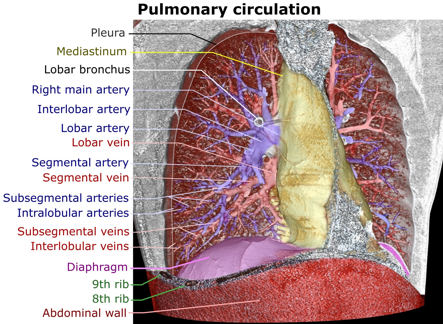
The bronchial veins are small vessels that return blood from the larger bronchi and structures at the roots of the lungs. The right side drains into the azygos vein, while the left side drains into the left superior intercostal vein or the accessory hemiazygos vein. Bronchial veins are thereby part of the bronchial circulation, carrying waste products away from the cells that constitute the lungs. The bronchial veins are counterparts to the bronchial arteries. However, they only carry ~13% of the blood flow of the bronchial arteries. The remaining blood is returned to the heart via the pulmonary veins. See also * Bronchial arteries * Pulmonary arteries * Pulmonary veins The pulmonary veins are the veins that transfer oxygenated blood from the lungs to the heart. The largest pulmonary veins are the four ''main pulmonary veins'', two from each lung that drain into the left atrium of the heart. The pulmonary vein ... References External links * Veins of the torso ... [...More Info...] [...Related Items...] OR: [Wikipedia] [Google] [Baidu] |
Azygos Vein
The azygos vein is a vein running up the right side of the thoracic vertebral column draining itself towards the superior vena cava. It connects the systems of superior vena cava and inferior vena cava and can provide an alternative path for blood to the right atrium when either of the venae cavae is blocked. Structure The azygos vein transports deoxygenated blood from the posterior walls of the thorax and abdomen into the superior vena cava. It is formed by the union of the ascending lumbar veins with the right subcostal veins at the level of the 12th thoracic vertebra, ascending to the right of the descending aorta and thoracic duct, passing behind the right crus of diaphragm, anterior to the vertebral bodies of T12 to T5 and right posterior intercostal arteries. At the level of T4 vertebrae, it arches over the root of the right lung from behind to the front to join the superior vena cava. The trachea and oesophagus is located medially to the arch of the azygous vein. The ... [...More Info...] [...Related Items...] OR: [Wikipedia] [Google] [Baidu] |
Bronchial Circulation
The bronchial circulation is the part of the circulatory system that supplies nutrients and oxygen to the cells that constitute the lungs, as well as carrying waste products away from them. It is complementary to the pulmonary circulation that brings deoxygenated blood to the lungs and carries oxygenated blood away from them in order to oxygenate the rest of the body. In the bronchial circulation, blood goes through the following steps: #Bronchial arteries that carry oxygenated blood to the lungs #Pulmonary capillaries, where there is exchange of water, oxygen, carbon dioxide, and many other nutrients and waste chemical substances between blood and the tissues #Veins, where only a minority of the blood goes through bronchial veins, and most of it through pulmonary veins. Blood reaches from the pulmonary circulation into the lungs for gas exchange to oxygenate the rest of the body tissues. But bronchial circulation supplies fully oxygenated arterial blood to the lung tissues the ... [...More Info...] [...Related Items...] OR: [Wikipedia] [Google] [Baidu] |
Pulmonary Arteries
A pulmonary artery is an artery in the pulmonary circulation that carries deoxygenated blood from the right side of the heart to the lungs. The largest pulmonary artery is the ''main pulmonary artery'' or ''pulmonary trunk'' from the heart, and the smallest ones are the arterioles, which lead to the capillaries that surround the pulmonary alveoli. Structure The pulmonary arteries are blood vessels that carry systemic venous blood from the right ventricle of the heart to the microcirculation of the lungs. Unlike in other organs where arteries supply oxygenated blood, the blood carried by the pulmonary arteries is deoxygenated, as it is venous blood returning to the heart. The main pulmonary arteries emerge from the right side of the heart, and then split into smaller arteries that progressively divide and become arterioles, eventually narrowing into the capillary microcirculation of the lungs where gas exchange occurs. Pulmonary trunk In order of blood flow, the pulmonary art ... [...More Info...] [...Related Items...] OR: [Wikipedia] [Google] [Baidu] |
Bronchial Arteries
In human anatomy, the bronchial arteries supply the lungs with nutrition and oxygenated blood. Although there is much variation, there are usually two bronchial arteries that run to the left lung, and one to the right lung and are a vital part of the respiratory system. Structure There are typically two left and one right bronchial arteries. The ''left bronchial arteries'' (superior and inferior) usually arise directly from the thoracic aorta. The single ''right bronchial artery'' usually arises from one of the following: * 1) the thoracic aorta at a common trunk with the right 3rd posterior intercostal artery * 2) the superior bronchial artery on the left side * 3) any number of the right intercostal arteries mostly the third right posterior. Function The bronchial arteries supply blood to the bronchi and connective tissue of the lungs. They travel with and branch with the bronchi, ending about at the level of the respiratory bronchioles. They anastomose with the branch ... [...More Info...] [...Related Items...] OR: [Wikipedia] [Google] [Baidu] |
Pulmonary Veins
The pulmonary veins are the veins that transfer oxygenated blood from the lungs to the heart. The largest pulmonary veins are the four ''main pulmonary veins'', two from each lung that drain into the left atrium of the heart. The pulmonary veins are part of the pulmonary circulation. Structure There are four main pulmonary veins, two from each lung – an inferior and a superior main vein, emerging from each hilum. The main pulmonary veins receive blood from three or four feeding veins in each lung, and drain into the left atrium. The peripheral feeding veins do not follow the bronchial tree. They run between the pulmonary segments from which they drain the blood. At the root of the lung, the right superior pulmonary vein lies in front of and a little below the pulmonary artery; the inferior is situated at the lowest part of the lung hilum. Behind the pulmonary artery is the bronchus. The right main pulmonary veins (contains oxygenated blood) pass behind the right atrium and ... [...More Info...] [...Related Items...] OR: [Wikipedia] [Google] [Baidu] |
Bronchial Arteries
In human anatomy, the bronchial arteries supply the lungs with nutrition and oxygenated blood. Although there is much variation, there are usually two bronchial arteries that run to the left lung, and one to the right lung and are a vital part of the respiratory system. Structure There are typically two left and one right bronchial arteries. The ''left bronchial arteries'' (superior and inferior) usually arise directly from the thoracic aorta. The single ''right bronchial artery'' usually arises from one of the following: * 1) the thoracic aorta at a common trunk with the right 3rd posterior intercostal artery * 2) the superior bronchial artery on the left side * 3) any number of the right intercostal arteries mostly the third right posterior. Function The bronchial arteries supply blood to the bronchi and connective tissue of the lungs. They travel with and branch with the bronchi, ending about at the level of the respiratory bronchioles. They anastomose with the branch ... [...More Info...] [...Related Items...] OR: [Wikipedia] [Google] [Baidu] |
Human Lungs
The lungs are the primary organs of the respiratory system in humans and most other animals, including some snails and a small number of fish. In mammals and most other vertebrates, two lungs are located near the backbone on either side of the heart. Their function in the respiratory system is to extract oxygen from the air and transfer it into the bloodstream, and to release carbon dioxide from the bloodstream into the atmosphere, in a process of gas exchange. Respiration is driven by different muscular systems in different species. Mammals, reptiles and birds use their different muscles to support and foster breathing. In earlier tetrapods, air was driven into the lungs by the pharyngeal muscles via buccal pumping, a mechanism still seen in amphibians. In humans, the main muscle of respiration that drives breathing is the diaphragm. The lungs also provide airflow that makes vocal sounds including human speech possible. Humans have two lungs, one on the left and on ... [...More Info...] [...Related Items...] OR: [Wikipedia] [Google] [Baidu] |
Cell (biology)
The cell is the basic structural and functional unit of life forms. Every cell consists of a cytoplasm enclosed within a membrane, and contains many biomolecules such as proteins, DNA and RNA, as well as many small molecules of nutrients and metabolites.Cell Movements and the Shaping of the Vertebrate Body in Chapter 21 of Molecular Biology of the Cell '' fourth edition, edited by Bruce Alberts (2002) published by Garland Science. The Alberts text discusses how the "cellular building blocks" move to shape developing embryos. It is also common to describe small molecules such as ... [...More Info...] [...Related Items...] OR: [Wikipedia] [Google] [Baidu] |
Accessory Hemiazygos Vein
The accessory hemiazygos vein, also called the superior hemiazygous vein, is a vein on the left side of the vertebral column that generally drains the fourth through eighth intercostal spaces on the left side of the body. Structure The accessory hemiazygos vein varies inversely in size with the left superior intercostal vein. It usually receives the posterior intercostal veins from the 4th, 5th, 6th, 7th, and 8th intercostal spaces between the left superior intercostal vein and highest tributary of the hemiazygos vein; the left bronchial vein sometimes opens into it. The vein usually crosses the body of the eighth thoracic vertebra to join the azygos vein The azygos vein is a vein running up the right side of the thoracic vertebral column draining itself towards the superior vena cava. It connects the systems of superior vena cava and inferior vena cava and can provide an alternative path for blo .... Alternatively, it ends in the hemiazygos vein. When this vein is small ... [...More Info...] [...Related Items...] OR: [Wikipedia] [Google] [Baidu] |
Hemiazygos Vein
The hemiazygos vein (vena azygos minor inferior) is a vein running superiorly in the lower thoracic region, just to the left side of the vertebral column. Structure The hemiazygos vein and the accessory hemiazygos vein, when taken together, essentially serve as the left-sided equivalent of the azygos vein. That is, the azygos vein serves to drain most of the posterior intercostal veins on the right side of the body, and the hemiazygos vein and the accessory hemiazygos vein drain most of the posterior intercostal veins on the left side of the body. Specifically, the hemiazygos vein mirrors the bottom part of the azygos vein. The structure of the hemiazygos vein is often variable. It usually begins in the left ascending lumbar vein or renal vein, and passes upward through the left crus of the diaphragm to enter the thorax. It continues ascending on the left side of the vertebral column, and around the level of the ninth thoracic vertebra, it passes rightward across the column, behin ... [...More Info...] [...Related Items...] OR: [Wikipedia] [Google] [Baidu] |
Left Superior Intercostal Vein
The superior intercostal veins are two veins that drain the 2nd, 3rd, and 4th intercostal spaces, one vein for each side of the body. Right superior intercostal vein The right superior intercostal vein drains the 2nd, 3rd, and 4th posterior intercostal veins on the right side of the body. It flows into the azygos vein. Left superior intercostal vein The left superior intercostal vein drains the 2nd and 3rd posterior intercostal veins on the left side of the body. It usually drains into the left brachiocephalic vein. It may also communicate with the accessory hemiazygos vein. As it passes posteriorly above the aortic arch, it crosses deep to the phrenic nerve and the pericardiacophrenic vessels and then superficial to the vagus nerve. See also * Supreme intercostal vein * Posterior intercostal veins The posterior intercostal veins are veins that drain the intercostal spaces posteriorly. They run with their corresponding posterior intercostal artery on the underside of the rib, t ... [...More Info...] [...Related Items...] OR: [Wikipedia] [Google] [Baidu] |
Azygos Vein
The azygos vein is a vein running up the right side of the thoracic vertebral column draining itself towards the superior vena cava. It connects the systems of superior vena cava and inferior vena cava and can provide an alternative path for blood to the right atrium when either of the venae cavae is blocked. Structure The azygos vein transports deoxygenated blood from the posterior walls of the thorax and abdomen into the superior vena cava. It is formed by the union of the ascending lumbar veins with the right subcostal veins at the level of the 12th thoracic vertebra, ascending to the right of the descending aorta and thoracic duct, passing behind the right crus of diaphragm, anterior to the vertebral bodies of T12 to T5 and right posterior intercostal arteries. At the level of T4 vertebrae, it arches over the root of the right lung from behind to the front to join the superior vena cava. The trachea and oesophagus is located medially to the arch of the azygous vein. The ... [...More Info...] [...Related Items...] OR: [Wikipedia] [Google] [Baidu] |


