|
Breast MRI
One alternative to mammography, breast MRI or contrast-enhanced magnetic resonance imaging (MRI), has shown substantial progress in the detection of breast cancer. Uses Some of the uses of MRI of the breasts are: screening for malignancy in women with greater than 20% lifetime risk of breast cancer (especially those with high risk genes such as BRCA1 and BRCA2), evaluate breast implants for rupture, screening the opposite side breast for malignancy in women with known one sided breast malignancy, extent of disease and the presence of multifocality and multicentricity in patients with invasive carcinoma and ductal carcinoma in situ (DCIS), and evaluate response to neoadjuvant chemotherapy. MRI breasts has the highest sensitivity to detect breast cancer when compared with other imaging modalities such as breast ultrasound or mammography. In the screening for breast cancer for high-risk women, Sensitivity and specificity, sensitivity of MRI range from 83 to 94% while specificity (the ... [...More Info...] [...Related Items...] OR: [Wikipedia] [Google] [Baidu] |
Mammography
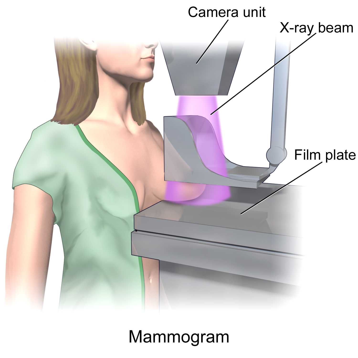
Mammography (also called mastography) is the process of using low-energy X-rays (usually around 30 kVp) to examine the human breast for diagnosis and screening. The goal of mammography is the early detection of breast cancer, typically through detection of characteristic masses or microcalcifications. As with all X-rays, mammograms use doses of ionizing radiation to create images. These images are then analyzed for abnormal findings. It is usual to employ lower-energy X-rays, typically Mo (K-shell X-ray energies of 17.5 and 19.6 keV) and Rh (20.2 and 22.7 keV) than those used for radiography of bones. Mammography may be 2D or 3D ( tomosynthesis), depending on the available equipment and/or purpose of the examination. Ultrasound, ductography, positron emission mammography (PEM), and magnetic resonance imaging (MRI) are adjuncts to mammography. Ultrasound is typically used for further evaluation of masses found on mammography or palpable masses that may or may not be seen o ... [...More Info...] [...Related Items...] OR: [Wikipedia] [Google] [Baidu] |
Magnetic Resonance Imaging
Magnetic resonance imaging (MRI) is a medical imaging technique used in radiology to form pictures of the anatomy and the physiological processes of the body. MRI scanners use strong magnetic fields, magnetic field gradients, and radio waves to generate images of the organs in the body. MRI does not involve X-rays or the use of ionizing radiation, which distinguishes it from CT and PET scans. MRI is a medical application of nuclear magnetic resonance (NMR) which can also be used for imaging in other NMR applications, such as NMR spectroscopy. MRI is widely used in hospitals and clinics for medical diagnosis, staging and follow-up of disease. Compared to CT, MRI provides better contrast in images of soft-tissues, e.g. in the brain or abdomen. However, it may be perceived as less comfortable by patients, due to the usually longer and louder measurements with the subject in a long, confining tube, though "Open" MRI designs mostly relieve this. Additionally, implants and ... [...More Info...] [...Related Items...] OR: [Wikipedia] [Google] [Baidu] |
Breast Cancer
Breast cancer is cancer that develops from breast tissue. Signs of breast cancer may include a lump in the breast, a change in breast shape, dimpling of the skin, milk rejection, fluid coming from the nipple, a newly inverted nipple, or a red or scaly patch of skin. In those with distant spread of the disease, there may be bone pain, swollen lymph nodes, shortness of breath, or yellow skin. Risk factors for developing breast cancer include obesity, a lack of physical exercise, alcoholism, hormone replacement therapy during menopause, ionizing radiation, an early age at first menstruation, having children late in life or not at all, older age, having a prior history of breast cancer, and a family history of breast cancer. About 5–10% of cases are the result of a genetic predisposition inherited from a person's parents, including BRCA1 and BRCA2 among others. Breast cancer most commonly develops in cells from the lining of milk ducts and the lobules that supply ... [...More Info...] [...Related Items...] OR: [Wikipedia] [Google] [Baidu] |
American College Of Radiology
The American College of Radiology (ACR), founded in 1923, is a professional medical society representing nearly 40,000 diagnostic radiologists, radiation oncologists, interventional radiologists, nuclear medicine physicians and medical physicists. The ACR has 54 chapters in the United States, Canada and the Council of Affiliated Regional Radiation Oncology Societies (CARROS). Medical imaging accreditation The ACR has accredited more than 39,000 medical imaging facilities in 10 imaging modalities since 1987, including: * Breast MRI * Breast Ultrasound * Computed Tomography *Mammography *Magnetic Resonance Imaging *Nuclear Medicine * Positron Emission Tomography *Radiation Oncology Practice * Stereotactic Breast Biopsy *Ultrasound ACR Appropriateness Criteria The ACR Appropriateness Criteria (ACR AC) are evidence-based guidelines that assist referring physicians and other providers in making the most appropriate imaging or treatment decision for a specific clinical condition. The AC ... [...More Info...] [...Related Items...] OR: [Wikipedia] [Google] [Baidu] |
Breast Ultrasound
Breast ultrasound is the use of medical ultrasonography to perform imaging of the breast. It can be considered either a diagnostic or a screening procedure. It may be used either with or without a mammogram. It may be useful in younger women, where the denser fibrous tissue of the breast may make mammograms more difficult to interpret. Automated whole-breast ultrasound (AWBU) is an ultrasound investigation of the breast that is largely independent of the operator skill and that allows the reconstruction of volumetric images of the breast. Using high-frequency ultrasound, a diagnostic evaluation of the lactiferous ducts by means of ultrasound (duct sonography) can be performed. In this manner, dilated ducts and intraductal masses can be made visible. Another technique for visualizing the system of lactiferous ducts is galactography, which allows a wider area of the lactiferous duct system to be visualized. A type of ultrasound examination to measure tissue stiffness, which is ... [...More Info...] [...Related Items...] OR: [Wikipedia] [Google] [Baidu] |
Sensitivity And Specificity
''Sensitivity'' and ''specificity'' mathematically describe the accuracy of a test which reports the presence or absence of a condition. Individuals for which the condition is satisfied are considered "positive" and those for which it is not are considered "negative". *Sensitivity (true positive rate) refers to the probability of a positive test, conditioned on truly being positive. *Specificity (true negative rate) refers to the probability of a negative test, conditioned on truly being negative. If the true condition can not be known, a " gold standard test" is assumed to be correct. In a diagnostic test, sensitivity is a measure of how well a test can identify true positives and specificity is a measure of how well a test can identify true negatives. For all testing, both diagnostic and screening, there is usually a trade-off between sensitivity and specificity, such that higher sensitivities will mean lower specificities and vice versa. If the goal is to return the ratio at w ... [...More Info...] [...Related Items...] OR: [Wikipedia] [Google] [Baidu] |
False Positive
A false positive is an error in binary classification in which a test result incorrectly indicates the presence of a condition (such as a disease when the disease is not present), while a false negative is the opposite error, where the test result incorrectly indicates the absence of a condition when it is actually present. These are the two kinds of errors in a binary test, in contrast to the two kinds of correct result (a and a ). They are also known in medicine as a false positive (or false negative) diagnosis, and in statistical classification as a false positive (or false negative) error. In statistical hypothesis testing the analogous concepts are known as type I and type II errors, where a positive result corresponds to rejecting the null hypothesis, and a negative result corresponds to not rejecting the null hypothesis. The terms are often used interchangeably, but there are differences in detail and interpretation due to the differences between medical testing and stati ... [...More Info...] [...Related Items...] OR: [Wikipedia] [Google] [Baidu] |
Nephrogenic Systemic Fibrosis
Nephrogenic systemic fibrosis is a rare syndrome that involves fibrosis of skin, joints, eyes, and internal organs. NSF is caused by exposure to gadolinium in gadolinium-based MRI contrast agents (GBCAs) in patients with impaired kidney function. Epidemiological studies suggest that the incidence of NSF is unrelated to gender or ethnicity and it is not thought to have a genetic basis. After GBCAs were identified as a cause of the disorder in 2006, and screening and prevention measures put in place, it is now considered rare. Signs and symptoms Clinical features of NSF develop within days to months and, in some cases, years following exposure to some GBCAs. The main symptoms are the thickening and hardening of the skin associated with brawny hyperpigmentation, typically presenting in a symmetric fashion. The skin gradually becomes fibrotic and adheres to the underlying fascia. The symptoms initiate distally in the limbs and progress proximally, sometimes involving the trunk. Jo ... [...More Info...] [...Related Items...] OR: [Wikipedia] [Google] [Baidu] |
MRI Contrast Agent
MRI contrast agents are contrast agents used to improve the visibility of internal body structures in magnetic resonance imaging (MRI). The most commonly used compounds for contrast enhancement are gadolinium-based. Such MRI contrast agents shorten the relaxation times of nuclei within body tissues following oral or intravenous administration. In MRI scanners, sections of the body are exposed to a strong magnetic field causing primarily the hydrogen nuclei ("spins") of water in tissues to be polarized in the direction of the magnetic field. An intense radiofrequency pulse is applied that tips the magnetization generated by the hydrogen nuclei in the direction of the receiver coil where the spin polarization can be detected. Random molecular rotational oscillations matching the resonance frequency of the nuclear spins provide the "relaxation" mechanisms that bring the net magnetization back to its equilibrium position in alignment with the applied magnetic field. The magnitude of t ... [...More Info...] [...Related Items...] OR: [Wikipedia] [Google] [Baidu] |
Scleromyxedema
Papular mucinosis (also known as scleromyxedema, "generalized lichen myxedematosus" and "sclerodermoid lichen myxedematosus") is a rare skin disease. Localized and disseminated cases are called papular mucinosis or lichen myxedematosus while generalized, confluent papular forms with sclerosis are called scleromyxedema. Frequently, all three forms are regarded as papular mucinosis. However, some authors restrict it to only mild cases. Another form, acral persistent papular mucinosis is regarded as a separate entity. Presentation Papular mucinosis is chronic and may be progressive. The dermal layer of the skin breaks out into small and solid bumps, usually conical in shape and measured from 2 to 4 mm or sometimes flat-topped papules. Unlike pustules, these bumps do not contain pus. Instead they contain mucin, a waxy substance of mucus, the body's natural and protective lubricant found in saliva and epithelial cells in lungs and the sensitive part of the nose. They usually come ... [...More Info...] [...Related Items...] OR: [Wikipedia] [Google] [Baidu] |
Scleroderma
Scleroderma is a group of autoimmune diseases that may result in changes to the skin, blood vessels, muscles, and internal organs. The disease can be either localized to the skin or involve other organs, as well. Symptoms may include areas of thickened skin, stiffness, feeling tired, and poor blood flow to the fingers or toes with cold exposure. One form of the condition, known as CREST syndrome, classically results in calcium deposits, Raynaud's syndrome, esophageal problems, thickening of the skin of the fingers and toes, and areas of small, dilated blood vessels. The cause is unknown, but it may be due to an abnormal immune response. Risk factors include family history, certain genetic factors, and exposure to silica. The underlying mechanism involves the abnormal growth of connective tissue, which is believed to be the result of the immune system attacking healthy tissues. Diagnosis is based on symptoms, supported by a skin biopsy or blood tests. While no cur ... [...More Info...] [...Related Items...] OR: [Wikipedia] [Google] [Baidu] |
Chronic Kidney Disease
Chronic kidney disease (CKD) is a type of kidney disease in which a gradual loss of kidney function occurs over a period of months to years. Initially generally no symptoms are seen, but later symptoms may include leg swelling, feeling tired, vomiting, loss of appetite, and confusion. Complications can relate to hormonal dysfunction of the kidneys and include (in chronological order) high blood pressure (often related to activation of the renin–angiotensin system system), bone disease, and anemia. Additionally CKD patients have markedly increased cardiovascular complications with increased risks of death and hospitalization. Causes of chronic kidney disease include diabetes, high blood pressure, glomerulonephritis, and polycystic kidney disease. Risk factors include a family history of chronic kidney disease. Diagnosis is by blood tests to measure the estimated glomerular filtration rate (eGFR), and a urine test to measure albumin. Ultrasound or kidney biopsy may be pe ... [...More Info...] [...Related Items...] OR: [Wikipedia] [Google] [Baidu] |


