|
Bovine Prolapsed Uterus
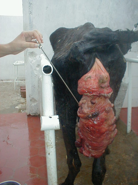
Bovine uterine prolapse occurs when the bovine uterus protrudes after calving. It is most common in dairy cattle and can occur in beef cows occasionally with hypocalcaemia. It is not as commonly seen in heifers, but occasionally can be seen in dairy heifers and most commonly Herefords. Uterine prolapse is considered a medical emergency that puts the cow at risk of shock or death by blood loss. Factors during calving that increase the risk of uterine prolapse include: calving complications that cause injury or irritation of the external birth canal, severe straining during labor, and excessive pressure when a calf is manually extracted. Non-calving factors include nutrition problems such as low blood calcium, magnesium, protein, or generally poor body conditions. In a complete uterine prolapse, the uterine horns also come out. When this happens, the uterus will hang below the hocks of the animal. When the uterus hangs below the hocks, the cow may lie on, step on or kick the ex ... [...More Info...] [...Related Items...] OR: [Wikipedia] [Google] [Baidu] |
Uterine Prolapse
Uterine prolapse is when the uterus descends towards or through the opening of the vagina. Symptoms may include vaginal fullness, pain with sex, trouble urinating, urinary incontinence, and constipation. Often it gets worse over time. Low back pain and vaginal bleeding may also occur. Risk factors include pregnancy, childbirth, obesity, constipation, and chronic coughing. Diagnosis is based on examination. It is a form of pelvic organ prolapse, together with bladder prolapse, large bowel prolapse, and small bowel prolapse. Preventive efforts include managing chronic breathing problems, not smoking, and maintaining a healthy weight. Mild cases may be treated with a pessary together with hormone replacement therapy. More severe cases may require surgery such as a vaginal hysterectomy. About 14% of women are affected. It occurs most commonly after menopause. Causes The most common cause of uterine prolapse is trauma during childbirth, in particular multiple or difficul ... [...More Info...] [...Related Items...] OR: [Wikipedia] [Google] [Baidu] |
Abdominal Cavity
The abdominal cavity is a large body cavity in humans and many other animals that contains many organs. It is a part of the abdominopelvic cavity. It is located below the thoracic cavity, and above the pelvic cavity. Its dome-shaped roof is the thoracic diaphragm, a thin sheet of muscle under the lungs, and its floor is the pelvic inlet, opening into the pelvis. Structure Organs Organs of the abdominal cavity include the stomach, liver, gallbladder, spleen, pancreas, small intestine, kidneys, large intestine, and adrenal glands. Peritoneum The abdominal cavity is lined with a protective membrane termed the peritoneum. The inside wall is covered by the parietal peritoneum. The kidneys are located behind the peritoneum, in the retroperitoneum, outside the abdominal cavity. The viscera are also covered by visceral peritoneum. Between the visceral and parietal peritoneum is the peritoneal cavity, which is a potential space. It contains a serous fluid called peritone ... [...More Info...] [...Related Items...] OR: [Wikipedia] [Google] [Baidu] |
Rumen
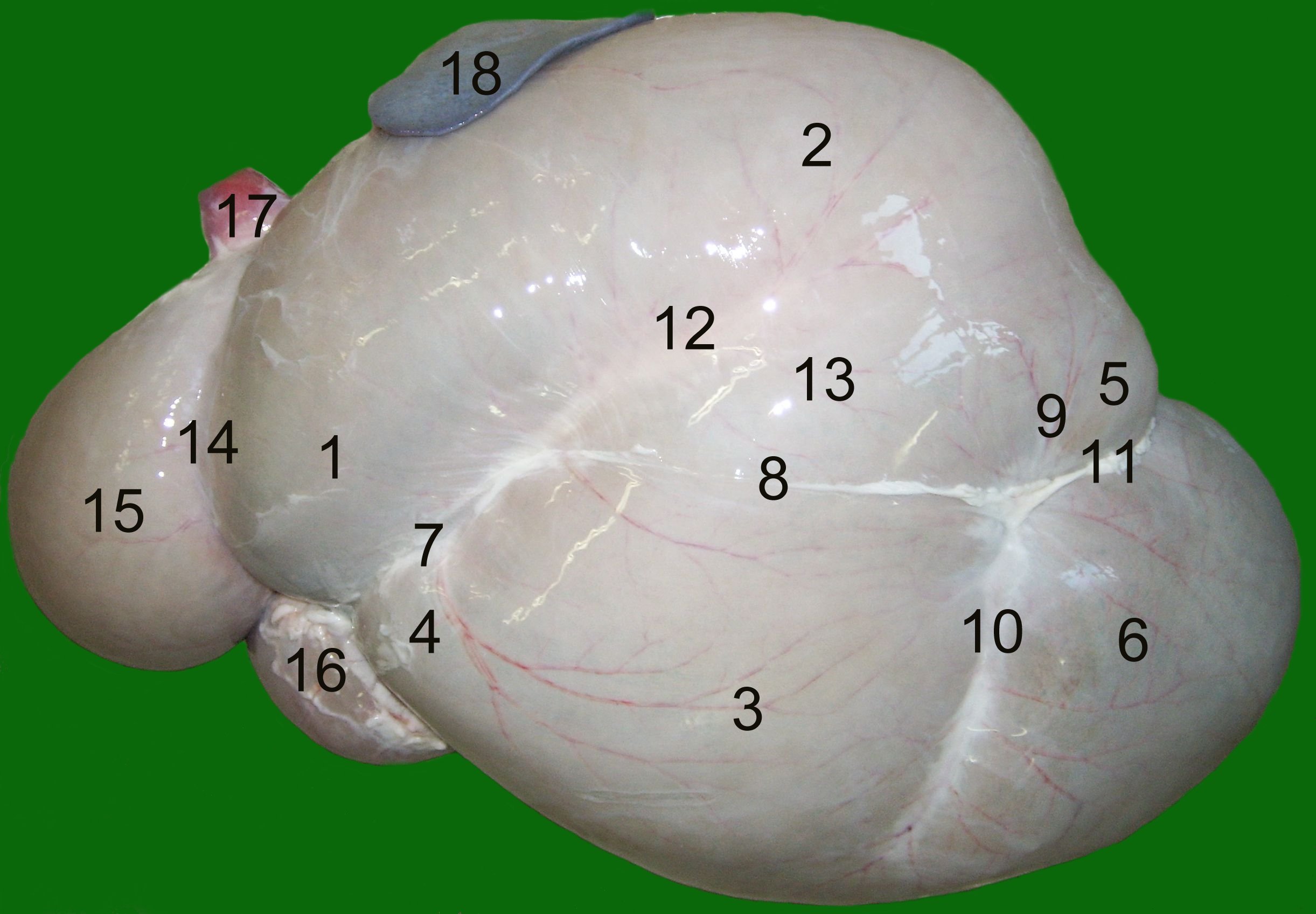
The rumen, also known as a paunch, is the largest stomach compartment in ruminants and the larger part of the reticulorumen, which is the first chamber in the alimentary canal of ruminant animals. The rumen's microbial favoring environment allows it to serve as the primary site for microbial fermentation of ingested feed. The smaller part of the reticulorumen is the reticulum, which is fully continuous with the rumen, but differs from it with regard to the texture of its lining. Brief anatomy The rumen is composed of several muscular sacs, the cranial sac, ventral sac, ventral blindsac, and reticulum. The lining of the rumen wall is covered in small fingerlike projections called papillae, which are flattened, approximately 5mm in length and 3mm wide in cattle. The reticulum is lined with ridges that form a hexagonal honeycomb pattern. The ridges are approximately 0.1–0.2mm wide and are raised 5mm above the reticulum wall. The hexagons in the reticulum are approxima ... [...More Info...] [...Related Items...] OR: [Wikipedia] [Google] [Baidu] |
Cotyledon (mammal)
The placenta of humans, and certain other mammals contains structures known as cotyledons, which transmit fetal blood and allow exchange of oxygen and nutrients with the maternal blood. Ruminants The Artiodactyla have a cotyledonary placenta. In this form of placenta the chorionic villi form a number of separate circular structures (''cotyledons'') which are distributed over the surface of the chorionic sac. Sheep, goats and cattle have between 72 and 125 cotyledons whereas deer have 4-6 larger cotyledons. Human The form of the human placenta is generally classified as a discoid placenta. Within this the ''cotyledons'' are the approximately 15-25 separations of the decidua basalis of the placenta, separated by placental septa The Southeastern Pennsylvania Transportation Authority (SEPTA) is a regional public transportation authority that operates bus, rapid transit, commuter rail, light rail, and electric trolleybus services for nearly 4 million people in five c ....Pag ... [...More Info...] [...Related Items...] OR: [Wikipedia] [Google] [Baidu] |
Placenta
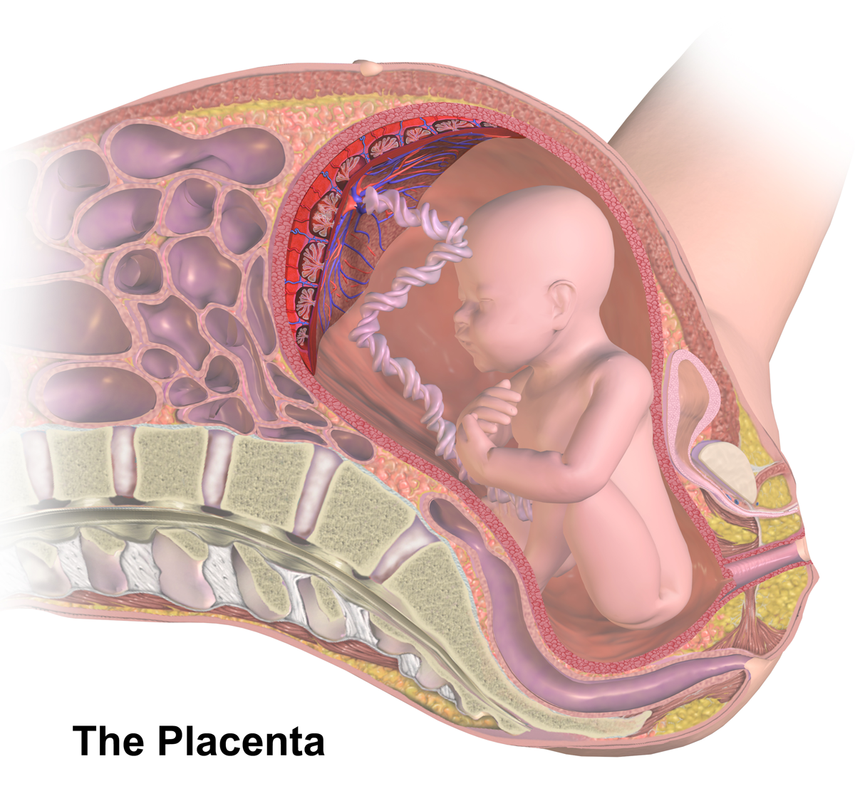
The placenta is a temporary embryonic and later fetal organ that begins developing from the blastocyst shortly after implantation. It plays critical roles in facilitating nutrient, gas and waste exchange between the physically separate maternal and fetal circulations, and is an important endocrine organ, producing hormones that regulate both maternal and fetal physiology during pregnancy. The placenta connects to the fetus via the umbilical cord, and on the opposite aspect to the maternal uterus in a species-dependent manner. In humans, a thin layer of maternal decidual ( endometrial) tissue comes away with the placenta when it is expelled from the uterus following birth (sometimes incorrectly referred to as the 'maternal part' of the placenta). Placentas are a defining characteristic of placental mammals, but are also found in marsupials and some non-mammals with varying levels of development. Mammalian placentas probably first evolved about 150 million to 200 millio ... [...More Info...] [...Related Items...] OR: [Wikipedia] [Google] [Baidu] |
Animal Euthanasia
Animal euthanasia ( euthanasia from el, εὐθανασία; "good death") is the act of killing an animal or allowing it to die by withholding extreme medical measures. Reasons for euthanasia include incurable (and especially painful) conditions or diseases, lack of resources to continue supporting the animal, or laboratory test procedures. Euthanasia methods are designed to cause minimal pain and distress. Euthanasia is distinct from animal slaughter and pest control although in some cases the procedure is the same. In domesticated animals, this process is commonly referred to by euphemisms such as "put down" or "put to sleep". Methods The methods of euthanasia can be divided into pharmacological and physical methods. Acceptable pharmacological methods include injected drugs and gases that first depress the central nervous system and then cardiovascular activity. Acceptable physical methods must first cause rapid loss of consciousness by disrupting the central nervous sys ... [...More Info...] [...Related Items...] OR: [Wikipedia] [Google] [Baidu] |
Hemorrhage
Bleeding, hemorrhage, haemorrhage or blood loss, is blood escaping from the circulatory system from damaged blood vessels. Bleeding can occur internally, or externally either through a natural opening such as the mouth, nose, ear, urethra, vagina or anus, or through a puncture in the skin. Hypovolemia is a massive decrease in blood volume, and death by excessive loss of blood is referred to as exsanguination. Typically, a healthy person can endure a loss of 10–15% of the total blood volume without serious medical difficulties (by comparison, blood donation typically takes 8–10% of the donor's blood volume). The stopping or controlling of bleeding is called hemostasis and is an important part of both first aid and surgery. Types * Upper head ** Intracranial hemorrhage – bleeding in the skull. ** Cerebral hemorrhage – a type of intracranial hemorrhage, bleeding within the brain tissue itself. ** Intracerebral hemorrhage – bleeding in the brain caused by the rup ... [...More Info...] [...Related Items...] OR: [Wikipedia] [Google] [Baidu] |
Endometrium
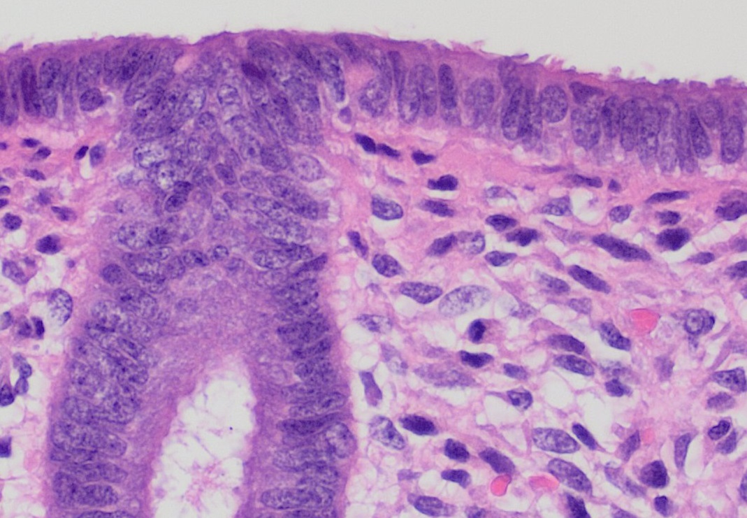
The endometrium is the inner epithelial layer, along with its mucous membrane, of the mammalian uterus. It has a basal layer and a functional layer: the basal layer contains stem cells which regenerate the functional layer. The functional layer thickens and then is shed during menstruation in humans and some other mammals, including apes, Old World monkeys, some species of bat, the elephant shrew and the Cairo spiny mouse. In most other mammals, the endometrium is reabsorbed in the estrous cycle. During pregnancy, the glands and blood vessels in the endometrium further increase in size and number. Vascular spaces fuse and become interconnected, forming the placenta, which supplies oxygen and nutrition to the embryo and fetus.Blue Histology - Female Reproductive System . School ... [...More Info...] [...Related Items...] OR: [Wikipedia] [Google] [Baidu] |
Amputation
Amputation is the removal of a limb by trauma, medical illness, or surgery. As a surgical measure, it is used to control pain or a disease process in the affected limb, such as malignancy or gangrene. In some cases, it is carried out on individuals as a preventive surgery for such problems. A special case is that of congenital amputation, a congenital disorder, where fetal limbs have been cut off by constrictive bands. In some countries, amputation is currently used to punish people who commit crimes. Amputation has also been used as a tactic in war and acts of terrorism; it may also occur as a war injury. In some cultures and religions, minor amputations or mutilations are considered a ritual accomplishment. When done by a person, the person executing the amputation is an amputator. The oldest evidence of this practice comes from a skeleton found buried in Liang Tebo cave, East Kalimantan, Indonesian Borneo dating back to at least 31,000 years ago, where it was done whe ... [...More Info...] [...Related Items...] OR: [Wikipedia] [Google] [Baidu] |
Evert
Evert is a Dutch and Swedish short form of the Germanic masculine name "Everhard" (alternative Eberhard). at the database of given names in the Netherlands. It is also used as surname. Notable people with the name include: Given name * (1602–1657), Dutch still life painter * (1772–1809), Norwegian naval officer *[...More Info...] [...Related Items...] OR: [Wikipedia] [Google] [Baidu] |


