|
Blob (visual System)
Blobs are sections of the visual cortex where groups of neurons that are sensitive to color assemble in cylindrical shapes. They were first identified in 1979 by Margaret Wong-Riley when she used a cytochrome oxidase stain, from which they get their name. These areas receive input from parvocellular cells in layer 4Cβ of the primary visual cortex and output to the thin stripes of area V2. Interblobs are areas between blobs which receive the same input, but are sensitive to orientation Orientation may refer to: Positioning in physical space * Map orientation, the relationship between directions on a map and compass directions * Orientation (housing), the position of a building with respect to the sun, a concept in building de ... instead of color. They output to the pale and thick stripes of area V2. Blobs are on the parvocellular pathway. This pathway begins at the photoreceptors which then relay signals to the 'P' ganglion cells in the retina. The pathway then continues out ... [...More Info...] [...Related Items...] OR: [Wikipedia] [Google] [Baidu] |
Cytochrome Oxidase
The enzyme cytochrome c oxidase or Complex IV, (was , now reclassified as a translocasEC 7.1.1.9 is a large transmembrane protein complex found in bacteria, archaea, and mitochondria of eukaryotes. It is the last enzyme in the respiratory electron transport chain of cells located in the membrane. It receives an electron from each of four cytochrome c molecules and transfers them to one oxygen molecule and four protons, producing two molecules of water. In addition to binding the four protons from the inner aqueous phase, it transports another four protons across the membrane, increasing the transmembrane difference of proton electrochemical potential, which the ATP synthase then uses to synthesize ATP. Structure The complex The complex is a large integral membrane protein composed of several metal prosthetic sites and 14 protein subunits in mammals. In mammals, eleven subunits are nuclear in origin, and three are synthesized in the mitochondria. The complex contains two ... [...More Info...] [...Related Items...] OR: [Wikipedia] [Google] [Baidu] |
Brain Research
''Brain Research'' is a peer-reviewed scientific journal focusing on several aspects of neuroscience. It publishes research reports and " minireviews". The editor-in-chief is Matthew J. LaVoie (University of Florida). Until 2011, full reviews were published in ''Brain Research Reviews'', which is now integrated into the main section, albeit with independent volume numbering. In 2006, four other previously established semi-independent journal sections ('' Cognitive Brain Research, Developmental Brain Research, Molecular Brain Research,'' and '' Brain Research Protocols'') were merged with ''Brain Research''. The journal has nine main subsections: * ''Cellular and Molecular Systems'' * ''Nervous System Development, Regeneration and Aging'' * ''Neurophysiology, Neuropharmacology and other forms of Intercellular Communication'' * ''Structural Organization of the Brain'' * ''Sensory and Motor Systems'' * ''Regulatory Systems'' * ''Cognitive and Behavioral Neuroscience'' * ''Disease-Re ... [...More Info...] [...Related Items...] OR: [Wikipedia] [Google] [Baidu] |
Parvocellular Cell
Parvocellular cells, also called P-cells, are neurons located within the parvocellular layers of the lateral geniculate nucleus (LGN) of the thalamus. "''Parvus''" is Latin for "small", and the name "parvocellular" refers to the small size of the cell compared to the larger magnocellular cells. Phylogenetically, parvocellular neurons are more modern than magnocellular ones. Function The parvocellular neurons of the visual system receive their input from midget cells, a type of retinal ganglion cell, whose axons are exiting the optic tract. These synapses occur in one of the four dorsal parvocellular layers of the lateral geniculate nucleus. The information from each eye is kept separate at this point, and continues to be segregated until processing in the visual cortex. The electrically-encoded visual information leaves the parvocellular cells via relay cells in the optic radiations, traveling to the primary visual cortex layer 4C-β. The parvocellular neurons are sensiti ... [...More Info...] [...Related Items...] OR: [Wikipedia] [Google] [Baidu] |
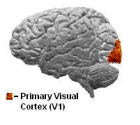
Primary Visual Cortex
The visual cortex of the brain is the area of the cerebral cortex that processes visual information. It is located in the occipital lobe. Sensory input originating from the eyes travels through the lateral geniculate nucleus in the thalamus and then reaches the visual cortex. The area of the visual cortex that receives the sensory input from the lateral geniculate nucleus is the primary visual cortex, also known as visual area 1 ( V1), Brodmann area 17, or the striate cortex. The extrastriate areas consist of visual areas 2, 3, 4, and 5 (also known as V2, V3, V4, and V5, or Brodmann area 18 and all Brodmann area 19). Both hemispheres of the brain include a visual cortex; the visual cortex in the left hemisphere receives signals from the right visual field, and the visual cortex in the right hemisphere receives signals from the left visual field. Introduction The primary visual cortex (V1) is located in and around the calcarine fissure in the occipital lobe. Each hemisphere's V1 ... [...More Info...] [...Related Items...] OR: [Wikipedia] [Google] [Baidu] |
Visual Cortex
The visual cortex of the brain is the area of the cerebral cortex that processes visual information. It is located in the occipital lobe. Sensory input originating from the eyes travels through the lateral geniculate nucleus in the thalamus and then reaches the visual cortex. The area of the visual cortex that receives the sensory input from the lateral geniculate nucleus is the primary visual cortex, also known as visual area 1 ( V1), Brodmann area 17, or the striate cortex. The extrastriate areas consist of visual areas 2, 3, 4, and 5 (also known as V2, V3, V4, and V5, or Brodmann area 18 and all Brodmann area 19). Both hemispheres of the brain include a visual cortex; the visual cortex in the left hemisphere receives signals from the right visual field, and the visual cortex in the right hemisphere receives signals from the left visual field. Introduction The primary visual cortex (V1) is located in and around the calcarine fissure in the occipital lobe. Each hemisphere's V1 ... [...More Info...] [...Related Items...] OR: [Wikipedia] [Google] [Baidu] |
Orientation (geometry)
In geometry, the orientation, angular position, attitude, bearing, or direction of an object such as a line, plane or rigid body is part of the description of how it is placed in the space it occupies. More specifically, it refers to the imaginary rotation that is needed to move the object from a reference placement to its current placement. A rotation may not be enough to reach the current placement. It may be necessary to add an imaginary translation, called the object's location (or position, or linear position). The location and orientation together fully describe how the object is placed in space. The above-mentioned imaginary rotation and translation may be thought to occur in any order, as the orientation of an object does not change when it translates, and its location does not change when it rotates. Euler's rotation theorem shows that in three dimensions any orientation can be reached with a single rotation around a fixed axis. This gives one common way of representing ... [...More Info...] [...Related Items...] OR: [Wikipedia] [Google] [Baidu] |
Cerebrum
The cerebrum, telencephalon or endbrain is the largest part of the brain containing the cerebral cortex (of the two cerebral hemispheres), as well as several subcortical structures, including the hippocampus, basal ganglia, and olfactory bulb. In the human brain, the cerebrum is the uppermost region of the central nervous system. The cerebrum prenatal development, develops prenatally from the forebrain (prosencephalon). In mammals, the Dorsum (biology), dorsal telencephalon, or Pallium (neuroanatomy), pallium, develops into the cerebral cortex, and the ventral telencephalon, or Pallium (neuroanatomy), subpallium, becomes the basal ganglia. The cerebrum is also divided into approximately symmetric Lateralization of brain function, left and right cerebral hemispheres. With the assistance of the cerebellum, the cerebrum controls all voluntary actions in the human body. Structure The cerebrum is the largest part of the brain. Depending upon the position of the animal it lies eithe ... [...More Info...] [...Related Items...] OR: [Wikipedia] [Google] [Baidu] |

