|
Beta Thymosins
Beta thymosins are a family of proteins which have in common a sequence of about 40 amino acids similar to the small protein thymosin β4. They are found almost exclusively in multicellular animals. Thymosin β4 was originally obtained from the thymus in company with several other small proteins which although named collectively "thymosins" are now known to be structurally and genetically unrelated and present in many different animal tissues. Single domain β-thymosins Distribution Monomeric β-thymosins, i.e. those of molecular weight similar to the peptides originally isolated from thymus by Goldstein, are found almost exclusively in cells of multicellular animals. Known exceptions are monomeric thymosins found in a few single-celled organisms, significantly those currently regarded as the closest relatives of multicellular animals: choanoflagellates and filastereans. Although found in very early-diverged animals such as sponges, monomeric thymosins are absent ... [...More Info...] [...Related Items...] OR: [Wikipedia] [Google] [Baidu] |
Ubiquitin
Ubiquitin is a small (8.6 kDa) regulatory protein found in most tissues of eukaryotic organisms, i.e., it is found ''ubiquitously''. It was discovered in 1975 by Gideon Goldstein and further characterized throughout the late 1970s and 1980s. Four genes in the human genome code for ubiquitin: UBB, UBC, UBA52 and RPS27A. The addition of ubiquitin to a substrate protein is called ubiquitylation (or, alternatively, ubiquitination or ubiquitinylation). Ubiquitylation affects proteins in many ways: it can mark them for degradation via the proteasome, alter their cellular location, affect their activity, and promote or prevent protein interactions. Ubiquitylation involves three main steps: activation, conjugation, and ligation, performed by ubiquitin-activating enzymes (E1s), ubiquitin-conjugating enzymes (E2s), and ubiquitin ligases (E3s), respectively. The result of this sequential cascade is to bind ubiquitin to lysine residues on the protein substrate via an isopeptide bond, ... [...More Info...] [...Related Items...] OR: [Wikipedia] [Google] [Baidu] |
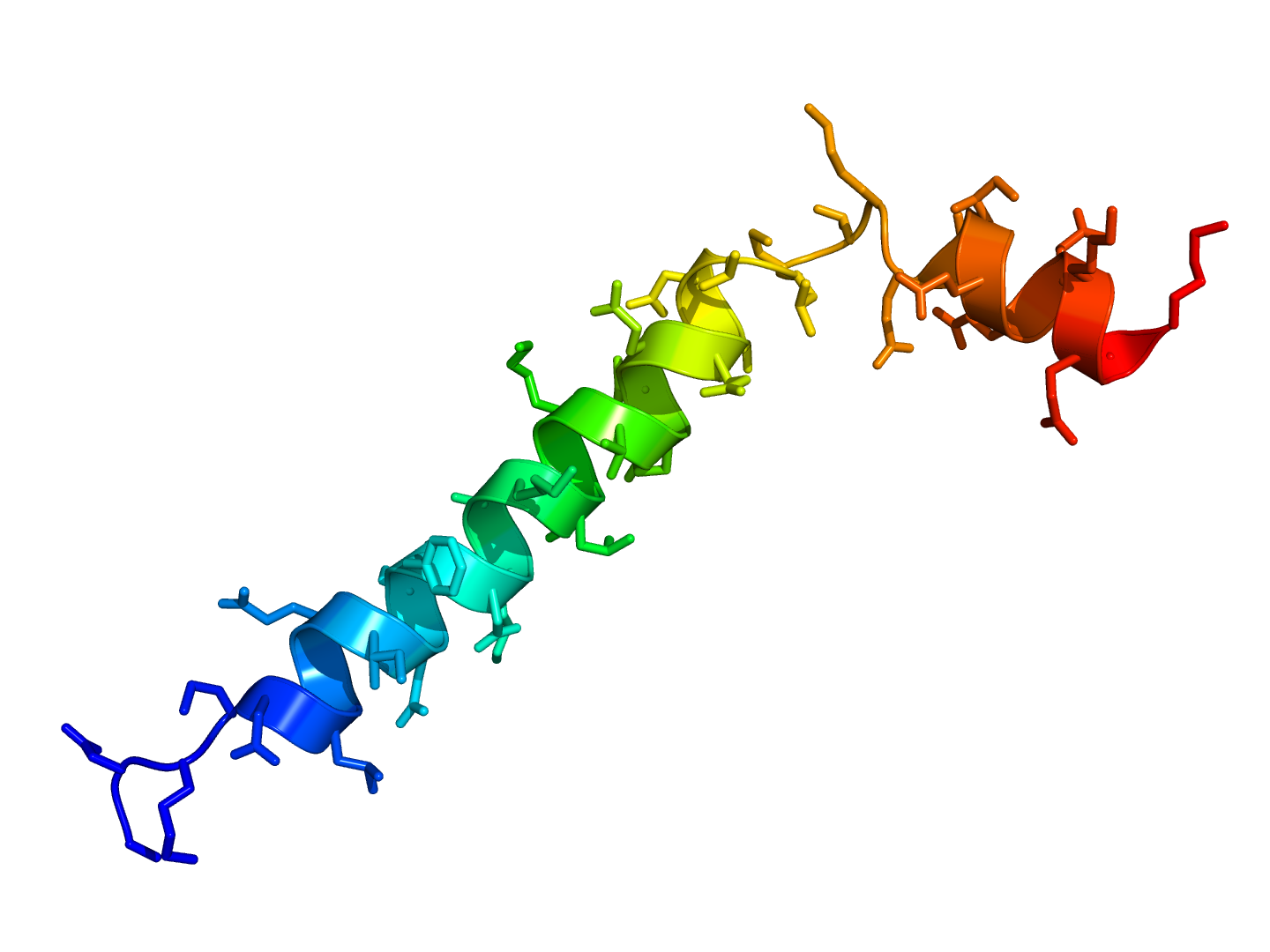
Thymosin 1HJ0
Thymosins are small proteins present in many animal tissues. They are named thymosins because they were originally isolated from the thymus, but most are now known to be present in many other tissues. Thymosins have diverse biological activities, and two in particular, thymosins α1 and β4, have potentially important uses in medicine, some of which have already progressed from the laboratory to the clinic. In relation to diseases, thymosins have been categorized as biological response modifiers. Thymosins are important for proper T-cell development and differentiation. Discovery The discovery of thymosins in the mid 1960s emerged from investigations of the role of the thymus in development of the vertebrate immune system. Begun by Allan L. Goldstein in the Laboratory of Abraham White at the Albert Einstein College of Medicine in New York, the work continued at University of Texas Medical Branch in Galveston and at The George Washington University School of Medicine and Health ... [...More Info...] [...Related Items...] OR: [Wikipedia] [Google] [Baidu] |
Prostate Cancer
Prostate cancer is cancer of the prostate. Prostate cancer is the second most common cancerous tumor worldwide and is the fifth leading cause of cancer-related mortality among men. The prostate is a gland in the male reproductive system that surrounds the urethra just below the bladder. It is located in the hypogastric region of the abdomen. To give an idea of where it is located, the bladder is superior to the prostate gland as shown in the image The rectum is posterior in perspective to the prostate gland and the ischial tuberosity of the pelvic bone is inferior. Only those who have male reproductive organs are able to get prostate cancer. Most prostate cancers are slow growing. Cancerous cells may spread to other areas of the body, particularly the bones and lymph nodes. It may initially cause no symptoms. In later stages, symptoms include pain or difficulty urinating, blood in the urine, or pain in the pelvis or back. Benign prostatic hyperplasia may produce similar symptoms ... [...More Info...] [...Related Items...] OR: [Wikipedia] [Google] [Baidu] |
Epidermolysis Bullosa Simplex
Epidermolysis bullosa simplex (EBS) is a disorder resulting from mutations in the genes encoding keratin 5 or keratin 14.Freedberg, et al. (2003). ''Fitzpatrick's Dermatology in General Medicine''. (6th ed.). McGraw-Hill. . Epidermolysis bullosa simplex (EBS) is one of the major forms of epidermolysis bullosa, a group of genetic conditions that cause the skin to be very fragile and to blister easily. Blister formation of EBS occurs at the dermal-epidermal junction. Cause Epidermolysis bullosa simplex is caused by genetic mutations that prevent the proper formation of protein structures in the skin’s epidermis. This results in skin that blisters easily, from even minor insults. The affected genes, KRT5 and the KRT14, which are responsible for the creation of keratin 5 and keratin 14 proteins respectively, are tied to the four major types of epidermolysis bullosa simplex. However, a small number of epidermolysis bullosa simplex patients do not have mutations in their KRT5 and ... [...More Info...] [...Related Items...] OR: [Wikipedia] [Google] [Baidu] |
Venostasis
Venostasis, or venous stasis, is a condition of slow blood flow in the veins, usually of the legs. Presentation Complications Potential complications of venous stasis are: * Venous ulcers * Blood clot formation in veins (venous thrombosis), that can occur in the deep veins of the legs (deep vein thrombosis, DVT) or in the superficial veins Causes Causes of venous stasis include: * Obesity * Pregnancy * Previous damage to leg * Blood clot * Smoking * Swelling and inflammation of a vein close to the skin * Congestive heart failure. * Long periods of immobility that can be encountered from driving, flying, bed rest/hospitalization, or having an orthopedic cast. Recommendations by clinicians to reduce venous stasis and DVT/PE often encourage increasing walking, calf exercises, and intermittent pneumatic compression when possible. Diagnosis See also * Virchow's triad Virchow's triad or the triad of Virchow () describes the three broad categories of factors that are thought to con ... [...More Info...] [...Related Items...] OR: [Wikipedia] [Google] [Baidu] |
Bed Sores
Pressure ulcers, also known as pressure sores, bed sores or pressure injuries, are localised damage to the skin and/or underlying tissue that usually occur over a bony prominence as a result of usually long-term pressure, or pressure in combination with shear or friction. The most common sites are the skin overlying the sacrum, coccyx, heels, and hips, though other sites can be affected, such as the elbows, knees, ankles, back of shoulders, or the back of the cranium. Pressure ulcers occur due to pressure applied to soft tissue resulting in completely or partially obstructed blood flow to the soft tissue. Shear is also a cause, as it can pull on blood vessels that feed the skin. Pressure ulcers most commonly develop in individuals who are not moving about, such as those who are on chronic bedrest or consistently use a wheelchair. It is widely believed that other factors can influence the tolerance of skin for pressure and shear, thereby increasing the risk of pressure ul ... [...More Info...] [...Related Items...] OR: [Wikipedia] [Google] [Baidu] |
Cytosol
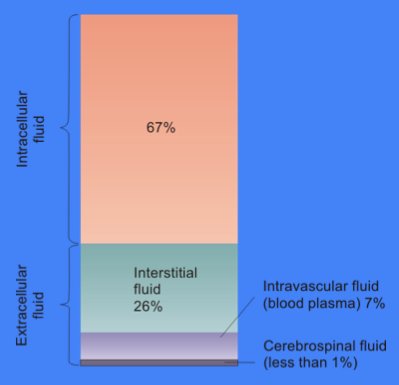
The cytosol, also known as cytoplasmic matrix or groundplasm, is one of the liquids found inside cells (intracellular fluid (ICF)). It is separated into compartments by membranes. For example, the mitochondrial matrix separates the mitochondrion into many compartments. In the eukaryotic cell, the cytosol is surrounded by the cell membrane and is part of the cytoplasm, which also comprises the mitochondria, plastids, and other organelles (but not their internal fluids and structures); the cell nucleus is separate. The cytosol is thus a liquid matrix around the organelles. In prokaryotes, most of the chemical reactions of metabolism take place in the cytosol, while a few take place in membranes or in the periplasmic space. In eukaryotes, while many metabolic pathways still occur in the cytosol, others take place within organelles. The cytosol is a complex mixture of substances dissolved in water. Although water forms the large majority of the cytosol, its structure and prope ... [...More Info...] [...Related Items...] OR: [Wikipedia] [Google] [Baidu] |
Thymosin α1
Thymosin α1 is a peptide fragment derived from prothymosin alpha, a protein that in humans is encoded by the ''PTMA'' gene. It was the first of the peptides from Thymosin Fraction 5 to be completely sequenced and synthesized. Unlike β thymosins, to which it is genetically and chemically unrelated, thymosin α1 is produced as a 28-amino acid fragment, from a longer, 113-amino acid precursor, prothymosin α. Function Thymosin α1 is believed to be a major component of Thymosin Fraction 5 responsible for the activity of that preparation in restoring immune function in animals lacking thymus glands. It has been found to enhance cell-mediated immunity in humans as well as experimental animals. Therapeutic application Thymosin α1 is approved in 35 under-developed or developing countries for the treatment of Hepatitis B and C, and it is also used to boost the immune response in the treatment of other diseases. Clinical studies Clinical trials suggest it may be useful in cys ... [...More Info...] [...Related Items...] OR: [Wikipedia] [Google] [Baidu] |
Convergent Evolution
Convergent evolution is the independent evolution of similar features in species of different periods or epochs in time. Convergent evolution creates analogous structures that have similar form or function but were not present in the last common ancestor of those groups. The cladistic term for the same phenomenon is homoplasy. The recurrent evolution of flight is a classic example, as flying insects, birds, pterosaurs, and bats have independently evolved the useful capacity of flight. Functionally similar features that have arisen through convergent evolution are ''analogous'', whereas '' homologous'' structures or traits have a common origin but can have dissimilar functions. Bird, bat, and pterosaur wings are analogous structures, but their forelimbs are homologous, sharing an ancestral state despite serving different functions. The opposite of convergence is divergent evolution, where related species evolve different traits. Convergent evolution is similar to parallel evo ... [...More Info...] [...Related Items...] OR: [Wikipedia] [Google] [Baidu] |
Actin
Actin is a family of globular multi-functional proteins that form microfilaments in the cytoskeleton, and the thin filaments in muscle fibrils. It is found in essentially all eukaryotic cells, where it may be present at a concentration of over 100 μM; its mass is roughly 42 kDa, with a diameter of 4 to 7 nm. An actin protein is the monomeric subunit of two types of filaments in cells: microfilaments, one of the three major components of the cytoskeleton, and thin filaments, part of the contractile apparatus in muscle cells. It can be present as either a free monomer called G-actin (globular) or as part of a linear polymer microfilament called F-actin (filamentous), both of which are essential for such important cellular functions as the mobility and contraction of cells during cell division. Actin participates in many important cellular processes, including muscle contraction, cell motility, cell division and cytokinesis, vesicle and organelle movement, cell sign ... [...More Info...] [...Related Items...] OR: [Wikipedia] [Google] [Baidu] |
X-ray Crystallography
X-ray crystallography is the experimental science determining the atomic and molecular structure of a crystal, in which the crystalline structure causes a beam of incident X-rays to diffract into many specific directions. By measuring the angles and intensities of these diffracted beams, a crystallographer can produce a three-dimensional picture of the density of electrons within the crystal. From this electron density, the mean positions of the atoms in the crystal can be determined, as well as their chemical bonds, their crystallographic disorder, and various other information. Since many materials can form crystals—such as salts, metals, minerals, semiconductors, as well as various inorganic, organic, and biological molecules—X-ray crystallography has been fundamental in the development of many scientific fields. In its first decades of use, this method determined the size of atoms, the lengths and types of chemical bonds, and the atomic-scale differences among various mat ... [...More Info...] [...Related Items...] OR: [Wikipedia] [Google] [Baidu] |


