|
Best Disease
Vitelliform macular dystrophy is an irregular autosomal dominant eye disorder which can cause progressive vision loss. This disorder affects the retina, specifically cells in a small area near the center of the retina called the macula. The macula is responsible for sharp central vision, which is needed for detailed tasks such as reading, driving, and recognizing faces. The condition is characterized by yellow (or orange), slightly elevated, round structures similar to the yolk (Latin ''vitellus'') of an egg. Genetics ''Best disease'' is inherited in an autosomal dominant pattern, which means one copy of the altered gene in each cell is sufficient to cause the disorder. In most cases, an affected person has one parent with the condition. The inheritance pattern of adult-onset vitelliform macular dystrophy is definitively autosomal dominant. Many affected people, however, have no history of the disorder in their family and only a small number of affected families have been repor ... [...More Info...] [...Related Items...] OR: [Wikipedia] [Google] [Baidu] |
Autosomal Dominant
In genetics, dominance is the phenomenon of one variant (allele) of a gene on a chromosome masking or overriding the effect of a different variant of the same gene on the other copy of the chromosome. The first variant is termed dominant and the second recessive. This state of having two different variants of the same gene on each chromosome is originally caused by a mutation in one of the genes, either new (''de novo'') or inherited. The terms autosomal dominant or autosomal recessive are used to describe gene variants on non-sex chromosomes ( autosomes) and their associated traits, while those on sex chromosomes (allosomes) are termed X-linked dominant, X-linked recessive or Y-linked; these have an inheritance and presentation pattern that depends on the sex of both the parent and the child (see Sex linkage). Since there is only one copy of the Y chromosome, Y-linked traits cannot be dominant or recessive. Additionally, there are other forms of dominance such as incomplete d ... [...More Info...] [...Related Items...] OR: [Wikipedia] [Google] [Baidu] |
Lipofuscin
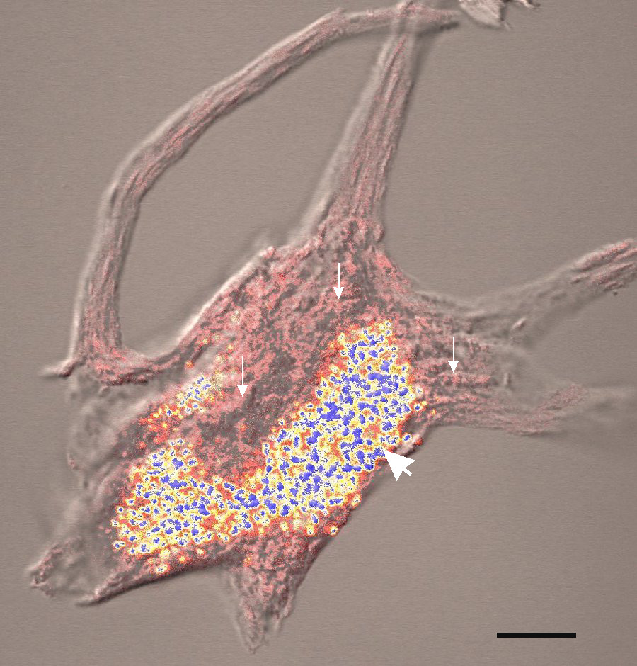
Lipofuscin is the name given to fine yellow-brown pigment granules composed of lipid-containing residues of lysosomal digestion. It is considered to be one of the aging or "wear-and-tear" pigments, found in the liver, kidney, heart muscle, retina, adrenals, nerve cells, and ganglion cells. Formation and turnover Lipofuscin appears to be the product of the oxidation of unsaturated fatty acids and may be symptomatic of membrane damage, or damage to mitochondria and lysosomes. Aside from a large lipid content, lipofuscin is known to contain sugars and metals, including mercury, aluminium, iron, copper and zinc.Chris Gaugler,Lipofuscin", ''Stanislaus Journal of Biochemical Reviews'' May 1997 Lipofuscin is also accepted as consisting of oxidized proteins (30–70%) as well as lipids (20–50%). It is a type of lipochrome and is specifically arranged around the nucleus. The accumulation of lipofuscin-like material may be the result of an imbalance between formation and disposal mech ... [...More Info...] [...Related Items...] OR: [Wikipedia] [Google] [Baidu] |
Autosomal Dominant Disorders
In genetics, dominance is the phenomenon of one variant (allele) of a gene on a chromosome masking or overriding the effect of a different variant of the same gene on the other copy of the chromosome. The first variant is termed dominant and the second recessive. This state of having two different variants of the same gene on each chromosome is originally caused by a mutation in one of the genes, either new (''de novo'') or inherited. The terms autosomal dominant or autosomal recessive are used to describe gene variants on non-sex chromosomes ( autosomes) and their associated traits, while those on sex chromosomes (allosomes) are termed X-linked dominant, X-linked recessive or Y-linked; these have an inheritance and presentation pattern that depends on the sex of both the parent and the child (see Sex linkage). Since there is only one copy of the Y chromosome, Y-linked traits cannot be dominant or recessive. Additionally, there are other forms of dominance such as incomplete d ... [...More Info...] [...Related Items...] OR: [Wikipedia] [Google] [Baidu] |
Electrooculography
Electrooculography (EOG) is a technique for measuring the corneo-retinal standing potential that exists between the front and the back of the human eye. The resulting signal is called the electrooculogram. Primary applications are in ophthalmological diagnosis and in recording eye movements. Unlike the electroretinogram, the EOG does not measure response to individual visual stimuli. To measure eye movement, pairs of electrodes are typically placed either above and below the eye or to the left and right of the eye. If the eye moves from center position toward one of the two electrodes, this electrode "sees" the positive side of the retina and the opposite electrode "sees" the negative side of the retina. Consequently, a potential difference occurs between the electrodes. Assuming that the resting potential is constant, the recorded potential is a measure of the eye's position. Principle The eye acts as a dipole in which the anterior pole is positive and the posterior pole is n ... [...More Info...] [...Related Items...] OR: [Wikipedia] [Google] [Baidu] |
Indocyanine Green Angiography
Indocyanine green angiography (ICGA) is a diagnostic procedure used to examine choroidal blood flow and associated pathology. Indocyanine green (ICG) is a water soluble cyanine dye which shows fluorescence in near-infrared (790–805 nm) range, with peak spectral absorption of 800-810 nm in blood. The near infrared light used in ICGA penetrates ocular pigments such as melanin and xanthophyll, as well as exudates and thin layers of sub-retinal vessels. Age-related macular degeneration is the third main cause of blindness worldwide, and it is the leading cause of blindness in industrialized countries. Indocyanine green angiography is widely used to study choroidal neovascularization in patients with exudative age-related macular degeneration. In nonexudative AMD, ICGA is used in classification of drusen and associated subretinal deposits. Indications Indications for indocyanine green angiography include: * Choroidal neovascularisation (CNV):Indocyanine green angiography is widely us ... [...More Info...] [...Related Items...] OR: [Wikipedia] [Google] [Baidu] |
Fluorescein Angiography
Fluorescein angiography (FA), fluorescent angiography (FAG), or fundus fluorescein angiography (FFA) is a technique for examining the circulation of the retina and choroid (parts of the fundus) using a fluorescent dye and a specialized camera. Sodium fluorescein is added into the systemic circulation, the retina is illuminated with blue light at a wavelength of 490 nanometers, and an angiogram is obtained by photographing the fluorescent green light that is emitted by the dye. The fluorescein is administered intravenously in intravenous fluorescein angiography (IVFA) and orally in oral fluorescein angiography (OFA). The test is a dye tracing method. The fluorescein dye also reappears in the patient urine, causing the urine to appear darker, and sometimes orange. It can also cause discolouration of the saliva. Fluorescein angiography is one of several health care applications of this dye, all of which have a risk of severe adverse effects. See fluorescein safety in health care a ... [...More Info...] [...Related Items...] OR: [Wikipedia] [Google] [Baidu] |
Eye Examination
An eye examination is a series of tests performed to assess vision and ability to focus on and discern objects. It also includes other tests and examinations pertaining to the eyes. Eye examinations are primarily performed by an optometrist, ophthalmologist, or an orthoptist. Health care professionals often recommend that all people should have periodic and thorough eye examinations as part of routine primary care, especially since many eye diseases are asymptomatic. Eye examinations may detect potentially treatable blinding eye diseases, ocular manifestations of systemic disease, or signs of tumours or other anomalies of the brain. A full eye examination consists of an external examination, followed by specific tests for visual acuity, pupil function, extraocular muscle motility, visual fields, intraocular pressure and ophthalmoscopy through a dilated pupil. A minimal eye examination consists of tests for visual acuity, pupil function, and extraocular muscle motility, as w ... [...More Info...] [...Related Items...] OR: [Wikipedia] [Google] [Baidu] |
Peripheral Vision
Peripheral vision, or ''indirect vision'', is vision as it occurs outside the point of fixation, i.e. away from the center of gaze or, when viewed at large angles, in (or out of) the "corner of one's eye". The vast majority of the area in the visual field is included in the notion of peripheral vision. "Far peripheral" vision refers to the area at the edges of the visual field, "mid-peripheral" vision refers to medium eccentricities, and "near-peripheral", sometimes referred to as "para-central" vision, exists adjacent to the center of gaze. Boundaries Inner boundaries The inner boundaries of peripheral vision can be defined in any of several ways depending on the context. In everyday language the term "peripheral vision" is often used to refer to what in technical usage would be called "far peripheral vision." This is vision outside of the range of stereoscopic vision. It can be conceived as bounded at the center by a circle 60° in radius or 120° in diameter, centered arou ... [...More Info...] [...Related Items...] OR: [Wikipedia] [Google] [Baidu] |
Scotoma
A scotoma is an area of partial alteration in the field of vision consisting of a partially diminished or entirely degenerated visual acuity that is surrounded by a field of normal – or relatively well-preserved – vision. Every normal mammalian eye has a scotoma in its field of vision, usually termed its blind spot. This is a location with no photoreceptor cells, where the retinal ganglion cell axons that compose the optic nerve exit the retina. This location is called the optic disc. There is no direct conscious awareness of visual scotomas. They are simply regions of reduced information within the visual field. Rather than recognizing an incomplete image, patients with scotomas report that things "disappear" on them. The presence of the blind spot scotoma can be demonstrated subjectively by covering one eye, carefully holding fixation with the open eye, and placing an object (such as one's thumb) in the lateral and horizontal visual field, about 15 degrees from fix ... [...More Info...] [...Related Items...] OR: [Wikipedia] [Google] [Baidu] |
Distorted Vision
Distorted may refer to: * Anything subject to distortion * Distorted (band), a progressive deathdoom metal band from Bat-Yam, Israel * ''Distorted'' (EP), an extended play by the band Distorted * ''Distorted'' (film) (2018) * Distorted Music Festival, a 2005 Australian music festival * ''Distorted'' (TV series), a 2017 South Korean television series * ''Distorted'', a 2001 demo album by Biomechanical * '' Beatmania IIDX 13: Distorted'', an arcade game See also * Distort (other) To distort is to alter the original shape of an object, image, sound, waveform or other form of information or representation. Distort may also refer to: * ''Distort'' (album), a 1998 industrial album by Collide * Distort Entertainment Distor ... * Distortion (other) {{disambig ... [...More Info...] [...Related Items...] OR: [Wikipedia] [Google] [Baidu] |
Cone Cell
Cone cells, or cones, are photoreceptor cells in the retinas of vertebrate eyes including the human eye. They respond differently to light of different wavelengths, and the combination of their responses is responsible for color vision. Cones function best in relatively bright light, called the photopic region, as opposed to rod cells, which work better in dim light, or the scotopic region. Cone cells are densely packed in the fovea centralis, a 0.3 mm diameter rod-free area with very thin, densely packed cones which quickly reduce in number towards the periphery of the retina. Conversely, they are absent from the optic disc, contributing to the blind spot. There are about six to seven million cones in a human eye (vs ~92 million rods), with the highest concentration being towards the macula. Cones are less sensitive to light than the rod cells in the retina (which support vision at low light levels), but allow the perception of color. They are also able to perceive ... [...More Info...] [...Related Items...] OR: [Wikipedia] [Google] [Baidu] |
Retinal Pigment Epithelium
The pigmented layer of retina or retinal pigment epithelium (RPE) is the pigmented cell layer just outside the neurosensory retina that nourishes retinal visual cells, and is firmly attached to the underlying choroid and overlying retinal visual cells. History The RPE was known in the 18th and 19th centuries as the pigmentum nigrum, referring to the observation that the RPE is dark (black in many animals, brown in humans); and as the tapetum nigrum, referring to the observation that in animals with a tapetum lucidum, in the region of the tapetum lucidum the RPE is not pigmented. Anatomy The RPE is composed of a single layer of hexagonal cells that are densely packed with pigment granules. When viewed from the outer surface, these cells are smooth and hexagonal in shape. When seen in section, each cell consists of an outer non-pigmented part containing a large oval nucleus and an inner pigmented portion which extends as a series of straight thread-like processes between the rods, ... [...More Info...] [...Related Items...] OR: [Wikipedia] [Google] [Baidu] |

_(14582377398).jpg)
