|
Basilar Plexus
The basilar plexus (transverse or basilar sinus) consists of several interlacing venous channels between the layers of the dura mater over the basilar part of the occipital bone (the clivus), and serves to connect the two inferior petrosal sinuses The inferior petrosal sinuses are two small sinuses situated on the inferior border of the petrous part of the temporal bone, one on each side. Each inferior petrosal sinus drains the cavernous sinus into the internal jugular vein. Structure The .... It communicates with the anterior vertebral venous plexus. References Veins of the head and neck {{circulatory-stub ... [...More Info...] [...Related Items...] OR: [Wikipedia] [Google] [Baidu] |
Base Of Skull
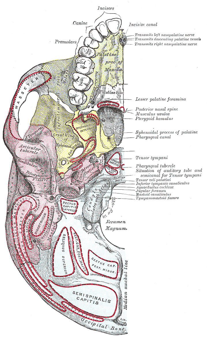
The base of skull, also known as the cranial base or the cranial floor, is the most inferior area of the skull. It is composed of the endocranium and the lower parts of the calvaria. Structure Structures found at the base of the skull are for example: Bones There are five bones that make up the base of the skull: *Ethmoid bone *Sphenoid bone *Occipital bone *Frontal bone *Temporal bone Sinuses *Occipital sinus *Superior sagittal sinus *Superior petrosal sinus Foramina of the skull * Foramen cecum *Optic foramen *Foramen lacerum *Foramen rotundum *Foramen magnum * Foramen ovale *Jugular foramen *Internal auditory meatus *Mastoid foramen *Sphenoidal emissary foramen *Foramen spinosum Sutures *Frontoethmoidal suture *Sphenofrontal suture *Sphenopetrosal suture *Sphenoethmoidal suture * Petrosquamous suture *Sphenosquamosal suture Other *Sphenoidal lingula *Subarcuate fossa *Dorsum sellae *Jugular process *Petro-occipital fissure *Condylar canal *Jugular tubercle *Tuberc ... [...More Info...] [...Related Items...] OR: [Wikipedia] [Google] [Baidu] |
Human Skull
The skull is a bone protective cavity for the brain. The skull is composed of four types of bone i.e., cranial bones, facial bones, ear ossicles and hyoid bone. However two parts are more prominent: the cranium and the mandible. In humans, these two parts are the neurocranium and the viscerocranium ( facial skeleton) that includes the mandible as its largest bone. The skull forms the anterior-most portion of the skeleton and is a product of cephalisation—housing the brain, and several sensory structures such as the eyes, ears, nose, and mouth. In humans these sensory structures are part of the facial skeleton. Functions of the skull include protection of the brain, fixing the distance between the eyes to allow stereoscopic vision, and fixing the position of the ears to enable sound localisation of the direction and distance of sounds. In some animals, such as horned ungulates (mammals with hooves), the skull also has a defensive function by providing the mount (on the front ... [...More Info...] [...Related Items...] OR: [Wikipedia] [Google] [Baidu] |
Venous
Veins are blood vessels in humans and most other animals that carry blood towards the heart. Most veins carry deoxygenated blood from the tissues back to the heart; exceptions are the pulmonary and umbilical veins, both of which carry oxygenated blood to the heart. In contrast to veins, arteries carry blood away from the heart. Veins are less muscular than arteries and are often closer to the skin. There are valves (called ''pocket valves'') in most veins to prevent backflow. Structure Veins are present throughout the body as tubes that carry blood back to the heart. Veins are classified in a number of ways, including superficial vs. deep, pulmonary vs. systemic, and large vs. small. *Superficial veins are those closer to the surface of the body, and have no corresponding arteries. *Deep veins are deeper in the body and have corresponding arteries. *Perforator veins drain from the superficial to the deep veins. These are usually referred to in the lower limbs and feet. *Communica ... [...More Info...] [...Related Items...] OR: [Wikipedia] [Google] [Baidu] |
Dura Mater
In neuroanatomy, dura mater is a thick membrane made of dense irregular connective tissue that surrounds the brain and spinal cord. It is the outermost of the three layers of membrane called the meninges that protect the central nervous system. The other two meningeal layers are the arachnoid mater and the pia mater. It envelops the arachnoid mater, which is responsible for keeping in the cerebrospinal fluid. It is derived primarily from the neural crest cell population, with postnatal contributions of the paraxial mesoderm. Structure The dura mater has several functions and layers. The dura mater is a membrane that envelops the arachnoid mater. It surrounds and supports the dural sinuses (also called dural venous sinuses, cerebral sinuses, or cranial sinuses) and carries blood from the brain toward the heart. Cranial dura mater has two layers called ''lamellae'', a superficial layer (also called the periosteal layer), which serves as the skull's inner periosteum, called the ... [...More Info...] [...Related Items...] OR: [Wikipedia] [Google] [Baidu] |
Occipital Bone
The occipital bone () is a neurocranium, cranial dermal bone and the main bone of the occiput (back and lower part of the skull). It is trapezoidal in shape and curved on itself like a shallow dish. The occipital bone overlies the occipital lobes of the cerebrum. At the base of skull in the occipital bone, there is a large oval opening called the foramen magnum, which allows the passage of the spinal cord. Like the other cranial bones, it is classed as a flat bone. Due to its many attachments and features, the occipital bone is described in terms of separate parts. From its front to the back is the basilar part of occipital bone, basilar part, also called the basioccipital, at the sides of the foramen magnum are the lateral parts of occipital bone, lateral parts, also called the exoccipitals, and the back is named as the squamous part of occipital bone, squamous part. The basilar part is a thick, somewhat quadrilateral piece in front of the foramen magnum and directed towards the ... [...More Info...] [...Related Items...] OR: [Wikipedia] [Google] [Baidu] |
Clivus (anatomy)
The clivus (, Latin for "slope"), or Blumenbach clivus, is a bony part of the cranium The skull is a bone protective cavity for the brain. The skull is composed of four types of bone i.e., cranial bones, facial bones, ear ossicles and hyoid bone. However two parts are more prominent: the cranium and the mandible. In humans, the ... at the base of the skull. It is a shallow depression behind the dorsum sellae of the sphenoid bone. It slopes gradually to the anterior part of the basilar occipital bone at its junction with the sphenoid bone. It extends to the foramen magnum. It is related to the pons and the abducens nerve (CN VI). Structure The clivus is a shallow depression behind the dorsum sellae of the sphenoid bone. It slopes gradually to the anterior part of the basilar occipital bone at its junction with the sphenoid bone. Synchondrosis of these two bones forms the clivus. The clivus extends inferiorly to the foramen magnum. On axial planes, it sits just posterior to ... [...More Info...] [...Related Items...] OR: [Wikipedia] [Google] [Baidu] |
Inferior Petrosal Sinuses
The inferior petrosal sinuses are two small sinuses situated on the inferior border of the petrous part of the temporal bone, one on each side. Each inferior petrosal sinus drains the cavernous sinus into the internal jugular vein. Structure The inferior petrosal sinus is situated in the inferior petrosal sulcus, formed by the junction of the petrous part of the temporal bone with the Basilar part of occipital bone, basilar part of the occipital bone. It begins below and behind the cavernous sinus and, passing through the anterior part of the jugular foramen, ends in the superior bulb of the internal jugular vein. Function The inferior petrosal sinus receives the internal auditory veins and also veins from the medulla oblongata, pons, and under surface of the cerebellum. Additional images File:Gray568.png, Sagittal section of the skull, showing the sinuses of the dura. See also * Dural venous sinuses * Inferior petrosal sinus sampling References * {{Authority contr ... [...More Info...] [...Related Items...] OR: [Wikipedia] [Google] [Baidu] |
Internal Vertebral Venous Plexuses
The internal vertebral venous plexuses (intraspinal veins) lie within the vertebral canal in the epidural space, and receive tributaries from the bones and from the spinal cord. They form a closer network than the external plexuses, and, running mainly in a vertical direction, form four longitudinal veins, two in front and two behind; they therefore may be divided into anterior and posterior groups. * The ''anterior internal plexuses'' consist of large veins which lie on the posterior surfaces of the vertebral bodies and intervertebral fibrocartilages on either side of the posterior longitudinal ligament; under cover of this ligament they are connected by transverse branches into which the basivertebral veins open. * The ''posterior internal plexuses'' are placed, one on either side of the middle line in front of the vertebral arches and ligamenta flava, and anastomose by veins passing through those ligaments with the posterior external plexuses. The anterior and posterior plexuses ... [...More Info...] [...Related Items...] OR: [Wikipedia] [Google] [Baidu] |


