|
Basidiobolus
''Basidiobolus ranarum'' is a filamentous fungus with worldwide distribution. The fungus was first isolated by Eidam in 1886. It can saprophytically live in the intestines of mainly cold-blooded vertebrates and on decaying fruits and soil. The fungus prefers glucose as a carbon source and grows rapidly at room temperature. ''Basidiobolus ranarum'' is also known as a cause of subcutaneous zygomycosis, usually causing granulomatous infections on a host's limbs. Infections are generally geographically limited to tropical and subtropical regions such as East and West Africa. Subcutaneous zygomycosis caused by ''B. ranarum'' is a rare disease and predominantly affects children and males. Common subcutaneous zygomycosis shows characteristic features and is relatively easy to be diagnosed; while, certain rare cases might show non-specific clinical features that might pose a difficulty on its identification. Although disease caused by this fungus is known to resolve spontaneously on its ... [...More Info...] [...Related Items...] OR: [Wikipedia] [Google] [Baidu] |
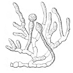
Basidiobolus Ranarum Linder Illustration
''Basidiobolus ranarum'' is a filamentous fungus with worldwide distribution. The fungus was first isolated by Eidam in 1886. It can saprophytically live in the intestines of mainly cold-blooded vertebrates and on decaying fruits and soil. The fungus prefers glucose as a carbon source and grows rapidly at room temperature. ''Basidiobolus ranarum'' is also known as a cause of subcutaneous zygomycosis, usually causing granulomatous infections on a host's limbs. Infections are generally geographically limited to tropical and subtropical regions such as East and West Africa. Subcutaneous zygomycosis caused by ''B. ranarum'' is a rare disease and predominantly affects children and males. Common subcutaneous zygomycosis shows characteristic features and is relatively easy to be diagnosed; while, certain rare cases might show non-specific clinical features that might pose a difficulty on its identification. Although disease caused by this fungus is known to resolve spontaneously on its ow ... [...More Info...] [...Related Items...] OR: [Wikipedia] [Google] [Baidu] |
Basidiobolomycosis
Basidiobolomycosis is a mycosis, fungal disease caused by ''Basidiobolus ranarum''. It may appear as one or more painless firm nodule (medicine), nodules in the skin which becomes purplish with an edge that appears to be slowly growing outwards. A serious but less common type affects the gastrointestinal tract, stomach and intestine, which usually presents with tummy ache, fever and a Abdominal mass, lump. ''B. ranarum'', can be found in soil, decaying vegetables and has been isolated from insects, some reptiles, amphibians, and mammals. The disease results from direct entry of the fungus through broken skin such as an Insect bites and stings, insect bite or trauma, or eating contaminated food. It generally affects people who are well. Diagnosis is by medical imaging, biopsy, microscopy, culture and histopathology. Treatment usually involves amphotericin B and surgery. Although ''B. ranarum'' is found around the world, the disease Basidiobolomycosis is generally reported in Tr ... [...More Info...] [...Related Items...] OR: [Wikipedia] [Google] [Baidu] |
Zygomycosis
Zygomycosis is the broadest term to refer to infections caused by ''bread mold fungi'' of the zygomycota phylum. However, because zygomycota has been identified as polyphyletic, and is not included in modern fungal classification systems, the diseases that zygomycosis can refer to are better called by their specific names: mucormycosis (after Mucorales), phycomycosis (after Phycomycetes) and basidiobolomycosis (after Basidiobolus). These rare yet serious and potentially life-threatening fungal infections usually affect the face or oropharyngeal (nose and mouth) cavity. Zygomycosis type infections are most often caused by common fungi found in soil and decaying vegetation. While most individuals are exposed to the fungi on a regular basis, those with immune disorders (immunocompromised) are more prone to fungal infection. These types of infections are also common after natural disasters, such as tornadoes or earthquakes, where people have open wounds that have become filled with s ... [...More Info...] [...Related Items...] OR: [Wikipedia] [Google] [Baidu] |
Filamentous Fungus
A mold () or mould () is one of the structures certain fungi can form. The dust-like, colored appearance of molds is due to the formation of spores containing fungal secondary metabolites. The spores are the dispersal units of the fungi. Not all fungi form molds. Some fungi form mushrooms; others grow as single cells and are called microfungi (for example yeasts). A large and taxonomically diverse number of fungal species form molds. The growth of hyphae results in discoloration and a fuzzy appearance, especially on food. The network of these tubular branching hyphae, called a mycelium, is considered a single organism. The hyphae are generally transparent, so the mycelium appears like very fine, fluffy white threads over the surface. Cross-walls (septa) may delimit connected compartments along the hyphae, each containing one or multiple, genetically identical nuclei. The dusty texture of many molds is caused by profuse production of asexual spores ( conidia) formed by di ... [...More Info...] [...Related Items...] OR: [Wikipedia] [Google] [Baidu] |
Antigen
In immunology, an antigen (Ag) is a molecule or molecular structure or any foreign particulate matter or a pollen grain that can bind to a specific antibody or T-cell receptor. The presence of antigens in the body may trigger an immune response. The term ''antigen'' originally referred to a substance that is an antibody generator. Antigens can be proteins, peptides (amino acid chains), polysaccharides (chains of monosaccharides/simple sugars), lipids, or nucleic acids. Antigens are recognized by antigen receptors, including antibodies and T-cell receptors. Diverse antigen receptors are made by cells of the immune system so that each cell has a specificity for a single antigen. Upon exposure to an antigen, only the lymphocytes that recognize that antigen are activated and expanded, a process known as clonal selection. In most cases, an antibody can only react to and bind one specific antigen; in some instances, however, antibodies may cross-react and bind more than one antigen. ... [...More Info...] [...Related Items...] OR: [Wikipedia] [Google] [Baidu] |
Immunofluorescence
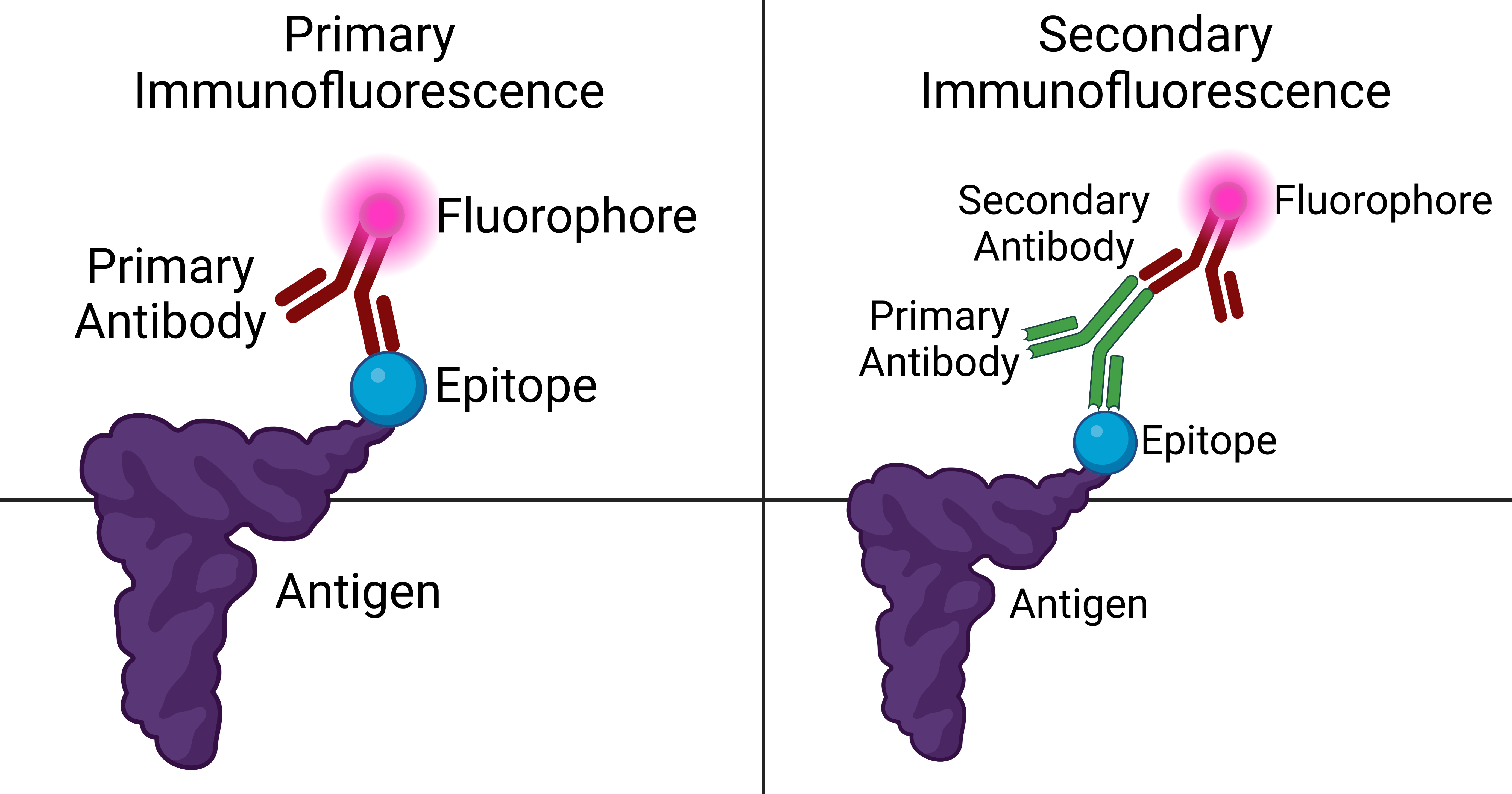
Immunofluorescence is a technique used for light microscopy with a fluorescence microscope and is used primarily on microbiological samples. This technique uses the specificity of antibodies to their antigen to target fluorescent dyes to specific biomolecule targets within a cell, and therefore allows visualization of the distribution of the target molecule through the sample. The specific region an antibody recognizes on an antigen is called an epitope. There have been efforts in epitope mapping since many antibodies can bind the same epitope and levels of binding between antibodies that recognize the same epitope can vary. Additionally, the binding of the fluorophore to the antibody itself cannot interfere with the immunological specificity of the antibody or the binding capacity of its antigen. Immunofluorescence is a widely used example of immunostaining (using antibodies to stain proteins) and is a specific example of immunohistochemistry (the use of the antibody-antigen rel ... [...More Info...] [...Related Items...] OR: [Wikipedia] [Google] [Baidu] |
Biopsychosocial Model
Biopsychosocial models are a class of trans-disciplinary models which look at the interconnection between biology, psychology, and socio-environmental factors. These models specifically examine how these aspects play a role in topics ranging from human development, to health and disease, to information processing, and to conflict. The term was first used to describe a model advocated by George L. Engel in 1977. It now also refers to any model which takes a similar approach, and has become an alternative to the biomedical and/or psychological dominance of many health care systems. History George L. Engel and Jon Romano of the University of Rochester in 1977, are widely credited with being the first to propose a biopsychosocial model. However, it had been proposed 100 years earlier and by others. Engel struggled with the then-prevailing biomedical approach to medicine as he strove for a more holistic approach by recognizing that each patient has their own thoughts, feelings, and ... [...More Info...] [...Related Items...] OR: [Wikipedia] [Google] [Baidu] |
Hypha
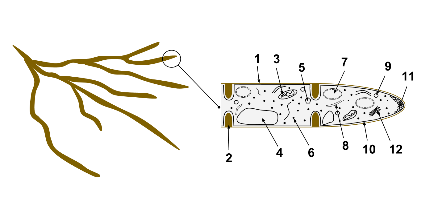
A hypha (; ) is a long, branching, filamentous structure of a fungus, oomycete, or actinobacterium. In most fungi, hyphae are the main mode of vegetative growth, and are collectively called a mycelium. Structure A hypha consists of one or more cells surrounded by a tubular cell wall. In most fungi, hyphae are divided into cells by internal cross-walls called "septa" (singular septum). Septa are usually perforated by pores large enough for ribosomes, mitochondria, and sometimes nuclei to flow between cells. The major structural polymer in fungal cell walls is typically chitin, in contrast to plants and oomycetes that have cellulosic cell walls. Some fungi have aseptate hyphae, meaning their hyphae are not partitioned by septa. Hyphae have an average diameter of 4–6 µm. Growth Hyphae grow at their tips. During tip growth, cell walls are extended by the external assembly and polymerization of cell wall components, and the internal production of new cell membrane. The S ... [...More Info...] [...Related Items...] OR: [Wikipedia] [Google] [Baidu] |
H&E Stain
Hematoxylin and eosin stain ( or haematoxylin and eosin stain or hematoxylin-eosin stain; often abbreviated as H&E stain or HE stain) is one of the principal tissue stains used in histology. It is the most widely used stain in medical diagnosis and is often the gold standard. For example, when a pathologist looks at a biopsy of a suspected cancer, the histological section is likely to be stained with H&E. H&E is the combination of two histological stains: hematoxylin and eosin. The hematoxylin stains cell nuclei a purplish blue, and eosin stains the extracellular matrix and cytoplasm pink, with other structures taking on different shades, hues, and combinations of these colors. Hence a pathologist can easily differentiate between the nuclear and cytoplasmic parts of a cell, and additionally, the overall patterns of coloration from the stain show the general layout and distribution of cells and provides a general overview of a tissue sample's structure. Thus, pattern recogniti ... [...More Info...] [...Related Items...] OR: [Wikipedia] [Google] [Baidu] |
Coenocyte
A coenocyte () is a multinucleate cell which can result from multiple nuclear divisions without their accompanying cytokinesis, in contrast to a syncytium, which results from cellular aggregation followed by dissolution of the cell membranes inside the mass. The word syncytium in animal embryology is used to refer to the coenocytic blastoderm of invertebrates. A coenocytic colony is referred to as a coenobium (plural coenobia), and most coenobia are composed of a distinct number of cells, often as a multiple of two (4, 8, etc.). Research suggests that coenobium formation may be a defense against grazing in some species. Physiological examples Protists Diplomonads, like '' Giardia'', have two nuclei. Ciliates have cells that contain two nuclei: a macronucleus and a micronucleus. The schizont of apicomplexan parasites is a form of a coenocyte (i.e. a plasmodium in the general sense) as well as the plasmodia of microsporidian (Fungi) and myxosporidian (Metazoa) parasites. The tro ... [...More Info...] [...Related Items...] OR: [Wikipedia] [Google] [Baidu] |
Hyaline
A hyaline substance is one with a glassy appearance. The word is derived from el, ὑάλινος, translit=hyálinos, lit=transparent, and el, ὕαλος, translit=hýalos, lit=crystal, glass, label=none. Histopathology Hyaline cartilage is named after its glassy appearance on fresh gross pathology. On light microscopy of H&E stained slides, the extracellular matrix of hyaline cartilage looks homogeneously pink, and the term "hyaline" is used to describe similarly homogeneously pink material besides the cartilage. Hyaline material is usually acellular and proteinaceous. For example, arterial hyaline is seen in aging, high blood pressure, diabetes mellitus and in association with some drugs (e.g. calcineurin inhibitors). It is bright pink with PAS staining. Ichthyology and entomology In ichthyology and entomology, ''hyaline'' denotes a colorless, transparent substance, such as unpigmented fins of fishes or clear insect wings. Resh, Vincent H. and R. T. Cardé, Eds. Encyclo ... [...More Info...] [...Related Items...] OR: [Wikipedia] [Google] [Baidu] |
Biopsy
A biopsy is a medical test commonly performed by a surgeon, interventional radiologist, or an interventional cardiologist. The process involves extraction of sample cells or tissues for examination to determine the presence or extent of a disease. The tissue is then fixed, dehydrated, embedded, sectioned, stained and mounted before it is generally examined under a microscope by a pathologist; it may also be analyzed chemically. When an entire lump or suspicious area is removed, the procedure is called an excisional biopsy. An incisional biopsy or core biopsy samples a portion of the abnormal tissue without attempting to remove the entire lesion or tumor. When a sample of tissue or fluid is removed with a needle in such a way that cells are removed without preserving the histological architecture of the tissue cells, the procedure is called a needle aspiration biopsy. Biopsies are most commonly performed for insight into possible cancerous or inflammatory conditions. History T ... [...More Info...] [...Related Items...] OR: [Wikipedia] [Google] [Baidu] |


