|
Basement Membrane
The basement membrane is a thin, pliable sheet-like type of extracellular matrix that provides cell and tissue support and acts as a platform for complex signalling. The basement membrane sits between Epithelium, epithelial tissues including mesothelium and endothelium, and the underlying connective tissue. Structure As seen with the electron microscope, the basement membrane is composed of two layers, the basal lamina and the reticular lamina. The underlying connective tissue attaches to the basal lamina with collagen VII anchoring fibrils and fibrillin microfibrils. The basal lamina layer can further be subdivided into two layers based on their visual appearance in electron microscopy. The lighter-colored layer closer to the epithelium is called the lamina lucida, while the denser-colored layer closer to the connective tissue is called the lamina densa. The Electron microscope, electron-dense lamina densa layer is about 30–70 nanometers thick and consists of an underlying ... [...More Info...] [...Related Items...] OR: [Wikipedia] [Google] [Baidu] |
Epithelium
Epithelium or epithelial tissue is one of the four basic types of animal tissue, along with connective tissue, muscle tissue and nervous tissue. It is a thin, continuous, protective layer of compactly packed cells with a little intercellular matrix. Epithelial tissues line the outer surfaces of organs and blood vessels throughout the body, as well as the inner surfaces of cavities in many internal organs. An example is the epidermis, the outermost layer of the skin. There are three principal shapes of epithelial cell: squamous (scaly), columnar, and cuboidal. These can be arranged in a singular layer of cells as simple epithelium, either squamous, columnar, or cuboidal, or in layers of two or more cells deep as stratified (layered), or ''compound'', either squamous, columnar or cuboidal. In some tissues, a layer of columnar cells may appear to be stratified due to the placement of the nuclei. This sort of tissue is called pseudostratified. All glands are made up of epithe ... [...More Info...] [...Related Items...] OR: [Wikipedia] [Google] [Baidu] |
Electron Microscope
An electron microscope is a microscope that uses a beam of accelerated electrons as a source of illumination. As the wavelength of an electron can be up to 100,000 times shorter than that of visible light photons, electron microscopes have a higher resolving power than light microscopes and can reveal the structure of smaller objects. A scanning transmission electron microscope has achieved better than 50 pm resolution in annular dark-field imaging mode and magnifications of up to about 10,000,000× whereas most light microscopes are limited by diffraction to about 200 nm resolution and useful magnifications below 2000×. Electron microscopes use shaped magnetic fields to form electron optical lens systems that are analogous to the glass lenses of an optical light microscope. Electron microscopes are used to investigate the ultrastructure of a wide range of biological and inorganic specimens including microorganisms, cells, large molecules, biopsy samples, ... [...More Info...] [...Related Items...] OR: [Wikipedia] [Google] [Baidu] |
Lamina Densa
The lamina densa is a component of the basement membrane zone between the epidermis and dermis of the skin, and is an electron-dense zone between the lamina lucida and dermis, synthesized by the basal cells of the epidermis, and composed of (1) type IV collagen, (2) anchoring fibrils made of type VII collagen, and (3) dermal microfibrils.James, William; Berger, Timothy; Elston, Dirk (2005). ''Andrews' Diseases of the Skin: Clinical Dermatology'' (10th ed.). Saunders. Page 5-6. . See also *Basal lamina The basal lamina is a layer of extracellular matrix secreted by the epithelial cells, on which the epithelium sits. It is often incorrectly referred to as the basement membrane, though it does constitute a portion of the basement membrane. The ba ... References Skin anatomy {{Dermatology-stub ... [...More Info...] [...Related Items...] OR: [Wikipedia] [Google] [Baidu] |
Nidogen
Nidogens, formerly known as entactins, are a family of sulfated monomeric glycoproteins located in the basal lamina of ParaHoxozoa, parahoxozoans. Two nidogens have been identified in humans: nidogen-1 (NID1) and nidogen-2 (NID2). Remarkably, vertebrates are still capable of stabilizing basement membrane in the absence of either identified nidogen. In contrast, those lacking both nidogen-1 and nidogen-2 typically die prematurely during embryonic development as a result of defects existing in the heart and lungs. Nidogen have been shown to play a crucial role during organogenesis in late embryonic development, particularly in cardiac and lung development. From an evolutionary perspective, nidogens are highly conserved across vertebrates and invertebrates, retaining their ability to bind laminin. In nematodes, nidogen-1 is necessary for axon guidance, but not for basement membrane assembly. References Human proteins Protein families Extracellular matrix proteins {{Biochemistry ... [...More Info...] [...Related Items...] OR: [Wikipedia] [Google] [Baidu] |
Lamina Lucida
The lamina lucida is a component of the basement membrane which is found between the epithelium and underlying connective tissue (e.g., epidermis and dermis of the skin). It is a roughly 40 nanometre wide electron-lucent zone between the plasma membrane of the basal cells and the (electron-dense) lamina densa of the basement membrane.James, William; Berger, Timothy; Elston, Dirk (2005). ''Andrews' Diseases of the Skin: Clinical Dermatology'' (10th ed.). Saunders. Page 5. . Similarly, electron-lucent and electron-dense zones can be seen between enamel of teeth and the junctional epithelium. The electron-lucent zone is adjacent to the cells of the junctional epithelium and might be considered a continuation of the lamina lucida as both are seen to harbour hemidesmosomes. However, unlike the lamina densa, the electron-dense zone adjacent to enamel show no signs of hemidesmosomes. Some theorize that the lamina lucida is an artifact created when preparing the tissue, and that the lam ... [...More Info...] [...Related Items...] OR: [Wikipedia] [Google] [Baidu] |
Basal Lamina
The basal lamina is a layer of extracellular matrix secreted by the epithelial cells, on which the epithelium sits. It is often incorrectly referred to as the basement membrane, though it does constitute a portion of the basement membrane. The basal lamina is visible only with the electron microscope, where it appears as an electron-dense layer that is 20–100 nm thick (with some exceptions that are thicker, such as basal lamina in lung alveoli and renal glomeruli). Structure The layers of the basal lamina ("BL") and those of the basement membrane ("BM") are described below: Anchoring fibrils composed of type VII collagen extend from the basal lamina into the underlying reticular lamina and loop around collagen bundles. Although found beneath all basal laminae, they are especially numerous in stratified squamous cells of the skin. These layers should not be confused with the lamina propria, which is found outside the basal lamina. Basement membrane The basement membra ... [...More Info...] [...Related Items...] OR: [Wikipedia] [Google] [Baidu] |
Hemidesmosome
Hemidesmosomes are very small stud-like structures found in keratinocytes of the epidermis of skin that attach to the extracellular matrix. They are similar in form to desmosomes when visualized by electron microscopy, however, desmosomes attach to adjacent cells. Hemidesmosomes are also comparable to focal adhesions, as they both attach cells to the extracellular matrix. Instead of desmogleins and desmocollins in the extracellular space, hemidesmosomes utilize integrins. Hemidesmosomes are found in epithelial cells connecting the basal epithelial cells to the lamina lucida, which is part of the basal lamina. Hemidesmosomes are also involved in signaling pathways, such as keratinocyte migration or carcinoma cell intrusion. Structure Hemidesmosomes can be categorized into two types based on their protein constituents. Type 1 hemidesmosomes are found in stratified and pseudo-stratified epithelium. Type 1 hemidesmosomes have five main elements: integrin α6 β4, plectin in its isof ... [...More Info...] [...Related Items...] OR: [Wikipedia] [Google] [Baidu] |
Dystroglycan
Dystroglycan is a protein that in humans is encoded by the ''DAG1'' gene. Dystroglycan is one of the dystrophin-associated glycoproteins, which is encoded by a 5.5 kb transcript in ''Homo sapiens'' on chromosome 3. There are two exons that are separated by a large intron. The spliced exons code for a protein product that is finally cleaved into two non-covalently associated subunits, lpha(N-terminal) and eta(C-terminal). Function In skeletal muscle the dystroglycan complex works as a transmembrane linkage between the extracellular matrix and the cytoskeleton. lphadystroglycan is extracellular and binds to merosin lpha2 laminin in the basement membrane, while etadystroglycan is a transmembrane protein and binds to dystrophin, which is a large rod-like cytoskeletal protein, absent in Duchenne muscular dystrophy patients. Dystrophin binds to intracellular actin cables. In this way, the dystroglycan complex, which links the extracellular matrix to the intracellular actin cab ... [...More Info...] [...Related Items...] OR: [Wikipedia] [Google] [Baidu] |
Entactin
Nidogen-1 (NID-1), formerly known as entactin, is a protein that in humans is encoded by the ''NID1'' gene. Both nidogen-1 and nidogen-2 are essential components of the basement membrane alongside other components such as type IV collagen, proteoglycans (heparan sulfate and glycosaminoglycans), laminin and fibronectin. Function Nidogen-1 is a member of the nidogen family of basement membrane glycoproteins. The protein interacts with several other components of basement membranes. Structurally it (along with perlecan) connects the networks formed by collagens and laminins to each other. It may also play a role in cell interactions with the extracellular matrix. Clinical significance Mutations in ''NID1'' cause autosomal dominant Dandy–Walker malformation with occipital encephalocele (ADDWOC). Interactions Nidogen-1 has been shown to interact with FBLN1 FBLN1 is the gene encoding fibulin-1, an extracellular matrix and plasma protein. Function Fibulin-1 is a secreted gl ... [...More Info...] [...Related Items...] OR: [Wikipedia] [Google] [Baidu] |
Integrin
Integrins are transmembrane receptors that facilitate cell-cell and cell-extracellular matrix (ECM) adhesion. Upon ligand binding, integrins activate signal transduction pathways that mediate cellular signals such as regulation of the cell cycle, organization of the intracellular cytoskeleton, and movement of new receptors to the cell membrane. The presence of integrins allows rapid and flexible responses to events at the cell surface (''e.g''. signal platelets to initiate an interaction with coagulation factors). Several types of integrins exist, and one cell generally has multiple different types on its surface. Integrins are found in all animals while integrin-like receptors are found in plant cells. Integrins work alongside other proteins such as cadherins, the immunoglobulin superfamily cell adhesion molecules, selectins and syndecans, to mediate cell–cell and cell–matrix interaction. Ligands for integrins include fibronectin, vitronectin, collagen and laminin. Stru ... [...More Info...] [...Related Items...] OR: [Wikipedia] [Google] [Baidu] |
Laminin
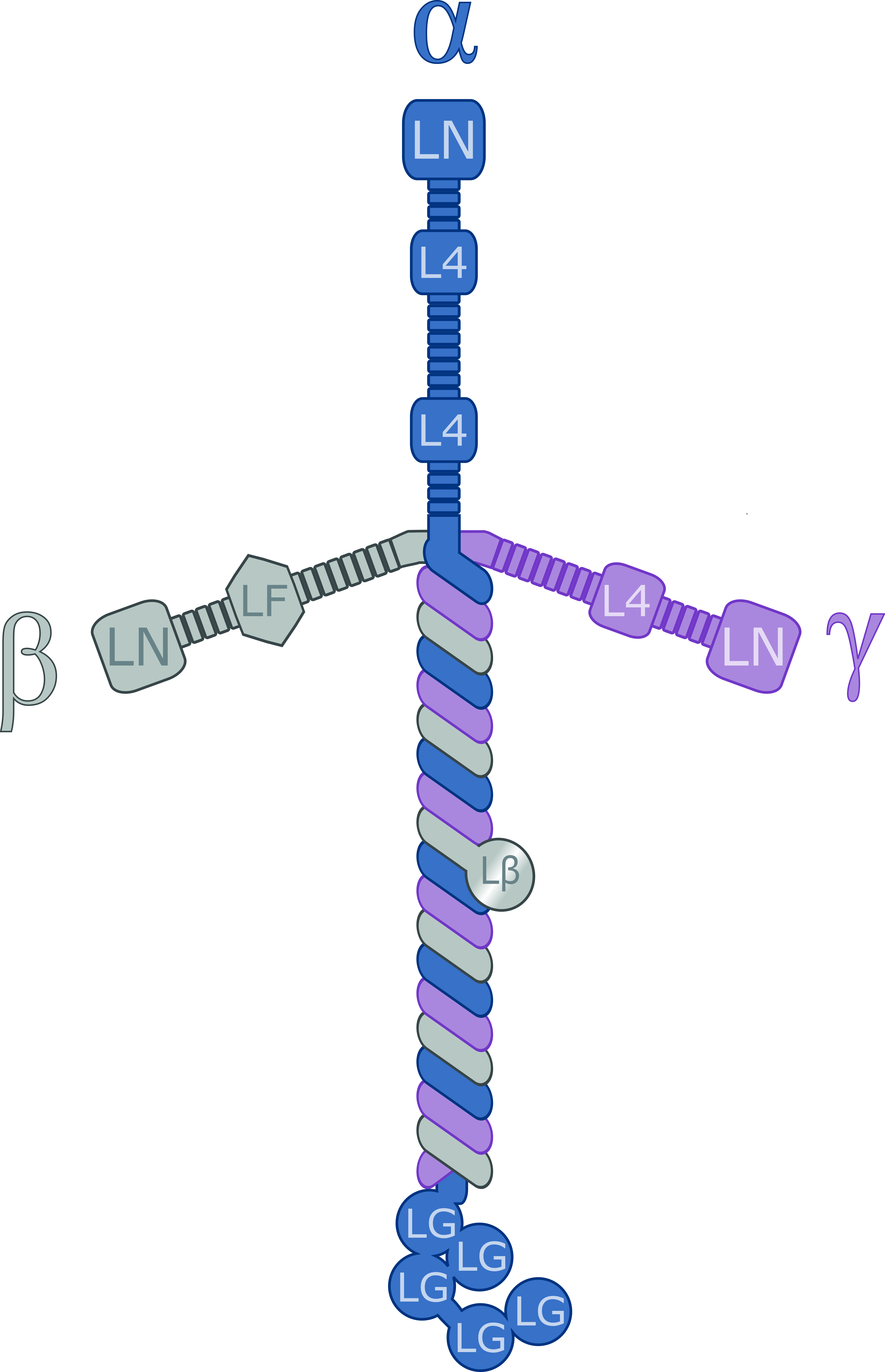
Laminins are a family of glycoproteins of the extracellular matrix of all animals. They are major components of the basal lamina (one of the layers of the basement membrane), the protein network foundation for most cells and organs. The laminins are an important and biologically active part of the basal lamina, influencing cell differentiation, migration, and adhesion. Laminins are heterotrimeric proteins with a high molecular mass (~400 to ~900 kDa). They contain three different chains (α, β and γ) encoded by five, four, and three paralogous genes in humans, respectively. The laminin molecules are named according to their chain composition. Thus, laminin-511 contains α5, β1, and γ1 chains. Fourteen other chain combinations have been identified ''in vivo''. The trimeric proteins intersect to form a cross-like structure that can bind to other cell membrane and extracellular matrix molecules. The three shorter arms are particularly good at binding to other laminin molecules, ... [...More Info...] [...Related Items...] OR: [Wikipedia] [Google] [Baidu] |
Perlecan
Perlecan (PLC) also known as basement membrane-specific heparan sulfate proteoglycan core protein (HSPG) or heparan sulfate proteoglycan 2 (HSPG2), is a protein that in humans is encoded by the ''HSPG2'' gene. The HSPG2 gene codes for a 4,391 amino acid protein with a molecular weight of 468,829. It is one of the largest known proteins. Perlecan was originally isolated from a tumor cell line and shown to be present in all native basement membranes. Perlecan is a large multidomain (five domains, labeled I-V) proteoglycan that binds to and cross-links many extracellular matrix (ECM) components and cell-surface molecules. Perlecan is synthesized by both vascular endothelial and smooth muscle cells and deposited in the extracellular matrix of parahoxozoans. Perlecan is highly conserved across species and the available data indicate that it has evolved from ancient ancestors by gene duplication and exon shuffling. Structure Perlecan consists of a core protein of molecular weight 4 ... [...More Info...] [...Related Items...] OR: [Wikipedia] [Google] [Baidu] |


