|
Barton's Fracture
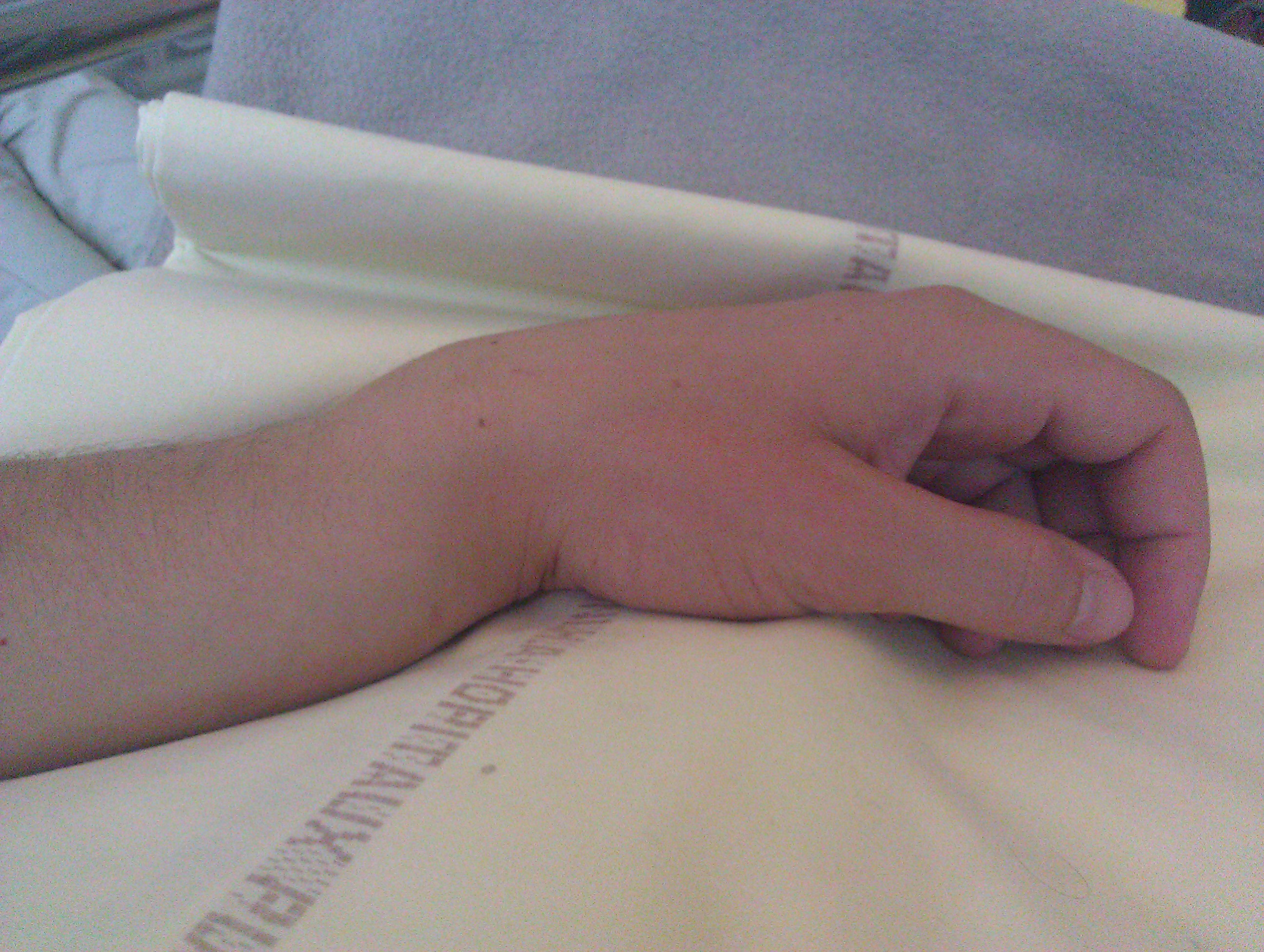
A Barton's fracture is a type of wrist injury where there is a broken bone associated with a dislocated bone in the wrist, typically occurring after falling on top of a bent wrist. It is an intra-articular fracture of the distal radius with dislocation of the radiocarpal joint. There exist two types of Barton's fracture – dorsal and palmar, the latter being more common. The Barton's fracture is caused by a fall on an extended and pronated wrist increasing carpal compression force on the dorsal rim. Intra-articular component distinguishes this fracture from a Smith's or a Colles' fracture. Treatment of this fracture is usually done by open reduction and internal fixation with a plate and screws, but occasionally the fracture can be treated conservatively. Eponym It is named after John Rhea Barton John Rhea Barton (April 1794 – January 1, 1871) was an American orthopedic surgeon remembered for describing Barton's fracture. Early life Barton was born in Lancaster, Penns ... [...More Info...] [...Related Items...] OR: [Wikipedia] [Google] [Baidu] |
Volume Rendering
In scientific visualization and computer graphics, volume rendering is a set of techniques used to display a 2D projection of a 3D discretely sampled data set, typically a 3D scalar field. A typical 3D data set is a group of 2D slice images acquired by a CT, MRI, or MicroCT scanner. Usually these are acquired in a regular pattern (e.g., one slice for each millimeter of depth) and usually have a regular number of image pixels in a regular pattern. This is an example of a regular volumetric grid, with each volume element, or voxel represented by a single value that is obtained by sampling the immediate area surrounding the voxel. To render a 2D projection of the 3D data set, one first needs to define a camera in space relative to the volume. Also, one needs to define the opacity and color of every voxel. This is usually defined using an RGBA (for red, green, blue, alpha) transfer function that defines the RGBA value for every possible voxel value. For example, a volu ... [...More Info...] [...Related Items...] OR: [Wikipedia] [Google] [Baidu] |
Fracture
Fracture is the separation of an object or material into two or more pieces under the action of stress. The fracture of a solid usually occurs due to the development of certain displacement discontinuity surfaces within the solid. If a displacement develops perpendicular to the surface, it is called a normal tensile crack or simply a crack; if a displacement develops tangentially, it is called a shear crack, slip band or dislocation. Brittle fractures occur with no apparent deformation before fracture. Ductile fractures occur after visible deformation. Fracture strength, or breaking strength, is the stress when a specimen fails or fractures. The detailed understanding of how a fracture occurs and develops in materials is the object of fracture mechanics. Strength Fracture strength, also known as breaking strength, is the stress at which a specimen fails via fracture. This is usually determined for a given specimen by a tensile test, which charts the stress–strain ... [...More Info...] [...Related Items...] OR: [Wikipedia] [Google] [Baidu] |
Bone Fracture
A bone fracture (abbreviated FRX or Fx, Fx, or #) is a medical condition in which there is a partial or complete break in the continuity of any bone in the body. In more severe cases, the bone may be broken into several fragments, known as a ''comminuted fracture''. A bone fracture may be the result of high force impact or stress, or a minimal trauma injury as a result of certain medical conditions that weaken the bones, such as osteoporosis, osteopenia, bone cancer, or osteogenesis imperfecta, where the fracture is then properly termed a pathologic fracture. Signs and symptoms Although bone tissue contains no pain receptors, a bone fracture is painful for several reasons: * Breaking in the continuity of the periosteum, with or without similar discontinuity in endosteum, as both contain multiple pain receptors. * Edema and hematoma of nearby soft tissues caused by ruptured bone marrow evokes pressure pain. * Involuntary muscle spasms trying to hold bone fragments in pla ... [...More Info...] [...Related Items...] OR: [Wikipedia] [Google] [Baidu] |
Distal Radius
The radius or radial bone is one of the two large bones of the forearm, the other being the ulna. It extends from the lateral side of the elbow to the thumb side of the wrist and runs parallel to the ulna. The ulna is usually slightly longer than the radius, but the radius is thicker. Therefore the radius is considered to be the larger of the two. It is a long bone, prism-shaped and slightly curved longitudinally. The radius is part of two joints: the elbow and the wrist. At the elbow, it joins with the capitulum of the humerus, and in a separate region, with the ulna at the radial notch. At the wrist, the radius forms a joint with the ulna bone. The corresponding bone in the lower leg is the fibula. Structure The long narrow medullary cavity is enclosed in a strong wall of compact bone. It is thickest along the interosseous border and thinnest at the extremities, same over the cup-shaped articular surface (fovea) of the head. The trabeculae of the spongy tissue are ... [...More Info...] [...Related Items...] OR: [Wikipedia] [Google] [Baidu] |
Radiocarpal Joint
In human anatomy, the wrist is variously defined as (1) the carpus or carpal bones, the complex of eight bones forming the proximal skeletal segment of the hand; "The wrist contains eight bones, roughly aligned in two rows, known as the carpal bones." (2) the wrist joint or radiocarpal joint, the joint between the radius and the carpus and; (3) the anatomical region surrounding the carpus including the distal parts of the bones of the forearm and the proximal parts of the metacarpus or five metacarpal bones and the series of joints between these bones, thus referred to as ''wrist joints''. "With the large number of bones composing the wrist (ulna, radius, eight carpas, and five metacarpals), it makes sense that there are many, many joints that make up the structure known as the wrist." This region also includes the carpal tunnel, the anatomical snuff box, bracelet lines, the flexor retinaculum, and the extensor retinaculum. As a consequence of these various definitions, frac ... [...More Info...] [...Related Items...] OR: [Wikipedia] [Google] [Baidu] |
Pronated
Motion, the process of movement, is described using specific anatomical terms. Motion includes movement of organs, joints, limbs, and specific sections of the body. The terminology used describes this motion according to its direction relative to the anatomical position of the body parts involved. Anatomists and others use a unified set of terms to describe most of the movements, although other, more specialized terms are necessary for describing unique movements such as those of the hands, feet, and eyes. In general, motion is classified according to the anatomical plane it occurs in. ''Flexion'' and ''extension'' are examples of ''angular'' motions, in which two axes of a joint are brought closer together or moved further apart. ''Rotational'' motion may occur at other joints, for example the shoulder, and are described as ''internal'' or ''external''. Other terms, such as ''elevation'' and ''depression'', describe movement above or below the horizontal plane. Many anatomi ... [...More Info...] [...Related Items...] OR: [Wikipedia] [Google] [Baidu] |
Smith's Fracture
A Smith's fracture, is a fracture of the distal radius. Although it can also be caused by a direct blow to the dorsal forearm or by a fall with the wrist flexed, the most common mechanism of injury for Smith's fracture occurs in a palmar fall with the wrist joint slightly dorsiflexed. Smith's fractures are less common than Colles' fractures. The distal fracture fragment is displaced volarly (ventrally), as opposed to a Colles' fracture which the fragment is displaced dorsally. Depending on the severity of the impact, there may be one or many fragments and it may or may not involve the articular surface of the wrist joint. Classification A commonly used classification of distal radial fractures is the Frykman Classification: * Type I: Extra-articular * Type II: Type I, with fracture of distal ulna * Type III: Radiocarpal joint involvement * Type IV: Type III with fracture of distal ulna * Type V: Distal radioulnar joint involved. * Type VI: Type V with fracture of distal ulna ... [...More Info...] [...Related Items...] OR: [Wikipedia] [Google] [Baidu] |
Colles' Fracture
A Colles' fracture is a type of fracture of the distal forearm in which the broken end of the radius is bent backwards. Symptoms may include pain, swelling, deformity, and bruising. Complications may include damage to the median nerve. It typically occurs as a result of a fall on an outstretched hand. Risk factors include osteoporosis. The diagnosis may be confirmed via X-rays. The tip of the ulna may also be broken. Treatment may include casting or surgery. Surgical reduction and casting is possible in the majority of cases in people over the age of 50. Pain management can be achieved during the reduction with procedural sedation and analgesia or a hematoma block. A year or two may be required for healing to occur. About 15% of people have a Colles' fracture at some point in their life. They occur more commonly in young adults and older people than in children and middle-aged adults. Women are more frequently affected than men. The fracture is named after Abraham Colle ... [...More Info...] [...Related Items...] OR: [Wikipedia] [Google] [Baidu] |
John Rhea Barton
John Rhea Barton (April 1794 – January 1, 1871) was an American orthopedic surgeon remembered for describing Barton's fracture. Early life Barton was born in Lancaster, Pennsylvania in April 1794. He was the son of Elizabeth ( née Rhea) Barton (b. 1759) and William Barton (1754–1817), a lawyer who designed the Great Seal of the United States. Among his siblings was older brother was William Paul Crillon Barton, the medical botanist, physician, professor, naval surgeon, and botanical illustrator. His uncle, Benjamin Smith Barton, was an eminent medical botanist and vice-president of the American Philosophical Society. Barton graduated from the School of Medicine (now known as the Perelman School of Medicine) at the University of Pennsylvania in 1818 and started teaching there soon after. Career He became surgeon at the Philadelphia Almshouse, working for Philip Syng Physick, and returned to the Pennsylvania Hospital as surgeon in 1823. He was said to be ambidex ... [...More Info...] [...Related Items...] OR: [Wikipedia] [Google] [Baidu] |
Radiograph Of Barton's Fracture
Radiography is an imaging technique using X-rays, gamma rays, or similar ionizing radiation and non-ionizing radiation to view the internal form of an object. Applications of radiography include medical radiography ("diagnostic" and "therapeutic") and industrial radiography. Similar techniques are used in airport security (where "body scanners" generally use backscatter X-ray). To create an image in conventional radiography, a beam of X-rays is produced by an X-ray generator and is projected toward the object. A certain amount of the X-rays or other radiation is absorbed by the object, dependent on the object's density and structural composition. The X-rays that pass through the object are captured behind the object by a detector (either photographic film or a digital detector). The generation of flat two dimensional images by this technique is called projectional radiography. In computed tomography (CT scanning) an X-ray source and its associated detectors rotate around the ... [...More Info...] [...Related Items...] OR: [Wikipedia] [Google] [Baidu] |


