|
Baker's Cyst
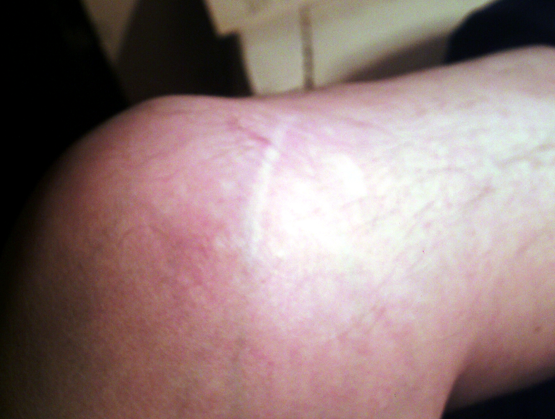
A Baker's cyst, also known as a popliteal cyst, is a type of fluid collection behind the knee. Often there are no symptoms. If symptoms do occur these may include swelling and pain behind the knee, or knee stiffness. If the cyst breaks open, pain may significantly increase with swelling of the calf. Rarely complications such as deep vein thrombosis, peripheral neuropathy, ischemia, or compartment syndrome may occur. Risk factors include other knee problems such as osteoarthritis, meniscal tears, or rheumatoid arthritis. The underlying mechanism involves the flow of synovial fluid from the knee joint to the gastrocnemio-semimembranosus bursa, resulting in its expansion. The diagnosis may be confirmed with ultrasound or magnetic resonance imaging (MRI). Treatment is initially with supportive care. If this is not effective aspiration and steroid injection or surgical removal may be carried out. Around 20% of people have a Baker's cyst. They occur most commonly in those 35 to 70 ... [...More Info...] [...Related Items...] OR: [Wikipedia] [Google] [Baidu] |
Deep Vein Thrombosis
Deep vein thrombosis (DVT) is a type of venous thrombosis involving the formation of a blood clot in a deep vein, most commonly in the legs or pelvis. A minority of DVTs occur in the arms. Symptoms can include pain, swelling, redness, and enlarged veins in the affected area, but some DVTs have no symptoms. The most common life-threatening concern with DVT is the potential for a clot to embolize (detach from the veins), travel as an embolus through the right side of the heart, and become lodged in a pulmonary artery that supplies blood to the lungs. This is called a pulmonary embolism (PE). DVT and PE comprise the cardiovascular disease of venous thromboembolism (VTE). About two-thirds of VTE manifests as DVT only, with one-third manifesting as PE with or without DVT. The most frequent long-term DVT complication is post-thrombotic syndrome, which can cause pain, swelling, a sensation of heaviness, itching, and in severe cases, ulcers. Recurrent VTE occurs in about 30% of those i ... [...More Info...] [...Related Items...] OR: [Wikipedia] [Google] [Baidu] |
Knee Joint
In humans and other primates, the knee joins the thigh with the leg and consists of two joints: one between the femur and tibia (tibiofemoral joint), and one between the femur and patella (patellofemoral joint). It is the largest joint in the human body. The knee is a modified hinge joint, which permits flexion and extension as well as slight internal and external rotation. The knee is vulnerable to injury and to the development of osteoarthritis. It is often termed a ''compound joint'' having tibiofemoral and patellofemoral components. (The fibular collateral ligament is often considered with tibiofemoral components.) Structure The knee is a modified hinge joint, a type of synovial joint, which is composed of three functional compartments: the patellofemoral articulation, consisting of the patella, or "kneecap", and the patellar groove on the front of the femur through which it slides; and the medial and lateral tibiofemoral articulations linking the femur, or thigh bone, ... [...More Info...] [...Related Items...] OR: [Wikipedia] [Google] [Baidu] |
Semimembranosus Muscle
The semimembranosus muscle () is the most medial of the three hamstring muscles in the thigh. It is so named because it has a flat tendon of origin. It lies posteromedially in the thigh, deep to the semitendinosus muscle. It extends the hip joint and flexes the knee joint. Structure The semimembranosus muscle, so called from its membranous tendon of origin, is situated at the back and medial side of the thigh. It is wider, flatter, and deeper than the semitendinosus (with which it shares very close insertion and attachment points). The muscle overlaps the upper part of the popliteal vessels. Origin The semimembranosus muscle originates by a thick tendon from the superolateral aspect of the ischial tuberosity. It arises above and medial to the biceps femoris muscle and semitendinosus muscle. The tendon of origin expands into an aponeurosis, which covers the upper part of the anterior surface of the muscle; from this aponeurosis, muscular fibers arise, and converge to another a ... [...More Info...] [...Related Items...] OR: [Wikipedia] [Google] [Baidu] |
Gastrocnemius Muscle
The gastrocnemius muscle (plural ''gastrocnemii'') is a superficial two-headed muscle that is in the back part of the lower leg of humans. It runs from its two heads just above the knee to the heel, a three joint muscle (knee, ankle and subtalar joints). The muscle is named via Latin, from Greek γαστήρ (''gaster'') 'belly' or 'stomach' and κνήμη (''knḗmē'') 'leg', meaning 'stomach of the leg' (referring to the bulging shape of the calf). Structure The gastrocnemius is located with the soleus in the posterior (back) compartment of the leg. The lateral head originates from the lateral condyle of the femur, while the medial head originates from the medial condyle of the femur. Its other end forms a common tendon with the soleus muscle; this tendon is known as the calcaneal tendon or Achilles tendon and inserts onto the posterior surface of the calcaneus, or heel bone. It is considered a superficial muscle as it is located directly under skin, and its shape may often ... [...More Info...] [...Related Items...] OR: [Wikipedia] [Google] [Baidu] |
Meniscus (anatomy)
A meniscus is a crescent-shaped fibrocartilaginous anatomical structure that, in contrast to an articular disc, only partly divides a joint cavity.Platzer (2004), p 208 In humans they are present in the knee, wrist, acromioclavicular, sternoclavicular, and temporomandibular joints; in other animals they may be present in other joints. Generally, the term "meniscus" is used to refer to the cartilage of the knee, either to the lateral or medial meniscus. Both are cartilaginous tissues that provide structural integrity to the knee when it undergoes tension and torsion. The menisci are also known as "semi-lunar" cartilages, referring to their half-moon, crescent shape. The term "meniscus" is from the Ancient Greek word (), meaning "crescent". Structure The menisci of the knee are two pads of fibrocartilaginous tissue which serve to disperse friction in the knee joint between the lower leg (tibia) and the thigh (femur). They are concave on the top and flat on the bottom, articula ... [...More Info...] [...Related Items...] OR: [Wikipedia] [Google] [Baidu] |
Cartilage
Cartilage is a resilient and smooth type of connective tissue. In tetrapods, it covers and protects the ends of long bones at the joints as articular cartilage, and is a structural component of many body parts including the rib cage, the neck and the bronchial tubes, and the intervertebral discs. In other taxa, such as chondrichthyans, but also in cyclostomes, it may constitute a much greater proportion of the skeleton. It is not as hard and rigid as bone, but it is much stiffer and much less flexible than muscle. The matrix of cartilage is made up of glycosaminoglycans, proteoglycans, collagen fibers and, sometimes, elastin. Because of its rigidity, cartilage often serves the purpose of holding tubes open in the body. Examples include the rings of the trachea, such as the cricoid cartilage and carina. Cartilage is composed of specialized cells called chondrocytes that produce a large amount of collagenous extracellular matrix, abundant ground substance that is rich in pro ... [...More Info...] [...Related Items...] OR: [Wikipedia] [Google] [Baidu] |
Arthritis
Arthritis is a term often used to mean any disorder that affects joints. Symptoms generally include joint pain and stiffness. Other symptoms may include redness, warmth, swelling, and decreased range of motion of the affected joints. In some types of arthritis, other organs are also affected. Onset can be gradual or sudden. There are over 100 types of arthritis. The most common forms are osteoarthritis (degenerative joint disease) and rheumatoid arthritis. Osteoarthritis usually occurs with age and affects the fingers, knees, and hips. Rheumatoid arthritis is an autoimmune disorder that often affects the hands and feet. Other types include gout, lupus, fibromyalgia, and septic arthritis. They are all types of rheumatic disease. Treatment may include resting the joint and alternating between applying ice and heat. Weight loss and exercise may also be useful. Recommended medications may depend on the form of arthritis. These may include pain medications such as ibuprofen ... [...More Info...] [...Related Items...] OR: [Wikipedia] [Google] [Baidu] |
Medial Malleolus
A malleolus is the bony prominence on each side of the human ankle. Each leg is supported by two bones, the tibia on the inner side (medial) of the leg and the fibula on the outer side (lateral) of the leg. The medial malleolus is the prominence on the inner side of the ankle, formed by the lower end of the tibia. The lateral malleolus is the prominence on the outer side of the ankle, formed by the lower end of the fibula. The word ''malleolus'' (), plural ''malleoli'' (), comes from Latin and means "small hammer". (It is cognate with ''mallet''.) Medial malleolus The medial malleolus is found at the foot end of the tibia. The medial surface of the lower extremity of tibia is prolonged downward to form a strong pyramidal process, flattened from without inward - the medial malleolus. * The ''medial surface'' of this process is convex and subcutaneous. * The ''lateral'' or ''articular surface'' is smooth and slightly concave, and articulates with the talus. * The ''anterior bo ... [...More Info...] [...Related Items...] OR: [Wikipedia] [Google] [Baidu] |
Ecchymosis
A bruise, also known as a contusion, is a type of hematoma of tissue, the most common cause being capillaries damaged by trauma, causing localized bleeding that extravasates into the surrounding interstitial tissues. Most bruises occur close enough to the epidermis such that the bleeding causes a visible discoloration. The bruise then remains visible until the blood is either absorbed by tissues or cleared by immune system action. Bruises which do not blanch under pressure can involve capillaries at the level of skin, subcutaneous tissue, muscle, or bone. Bruises are not to be confused with other similar-looking lesions. (Such lesions include petechia (less than , resulting from numerous and diverse etiologies such as adverse reactions from medications such as warfarin, straining, asphyxiation, platelet disorders and diseases such as ''cytomegalovirus''), purpura (, classified as palpable purpura or non-palpable purpura and indicates various pathologic conditions such as thromb ... [...More Info...] [...Related Items...] OR: [Wikipedia] [Google] [Baidu] |
Who Named It
''Whonamedit?'' is an online English-language dictionary of medical eponyms and the people associated with their identification. Though it is a dictionary, many eponyms and persons are presented in extensive articles with comprehensive bibliographies. The dictionary is hosted in Norway and maintained by medical historian Ole Daniel Enersen Ole Daniel Enersen (born March 14, 1943, in Oslo, Norway) is a Norwegian climber, photographer, journalist, writer, and medical historian. In 1965 he made the first ascent of the Trollveggen mountain in Romsdalen, Norway, along with Leif Norman .... References External links * Medical websites Medical dictionaries Eponyms {{online-dict-stub ... [...More Info...] [...Related Items...] OR: [Wikipedia] [Google] [Baidu] |
William Morrant Baker
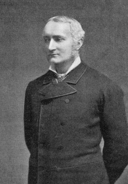
William Morrant Baker (20 October 1839, Andover, Hampshire, England – 3 October 1896, Pulborough, Sussex) was an English physician and surgeon. He first described the condition now known as Baker's cyst. Life William Morrant Baker was the son of a solicitor in the Hampshire town of Andover. He was apprenticed to the local surgeon, Mr. Payne. In 1858 he entered St. Bartholomew's Hospital Medical School in London and qualified in 1861. Baker became Sir James Paget's assistant for many years. From 1869 until 1885, he was lecturer in general anatomy and physiology at St. Bartholomew's Hospital. He was elected an assistant surgeon to the hospital in 1871 and a surgeon in 1882. He resigned his post as surgeon in 1892 due to his own locomotor ataxia condition. He was then appointed a governor of the hospital. Baker was also surgeon, later consultant surgeon, to the Evelina Children's Hospital, London and was examinator of general anatomy and physiology at the Royal College of Surge ... [...More Info...] [...Related Items...] OR: [Wikipedia] [Google] [Baidu] |
Corticosteroid
Corticosteroids are a class of steroid hormones that are produced in the adrenal cortex of vertebrates, as well as the synthetic analogues of these hormones. Two main classes of corticosteroids, glucocorticoids and mineralocorticoids, are involved in a wide range of physiological processes, including stress response, immune response, and regulation of inflammation, carbohydrate metabolism, protein catabolism, blood electrolyte levels, and behavior. Some common naturally occurring steroid hormones are cortisol (), corticosterone (), cortisone () and aldosterone (). (Note that cortisone and aldosterone are isomers.) The main corticosteroids produced by the adrenal cortex are cortisol and aldosterone. Classes * Glucocorticoids such as cortisol affect carbohydrate, fat, and protein metabolism, and have anti-inflammatory, immunosuppressive, anti-proliferative, and vasoconstrictive effects. Anti-inflammatory effects are mediated by blocking the action of inflammatory medi ... [...More Info...] [...Related Items...] OR: [Wikipedia] [Google] [Baidu] |


