|
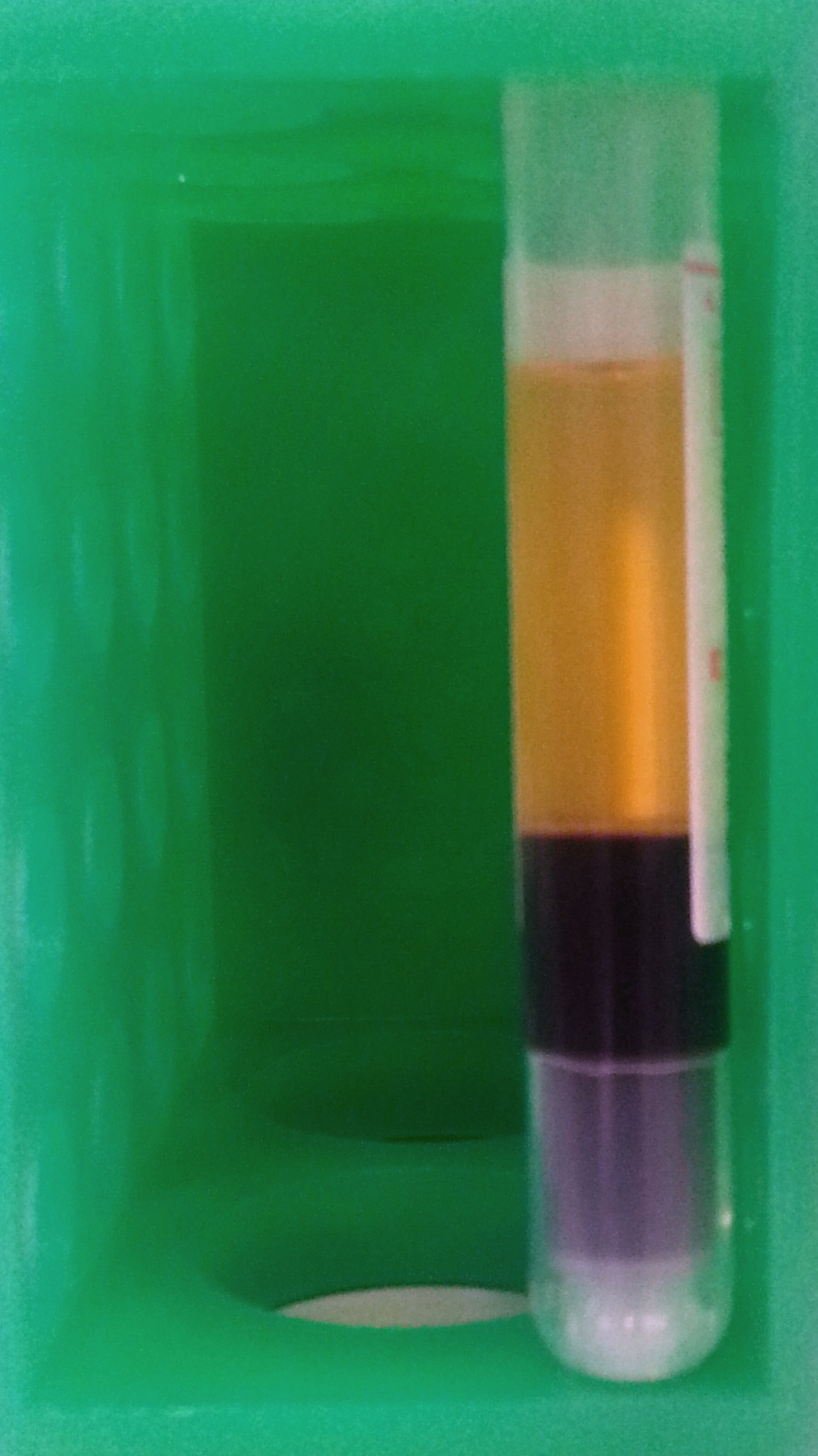
Buffy Coat
The buffy coat is the fraction of an anticoagulated blood sample that contains most of the white blood cells and platelets following centrifugation. It is rich in a number of immune cells including leukocytes, granulocytes. Description After centrifugation, one can distinguish a layer of clear fluid (the plasma), a layer of red fluid containing most of the red blood cells, and a thin layer in between. Composing less than 1% of the total volume of the blood sample, the buffy coat (so-called because it is usually buff in hue), contains most of the white blood cells and platelets. The buffy coat is usually whitish in color, but is sometimes green if the blood sample contains large amounts of neutrophils, which are high in green-colored myeloperoxidase. The layer beneath the buffy coat contains granulocytes and red blood cells. The buffy coat is commonly used for DNA extraction, with white blood cells providing approximately 10 times more concentrated sources of nucleated c ... [...More Info...] [...Related Items...] OR: [Wikipedia] [Google] [Baidu] |
Red Blood Cells
Red blood cells (RBCs), also referred to as red cells, red blood corpuscles (in humans or other animals not having nucleus in red blood cells), haematids, erythroid cells or erythrocytes (from Greek language, Greek ''erythros'' for "red" and ''kytos'' for "hollow vessel", with ''-cyte'' translated as "cell" in modern usage), are the most common type of blood cell and the vertebrate's principal means of delivering oxygen (O2) to the body tissue (biology), tissues—via blood flow through the circulatory system. RBCs take up oxygen in the lungs, or in fish the gills, and release it into tissues while squeezing through the body's capillary, capillaries. The cytoplasm of a red blood cell is rich in hemoglobin, an iron-containing biomolecule that can bind oxygen and is responsible for the red color of the cells and the blood. Each human red blood cell contains approximately 270 million hemoglobin molecules. The cell membrane is composed of proteins and lipids, and this structure ... [...More Info...] [...Related Items...] OR: [Wikipedia] [Google] [Baidu] |
Malaria
Malaria is a mosquito-borne infectious disease that affects humans and other animals. Malaria causes symptoms that typically include fever, tiredness, vomiting, and headaches. In severe cases, it can cause jaundice, seizures, coma, or death. Symptoms usually begin ten to fifteen days after being bitten by an infected mosquito. If not properly treated, people may have recurrences of the disease months later. In those who have recently survived an infection, reinfection usually causes milder symptoms. This partial resistance disappears over months to years if the person has no continuing exposure to malaria. Malaria is caused by single-celled microorganisms of the ''Plasmodium'' group. It is spread exclusively through bites of infected ''Anopheles'' mosquitoes. The mosquito bite introduces the parasites from the mosquito's saliva into a person's blood. The parasites travel to the liver where they mature and reproduce. Five species of ''Plasmodium'' can infect and be spread by h ... [...More Info...] [...Related Items...] OR: [Wikipedia] [Google] [Baidu] |
Leukocytes
White blood cells, also called leukocytes or leucocytes, are the cells of the immune system that are involved in protecting the body against both infectious disease and foreign invaders. All white blood cells are produced and derived from multipotent cells in the bone marrow known as hematopoietic stem cells. Leukocytes are found throughout the body, including the blood and lymphatic system. All white blood cells have nuclei, which distinguishes them from the other blood cells, the anucleated red blood cells (RBCs) and platelets. The different white blood cells are usually classified by cell lineage ( myeloid cells or lymphoid cells). White blood cells are part of the body's immune system. They help the body fight infection and other diseases. Types of white blood cells are granulocytes (neutrophils, eosinophils, and basophils), and agranulocytes (monocytes, and lymphocytes (T cells and B cells)). Myeloid cells (myelocytes) include neutrophils, eosinophils, mast cells, ... [...More Info...] [...Related Items...] OR: [Wikipedia] [Google] [Baidu] |
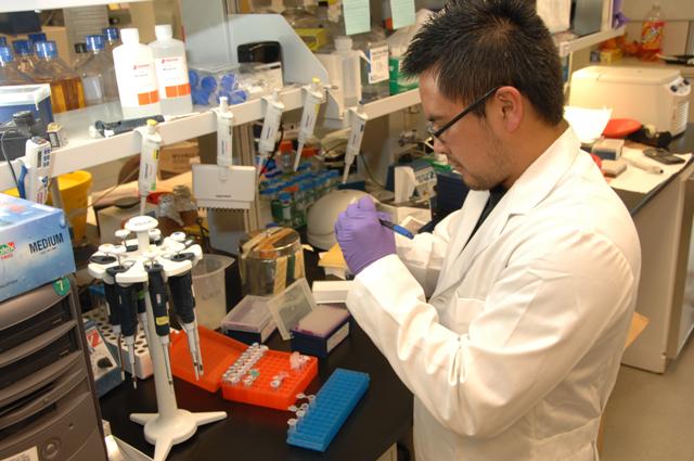
Medical Technologist
A medical laboratory scientist (MLS) or clinical laboratory scientist (CLS) or medical technologist (MT) performs diagnostic testing of blood and body fluids in clinical laboratories. The scope of a medical laboratory scientist's work begins with the receipt of patient or client specimens and terminates with the delivery of test results to physicians and other healthcare providers. The utility of clinical diagnostic testing relies squarely on the validity of test methodology. To this end, much of the work done by medical laboratory scientists involves ensuring specimen quality, interpreting test results, data-logging, testing control products, performing calibration, maintenance, validation, and troubleshooting of instrumentation as well as performing statistical analyses to verify the accuracy and repeatability of testing. Medical laboratory scientists may also assist healthcare providers with test selection and specimen collection and are responsible for prompt verbal delivery o ... [...More Info...] [...Related Items...] OR: [Wikipedia] [Google] [Baidu] |
Thick Smear
A blood smear, peripheral blood smear or blood film is a thin layer of blood smeared on a glass microscope slide and then stained in such a way as to allow the various blood cells to be examined microscopically. Blood smears are examined in the investigation of hematological (blood) disorders and are routinely employed to look for blood parasites, such as those of malaria and filariasis. Preparation A blood smear is made by placing a drop of blood on one end of a slide, and using a ''spreader slide'' to disperse the blood over the slide's length. The aim is to get a region, called a monolayer, where the cells are spaced far enough apart to be counted and differentiated. The monolayer is found in the "feathered edge" created by the spreader slide as it draws the blood forward. The slide is left to air dry, after which the blood is fixed to the slide by immersing it briefly in methanol. The fixative is essential for good staining and presentation of cellular detail. After fixat ... [...More Info...] [...Related Items...] OR: [Wikipedia] [Google] [Baidu] |
Ultraviolet
Ultraviolet (UV) is a form of electromagnetic radiation with wavelength from 10 nanometer, nm (with a corresponding frequency around 30 Hertz, PHz) to 400 nm (750 Hertz, THz), shorter than that of visible light, but longer than X-rays. UV radiation is present in sunlight, and constitutes about 10% of the total electromagnetic radiation output from the Sun. It is also produced by electric arcs and specialized lights, such as mercury-vapor lamps, tanning lamps, and black lights. Although long-wavelength ultraviolet is not considered an ionizing radiation because its photons lack the energy to ionization, ionize atoms, it can cause chemical reactions and causes many substances to glow or fluorescence, fluoresce. Consequently, the chemical and biological effects of UV are greater than simple heating effects, and many practical applications of UV radiation derive from its interactions with organic molecules. Short-wave ultraviolet light damages DNA and sterilizes surf ... [...More Info...] [...Related Items...] OR: [Wikipedia] [Google] [Baidu] |
Fluorescence Microscopy
A fluorescence microscope is an optical microscope that uses fluorescence instead of, or in addition to, scattering, reflection, and attenuation or absorption, to study the properties of organic or inorganic substances. "Fluorescence microscope" refers to any microscope that uses fluorescence to generate an image, whether it is a simple set up like an epifluorescence microscope or a more complicated design such as a confocal microscope, which uses optical sectioning to get better resolution of the fluorescence image. Principle The specimen is illuminated with light of a specific wavelength (or wavelengths) which is absorbed by the fluorophores, causing them to emit light of longer wavelengths (i.e., of a different color than the absorbed light). The illumination light is separated from the much weaker emitted fluorescence through the use of a spectral emission filter. Typical components of a fluorescence microscope are a light source (xenon arc lamp or mercury-vapor lamp are ... [...More Info...] [...Related Items...] OR: [Wikipedia] [Google] [Baidu] |
Fluorescence
Fluorescence is the emission of light by a substance that has absorbed light or other electromagnetic radiation. It is a form of luminescence. In most cases, the emitted light has a longer wavelength, and therefore a lower photon energy, than the absorbed radiation. A perceptible example of fluorescence occurs when the absorbed radiation is in the ultraviolet region of the electromagnetic spectrum (invisible to the human eye), while the emitted light is in the visible region; this gives the fluorescent substance a distinct color that can only be seen when the substance has been exposed to UV light. Fluorescent materials cease to glow nearly immediately when the radiation source stops, unlike phosphorescent materials, which continue to emit light for some time after. Fluorescence has many practical applications, including mineralogy, gemology, medicine, chemical sensors (fluorescence spectroscopy), fluorescent labelling, dyes, biological detectors, cosmic-ray detection, vacu ... [...More Info...] [...Related Items...] OR: [Wikipedia] [Google] [Baidu] |
Acridine Orange
Acridine orange is an organic compound that serves as a nucleic acid-selective fluorescent dye with cationic properties useful for cell cycle determination. Acridine orange is cell-permeable, which allows the dye to interact with DNA by intercalation, or RNA via electrostatic attractions. When bound to DNA, acridine orange is very similar spectrally to an organic compound known as fluorescein. Acridine orange and fluorescein have a maximum excitation at 502 nm and 525 nm (green). When acridine orange associates with RNA, the fluorescent dye experiences a maximum excitation shift from 525 nm (green) to 460 nm (blue). The shift in maximum excitation also produces a maximum emission of 650 nm (red). Acridine orange is able to withstand low pH environments, allowing the fluorescent dye to penetrate acidic organelles such as lysosomes and phagolysosomes that are membrane-bound organelles essential for acid hydrolysis or for producing products of phagocytosis of apopt ... [...More Info...] [...Related Items...] OR: [Wikipedia] [Google] [Baidu] |
Parasite
Parasitism is a close relationship between species, where one organism, the parasite, lives on or inside another organism, the host, causing it some harm, and is adapted structurally to this way of life. The entomologist E. O. Wilson has characterised parasites as "predators that eat prey in units of less than one". Parasites include single-celled protozoans such as the agents of malaria, sleeping sickness, and amoebic dysentery; animals such as hookworms, lice, mosquitoes, and vampire bats; fungi such as Armillaria mellea, honey fungus and the agents of ringworm; and plants such as mistletoe, dodder, and the Orobanchaceae, broomrapes. There are six major parasitic Behavioral ecology#Evolutionarily stable strategy, strategies of exploitation of animal hosts, namely parasitic castration, directly transmitted parasitism (by contact), wikt:trophic, trophicallytransmitted parasitism (by being eaten), Disease vector, vector-transmitted parasitism, parasitoidism, and micropreda ... [...More Info...] [...Related Items...] OR: [Wikipedia] [Google] [Baidu] |
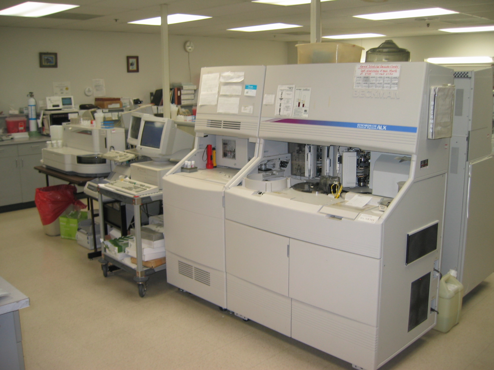
Medical Laboratory
A medical laboratory or clinical laboratory is a laboratory where tests are conducted out on clinical specimens to obtain information about the health of a patient to aid in diagnosis, treatment, and prevention of disease. Clinical Medical laboratories are an example of applied science, as opposed to research laboratory, research laboratories that focus on basic science, such as found in some academia, academic institutions. Medical laboratories vary in size and complexity and so offer a variety of testing services. More comprehensive services can be found in acute-care hospitals and medical centers, where 70% of clinical decisions are based on laboratory testing. Doctors offices and clinics, as well as skilled nursing and Nursing home, long-term care facilities, may have laboratories that provide more basic testing services. Commercial medical laboratories operate as independent businesses and provide testing that is otherwise not provided in other settings due to low test vol ... [...More Info...] [...Related Items...] OR: [Wikipedia] [Google] [Baidu] |


