|
Brasilitherium Riograndensis
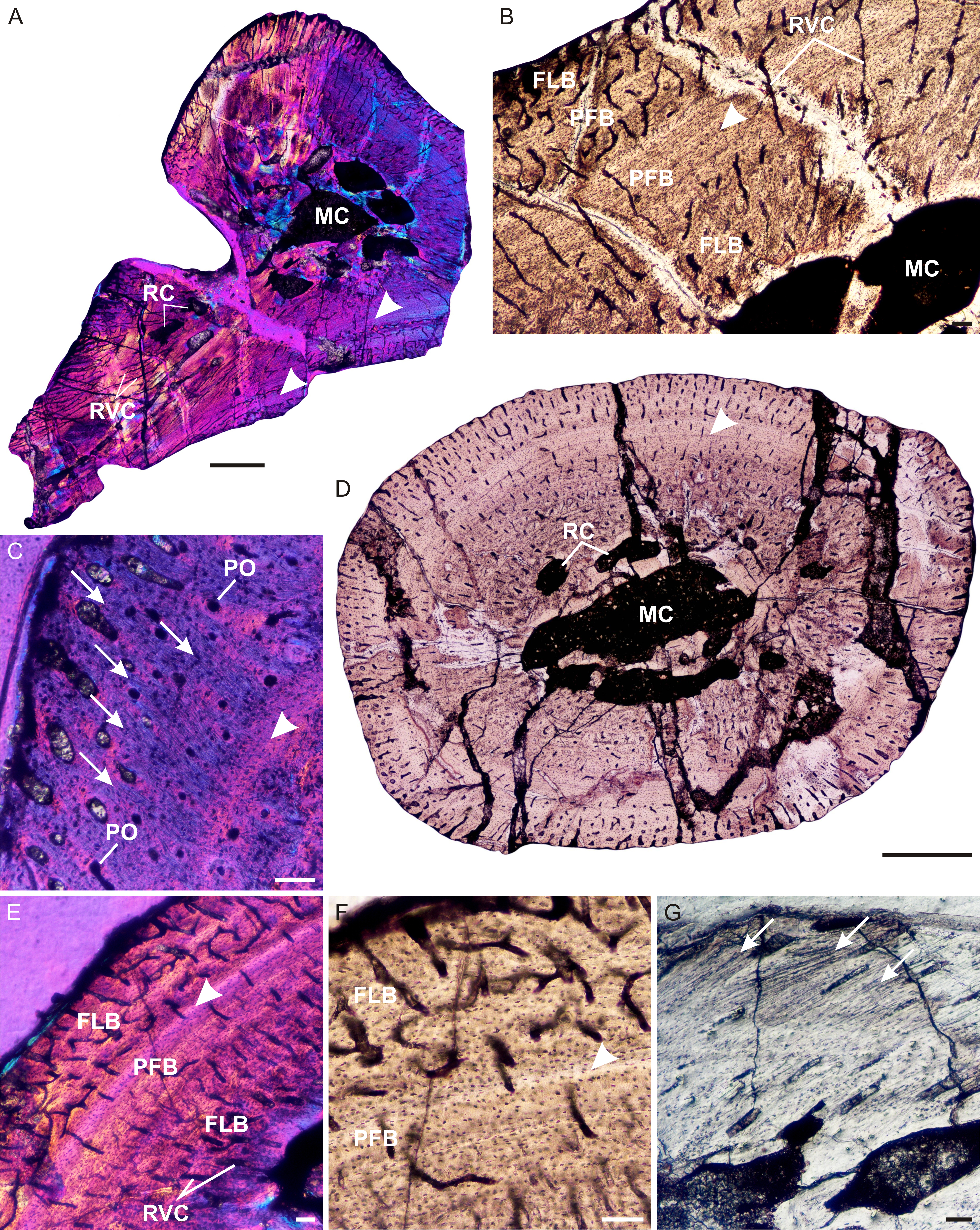
''Brasilodon'' ("tooth from Brazil") is an extinct genus of small, mammal-like cynodonts that lived in what is now Brazil during the Norian age of the Late Triassic epoch, about 225.42 million years ago. While no complete skeletons have been found, the length of ''Brasilodon'' has been estimated at around . Its dentition shows that it was most likely an insectivore. The genus is monotypic, containing only the species ''B. quadrangularis''. ''Brasilodon'' belongs to the family Brasilodontidae, whose members were some of the closest relatives of mammals, the only cynodonts alive today. Two other brasilodontid genera, ''Brasilitherium'' and ''Minicynodon'', are now considered to be junior synonyms of ''Brasilodon''. Discovery and naming The first three specimens referred to ''Brasilodon quadrangularis'' were found at the Linha São Luiz site, a quarry near the town of Faxinal do Soturno in the state of Rio Grande do Sul. The rocks where ''Brasilodon'' was found belong to t ... [...More Info...] [...Related Items...] OR: [Wikipedia] [Google] [Baidu] |
Late Triassic
The Late Triassic is the third and final epoch (geology), epoch of the Triassic geologic time scale, Period in the geologic time scale, spanning the time between annum, Ma and Ma (million years ago). It is preceded by the Middle Triassic Epoch and followed by the Early Jurassic Epoch. The corresponding series (stratigraphy), series of rock beds is known as the Upper Triassic. The Late Triassic is divided into the Carnian, Norian and Rhaetian Geologic time scale, Ages. Many of the first dinosaurs evolved during the Late Triassic, including ''Plateosaurus'', ''Coelophysis'', and ''Eoraptor''. The Triassic–Jurassic extinction event began during this epoch and is one of the five major mass extinction events of the Earth. Etymology The Triassic was named in 1834 by Friedrich August von Namoh, Friedrich von Alberti, after a succession of three distinct rock layers (Greek meaning 'triad') that are widespread in southern Germany: the lower Buntsandstein (colourful sandstone'')'', t ... [...More Info...] [...Related Items...] OR: [Wikipedia] [Google] [Baidu] |
Brasilitherium
''Brasilodon'' ("tooth from Brazil") is an extinct genus of small, mammal-like cynodonts that lived in what is now Brazil during the Norian age of the Late Triassic epoch, about 225.42 million years ago. While no complete skeletons have been found, the length of ''Brasilodon'' has been estimated at around . Its dentition shows that it was most likely an insectivore. The genus is monotypic, containing only the species ''B. quadrangularis''. ''Brasilodon'' belongs to the family Brasilodontidae, whose members were some of the closest relatives of mammals, the only cynodonts alive today. Two other brasilodontid genera, ''Brasilitherium'' and ''Minicynodon'', are now considered to be junior synonyms of ''Brasilodon''. Discovery and naming The first three specimens referred to ''Brasilodon quadrangularis'' were found at the Linha São Luiz site, a quarry near the town of Faxinal do Soturno in the state of Rio Grande do Sul. The rocks where ''Brasilodon'' was found belong to the ... [...More Info...] [...Related Items...] OR: [Wikipedia] [Google] [Baidu] |
Meckelian Groove
The Meckelian groove (or Meckel's groove, Meckelian fossa, or Meckelian foramen, or Meckelian canal) is an opening in the medial (inner) surface of the mandible (lower jaw) which exposes the Meckelian cartilage. Modern mammals (which includes placental
Placental mammals (infraclass Placentalia ) are one of the three extant subdivisions of the class Mammalia, the other two being Monotremata and Marsupialia. Placentalia contains the vast majority of extant mammals, which are partly distinguishe ... mammals) do not have a Meckeli ...
[...More Info...] [...Related Items...] OR: [Wikipedia] [Google] [Baidu] |
Ligament
A ligament is the fibrous connective tissue that connects bones to other bones. It is also known as ''articular ligament'', ''articular larua'', ''fibrous ligament'', or ''true ligament''. Other ligaments in the body include the: * Peritoneal ligament: a fold of peritoneum or other membranes. * Fetal remnant ligament: the remnants of a fetal tubular structure. * Periodontal ligament: a group of fibers that attach the cementum of teeth to the surrounding alveolar bone. Ligaments are similar to tendons and fasciae as they are all made of connective tissue. The differences among them are in the connections that they make: ligaments connect one bone to another bone, tendons connect muscle to bone, and fasciae connect muscles to other muscles. These are all found in the skeletal system of the human body. Ligaments cannot usually be regenerated naturally; however, there are periodontal ligament stem cells located near the periodontal ligament which are involved in the adult regener ... [...More Info...] [...Related Items...] OR: [Wikipedia] [Google] [Baidu] |
Mandibular Symphysis
In human anatomy, the facial skeleton of the skull the external surface of the mandible is marked in the median line by a faint ridge, indicating the mandibular symphysis (Latin: ''symphysis menti'') or line of junction where the two lateral halves of the mandible typically fuse at an early period of life (1-2 years). It is not a true symphysis as there is no cartilage between the two sides of the mandible. This ridge divides below and encloses a triangular eminence, the mental protuberance, the base of which is depressed in the center but raised on either side to form the mental tubercle. The lowest (most inferior) end of the mandibular symphysis — the point of the chin — is called the "menton". It serves as the origin for the geniohyoid and the genioglossus muscles. Other animals Solitary mammalian carnivores that rely on a powerful canine bite to subdue their prey have a strong mandibular symphysis, while pack hunters delivering shallow bites have a weaker one. When filter ... [...More Info...] [...Related Items...] OR: [Wikipedia] [Google] [Baidu] |
Dentary Bone
In anatomy, the mandible, lower jaw or jawbone is the largest, strongest and lowest bone in the human facial skeleton. It forms the lower jaw and holds the lower teeth in place. The mandible sits beneath the maxilla. It is the only movable bone of the skull (discounting the ossicles of the middle ear). It is connected to the temporal bones by the temporomandibular joints. The bone is formed in the fetus from a fusion of the left and right mandibular prominences, and the point where these sides join, the mandibular symphysis, is still visible as a faint ridge in the midline. Like other symphyses in the body, this is a midline articulation where the bones are joined by fibrocartilage, but this articulation fuses together in early childhood.Illustrated Anatomy of the Head and Neck, Fehrenbach and Herring, Elsevier, 2012, p. 59 The word "mandible" derives from the Latin word ''mandibula'', "jawbone" (literally "one used for chewing"), from '' mandere'' "to chew" and ''-bula'' ... [...More Info...] [...Related Items...] OR: [Wikipedia] [Google] [Baidu] |
Zygomatic Arch
In anatomy, the zygomatic arch, or cheek bone, is a part of the skull formed by the zygomatic process of the temporal bone (a bone extending forward from the side of the skull, over the opening of the ear) and the temporal process of the zygomatic bone (the side of the cheekbone), the two being united by an oblique suture (the zygomaticotemporal suture); the tendon of the temporal muscle passes medial to (i.e. through the middle of) the arch, to gain insertion into the coronoid process of the mandible (jawbone). The jugal point is the point at the anterior (towards face) end of the upper border of the zygomatic arch where the masseteric and maxillary edges meet at an angle, and where it meets the process of the zygomatic bone. The arch is typical of '' Synapsida'' (“fused arch”), a clade of amniotes that includes mammals and their extinct relatives, such as ''Moschops'' and '' Dimetrodon''. Structure The zygomatic process of the temporal arises by two roots: * an ''anter ... [...More Info...] [...Related Items...] OR: [Wikipedia] [Google] [Baidu] |
Eye Socket
In anatomy, the orbit is the cavity or socket of the skull in which the eye and its appendages are situated. "Orbit" can refer to the bony socket, or it can also be used to imply the contents. In the adult human, the volume of the orbit is , of which the eye occupies . The orbital contents comprise the eye, the orbital and retrobulbar fascia, extraocular muscles, cranial nerves II, III, IV, V, and VI, blood vessels, fat, the lacrimal gland with its sac and duct, the eyelids, medial and lateral palpebral ligaments, cheek ligaments, the suspensory ligament, septum, ciliary ganglion and short ciliary nerves. Structure The orbits are conical or four-sided pyramidal cavities, which open into the midline of the face and point back into the head. Each consists of a base, an apex and four walls."eye, human."Encyclopædia Britannica from Encyclopædia Britannica 2006 Ultimate Reference Suite DVD 2009 Openings There are two important foramina, or windows, two important fis ... [...More Info...] [...Related Items...] OR: [Wikipedia] [Google] [Baidu] |
Postorbital Bar
The postorbital bar (or postorbital bone) is a bony arched structure that connects the frontal bone of the skull to the zygomatic arch, which runs laterally around the eye socket. It is a trait that only occurs in mammalian taxa, such as most strepsirrhine primates and the hyrax, while haplorhine primates have evolved fully enclosed sockets. One theory for this evolutionary difference is the relative importance of vision to both orders. As haplorrhines (tarsiers and simians) tend to be diurnal, and rely heavily on visual input, many strepsirrhines are nocturnal and have a decreased reliance on visual input. Postorbital bars evolved several times independently during mammalian evolution and the evolutionary histories of several other clades. Some species, such as Tarsiers, have a postorbital septum. This septum can be considered as joined processes with a small articulation between the frontal bone, the zygomatic bone and the alisphenoid bone and is therefore different from the posto ... [...More Info...] [...Related Items...] OR: [Wikipedia] [Google] [Baidu] |
Prozostrodon
''Prozostrodon'' is an extinct genus of advanced cynodonts that was closely related to the ancestors of mammals. The remains were found in Brazil and are dated middle to late Triassic. It was originally described as a species of ''Thrinaxodon'' and was probably fairly similar to that genus in overall build. The holotype has a skull length of 6.7 cm, indicating the whole animal may have been the size of a cat, though there is some doubt as to whether the find represents an adult individual. The teeth were typical of advanced cynodonts, and the animal was probably a small carnivore hunting reptiles and other small prey. Later analysis indicated ''Prozostrodon'' was more closely related to the mammals than to the ''Thrinaxodon'' species, and it was given its own genus. Cladistic analysis indicates its closest relatives gave rise to the first mammaliaforms and therefore to the crown group mammals. The holotype of ''Prozostrodon'' was found in the Geopark of Paleorrota, Santa ... [...More Info...] [...Related Items...] OR: [Wikipedia] [Google] [Baidu] |
Postorbital Bone
The ''postorbital'' is one of the bones in vertebrate skulls which forms a portion of the dermal skull roof and, sometimes, a ring about the orbit. Generally, it is located behind the postfrontal and posteriorly to the orbital fenestra. In some vertebrates, the postorbital is fused with the postfrontal to create a postorbitofrontal. Birds have a separate postorbital as an embryo, but the bone fuses with the frontal Front may refer to: Arts, entertainment, and media Films * ''The Front'' (1943 film), a 1943 Soviet drama film * ''The Front'', 1976 film Music * The Front (band), an American rock band signed to Columbia Records and active in the 1980s and e ... before it hatches. References * Roemer, A. S. 1956. ''Osteology of the Reptiles''. University of Chicago Press. 772 pp. Skull {{Vertebrate anatomy-stub ... [...More Info...] [...Related Items...] OR: [Wikipedia] [Google] [Baidu] |
Prefrontal Bone
The prefrontal bone is a bone separating the lacrimal and frontal bones in many tetrapod skulls. It first evolved in the sarcopterygian clade Rhipidistia, which includes lungfish and the Tetrapodomorpha. The prefrontal is found in most modern and extinct lungfish, amphibians and reptiles. The prefrontal is lost in early mammaliaforms and so is not present in modern mammals either. In dinosaurs The prefrontal bone is a very small bone near the top of the skull, which is lost in many groups of coelurosaurian theropod dinosaurs and is completely absent in their modern descendants, the birds. Conversely, a well developed prefrontal is considered to be a primitive feature in dinosaurs. The prefrontal makes contact with several other bones in the skull. The anterior part of the bone articulates with the nasal bone and the lacrimal bone. The posterior part of the bone articulates with the frontal bone and more rarely the palpebral bone The palpebral bone is a small dermal bone found ... [...More Info...] [...Related Items...] OR: [Wikipedia] [Google] [Baidu] |


