|
Beam's Eye View
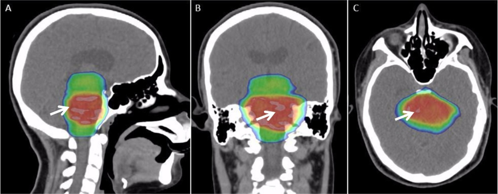
Beam's eye view (abbreviated BEV) is an imaging technique used in radiation therapy for quality assurance and planning of external beam radiotherapy (EBRT). These are primarily used to ensure that the relative orientation of the patient and the treatment machine are correct. The BEV image will typically include the images of the patient's anatomy and the beam modifiers, such as jaws or multi-leaf collimators (MLCs). Generation of beam's eye views * Physical construction: ** BEVs can be generated by exposing a high energy film (similar to photographic film) or an electronic portal imaging device (EPID) with the treatment beam itself after it passes through the patient and any beam modifiers (such as blocks). Although this type of image is an excellent indication of the basic quality of the treatment plan, the quality of film images can be poor. ** A BEV can be created using a radiation therapy simulator that mimics the treatment geometry (couch angle, gantry angle, etc.) using an X-r ... [...More Info...] [...Related Items...] OR: [Wikipedia] [Google] [Baidu] |
Radiation Therapy
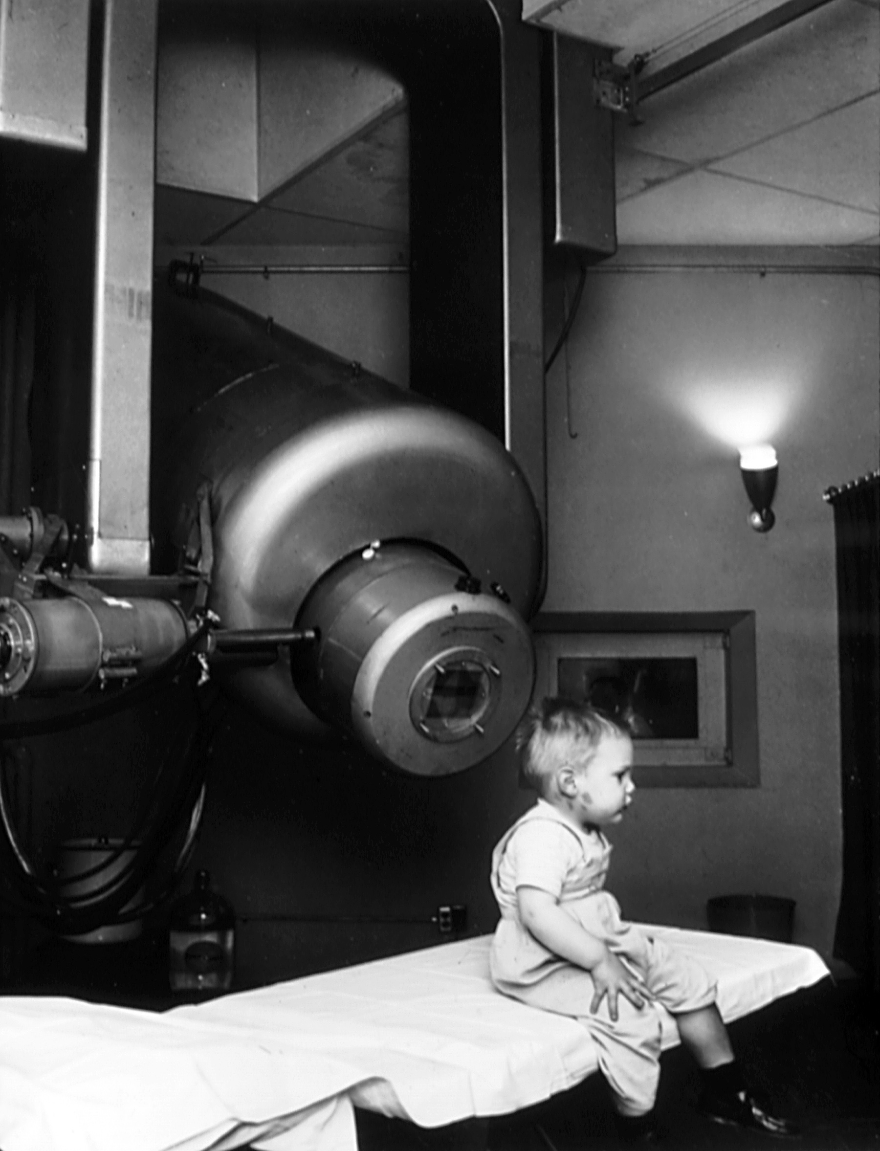
Radiation therapy or radiotherapy (RT, RTx, or XRT) is a therapy, treatment using ionizing radiation, generally provided as part of treatment of cancer, cancer therapy to either kill or control the growth of malignancy, malignant cell (biology), cells. It is normally delivered by a linear particle accelerator. Radiation therapy may be cure, curative in a number of types of cancer if they are localized to one area of the body, and have not metastasis, spread to other parts. It may also be used as part of adjuvant therapy, to prevent tumor recurrence after surgery to remove a primary malignant tumor (for example, early stages of breast cancer). Radiation therapy is synergistic with chemotherapy, and has been used before, during, and after chemotherapy in susceptible cancers. The subspecialty of oncology concerned with radiotherapy is called radiation oncology. A physician who practices in this subspecialty is a radiation oncologist. Radiation therapy is commonly applied to the canc ... [...More Info...] [...Related Items...] OR: [Wikipedia] [Google] [Baidu] |
External Beam Radiotherapy
External beam radiation therapy (EBRT) is a form of radiotherapy that utilizes a high-energy collimated beam of ionizing radiation, from a source outside the body, to target and kill cancer cells. The radiotherapy beam is composed of particles, which are focussed in a particular direction of travel using collimators. Each radiotherapy beam consists of one type of particle intended for use in treatment, though most beams contain some contamination by other particle types. Radiotherapy beams are classified by the particle they are intended to deliver, such as photons (as x-rays or gamma rays), electrons, and heavy ions; x-rays and electron beams are by far the most widely used sources for external beam radiotherapy. Orthovoltage ("superficial") X-rays are used for treating skin cancer and superficial structures. Megavoltage X-rays are used to treat deep-seated tumors (e.g. bladder, bowel, prostate, lung, or brain), whereas megavoltage electron beams are typically used to tre ... [...More Info...] [...Related Items...] OR: [Wikipedia] [Google] [Baidu] |
Photographic Film
Photographic film is a strip or sheet of transparent film base coated on one side with a gelatin photographic emulsion, emulsion containing microscopically small light-sensitive silver halide crystals. The sizes and other characteristics of the crystals determine the sensitivity, contrast, and image resolution, resolution of the film. Film is typically segmented in ''frames'', that give rise to separate photographs. The emulsion will gradually darken if left exposed to light, but the process is too slow and incomplete to be of any practical use. Instead, a very short exposure (photography), exposure to the image formed by a camera lens is used to produce only a very slight chemical change, proportional to the amount of light absorbed by each crystal. This creates an invisible latent image in the emulsion, which can be chemically photographic processing, developed into a visible photograph. In addition to visible light, all films are sensitive to ultraviolet light, X-rays, gamma ... [...More Info...] [...Related Items...] OR: [Wikipedia] [Google] [Baidu] |
X-ray
An X-ray (also known in many languages as Röntgen radiation) is a form of high-energy electromagnetic radiation with a wavelength shorter than those of ultraviolet rays and longer than those of gamma rays. Roughly, X-rays have a wavelength ranging from 10 Nanometre, nanometers to 10 Picometre, picometers, corresponding to frequency, frequencies in the range of 30 Hertz, petahertz to 30 Hertz, exahertz ( to ) and photon energies in the range of 100 electronvolt, eV to 100 keV, respectively. X-rays were discovered in 1895 in science, 1895 by the German scientist Wilhelm Röntgen, Wilhelm Conrad Röntgen, who named it ''X-radiation'' to signify an unknown type of radiation.Novelline, Robert (1997). ''Squire's Fundamentals of Radiology''. Harvard University Press. 5th edition. . X-rays can penetrate many solid substances such as construction materials and living tissue, so X-ray radiography is widely used in medical diagnostics (e.g., checking for Bo ... [...More Info...] [...Related Items...] OR: [Wikipedia] [Google] [Baidu] |
Digitally Reconstructed Radiograph
A computed tomography scan (CT scan), formerly called computed axial tomography scan (CAT scan), is a medical imaging technique used to obtain detailed internal images of the body. The personnel that perform CT scans are called radiographers or radiology technologists. CT scanners use a rotating X-ray tube and a row of detectors placed in a gantry to measure X-ray attenuations by different tissues inside the body. The multiple X-ray measurements taken from different angles are then processed on a computer using tomographic reconstruction algorithms to produce tomographic (cross-sectional) images (virtual "slices") of a body. CT scans can be used in patients with metallic implants or pacemakers, for whom magnetic resonance imaging (MRI) is contraindicated. Since its development in the 1970s, CT scanning has proven to be a versatile imaging technique. While CT is most prominently used in medical diagnosis, it can also be used to form images of non-living objects. The 1979 Nobel ... [...More Info...] [...Related Items...] OR: [Wikipedia] [Google] [Baidu] |
Radiobiology
Radiobiology (also known as radiation biology, and uncommonly as actinobiology) is a field of clinical and basic medical sciences that involves the study of the effects of radiation on living tissue (including ionizing radiation, ionizing and non-ionizing radiation), in particular health effects of radiation. Ionizing radiation is generally harmful and potentially lethal to living things but can have health benefits in radiation therapy for the treatment of cancer and thyrotoxicosis. Its most common impact is the radiation-induced cancer, induction of cancer with a Incubation period, latent period of years or decades after exposure. High doses can cause visually dramatic radiation burns, and/or rapid fatality through acute radiation syndrome. Controlled doses are used for medical imaging and radiotherapy. Health effects In general, ionizing radiation is harmful and potentially lethal to living beings but can have health benefits in radiation therapy for the treatment of cancer an ... [...More Info...] [...Related Items...] OR: [Wikipedia] [Google] [Baidu] |


