|
Basal Plate (neural Tube)
In the developing nervous system, the basal plate is the region of the neural tube ventral to the sulcus limitans. It extends from the rostral mesencephalon to the end of the spinal cord and contains primarily motor neurons, whereas neurons found in the alar plate are primarily associated with sensory functions. The cell types of the basal plate include lower motor neurons and four types of interneuron. Initially, the left and right sides of the basal plate are continuous, but during neurulation they become separated by the floor plate, and this process is directed by the notochord. Differentiation of neurons in the basal plate is under the influence of the protein Sonic hedgehog released by ventralizing structures, such as the notochord and floor plate. Gray642.png, The basal plate (basal lamina) is separated from the alar plate (alar lamina) by the sulcus limitans The sulcus limitans is found in the fourth ventricle of the brain. It separates the cranial nerve motor nuc ... [...More Info...] [...Related Items...] OR: [Wikipedia] [Google] [Baidu] |
Nervous System
In biology, the nervous system is the highly complex part of an animal that coordinates its actions and sensory information by transmitting signals to and from different parts of its body. The nervous system detects environmental changes that impact the body, then works in tandem with the endocrine system to respond to such events. Nervous tissue first arose in wormlike organisms about 550 to 600 million years ago. In vertebrates it consists of two main parts, the central nervous system (CNS) and the peripheral nervous system (PNS). The CNS consists of the brain and spinal cord. The PNS consists mainly of nerves, which are enclosed bundles of the long fibers or axons, that connect the CNS to every other part of the body. Nerves that transmit signals from the brain are called motor nerves or '' efferent'' nerves, while those nerves that transmit information from the body to the CNS are called sensory nerves or '' afferent''. Spinal nerves are mixed nerves that serve both fu ... [...More Info...] [...Related Items...] OR: [Wikipedia] [Google] [Baidu] |
Neuron
A neuron, neurone, or nerve cell is an electrically excitable cell that communicates with other cells via specialized connections called synapses. The neuron is the main component of nervous tissue in all animals except sponges and placozoa. Non-animals like plants and fungi do not have nerve cells. Neurons are typically classified into three types based on their function. Sensory neurons respond to stimuli such as touch, sound, or light that affect the cells of the sensory organs, and they send signals to the spinal cord or brain. Motor neurons receive signals from the brain and spinal cord to control everything from muscle contractions to glandular output. Interneurons connect neurons to other neurons within the same region of the brain or spinal cord. When multiple neurons are connected together, they form what is called a neural circuit. A typical neuron consists of a cell body (soma), dendrites, and a single axon. The soma is a compact structure, and the axon and dend ... [...More Info...] [...Related Items...] OR: [Wikipedia] [Google] [Baidu] |
Sonic Hedgehog
Sonic hedgehog protein (SHH) is encoded for by the ''SHH'' gene. The protein is named after the character ''Sonic the Hedgehog''. This signaling molecule is key in regulating embryonic morphogenesis in all animals. SHH controls organogenesis and the organization of the central nervous system, limbs, digits and many other parts of the body. Sonic hedgehog is a morphogen that patterns the developing embryo using a concentration gradient characterized by the French flag model. This model has a non-uniform distribution of SHH molecules which governs different cell fates according to concentration. Mutations in this gene can cause holoprosencephaly, a failure of splitting in the cerebral hemispheres, as demonstrated in an experiment using SHH knock-out mice in which the forebrain midline failed to develop and instead only a single fused telencephalic vesicle resulted. Sonic hedgehog still plays a role in differentiation, proliferation, and maintenance of adult tissues. Abnormal activ ... [...More Info...] [...Related Items...] OR: [Wikipedia] [Google] [Baidu] |
Notochord
In anatomy, the notochord is a flexible rod which is similar in structure to the stiffer cartilage. If a species has a notochord at any stage of its life cycle (along with 4 other features), it is, by definition, a chordate. The notochord consists of inner, vacuolated cells covered by fibrous and elastic sheaths, lies along the anteroposterior axis (''front to back''), is usually closer to the dorsal than the ventral surface of the embryo, and is composed of cells derived from the mesoderm. The most commonly cited functions of the notochord are: as a midline tissue that provides directional signals to surrounding tissue during development, as a skeletal (structural) element, and as a vertebral precursor. In lancelets the notochord persists throughout life as the main structural support of the body. In tunicates the notochord is present only in the larval stage, being completely absent in the adult animal. In these invertebrate chordates, the notochord is not vacuolated. In all ... [...More Info...] [...Related Items...] OR: [Wikipedia] [Google] [Baidu] |
Floor Plate
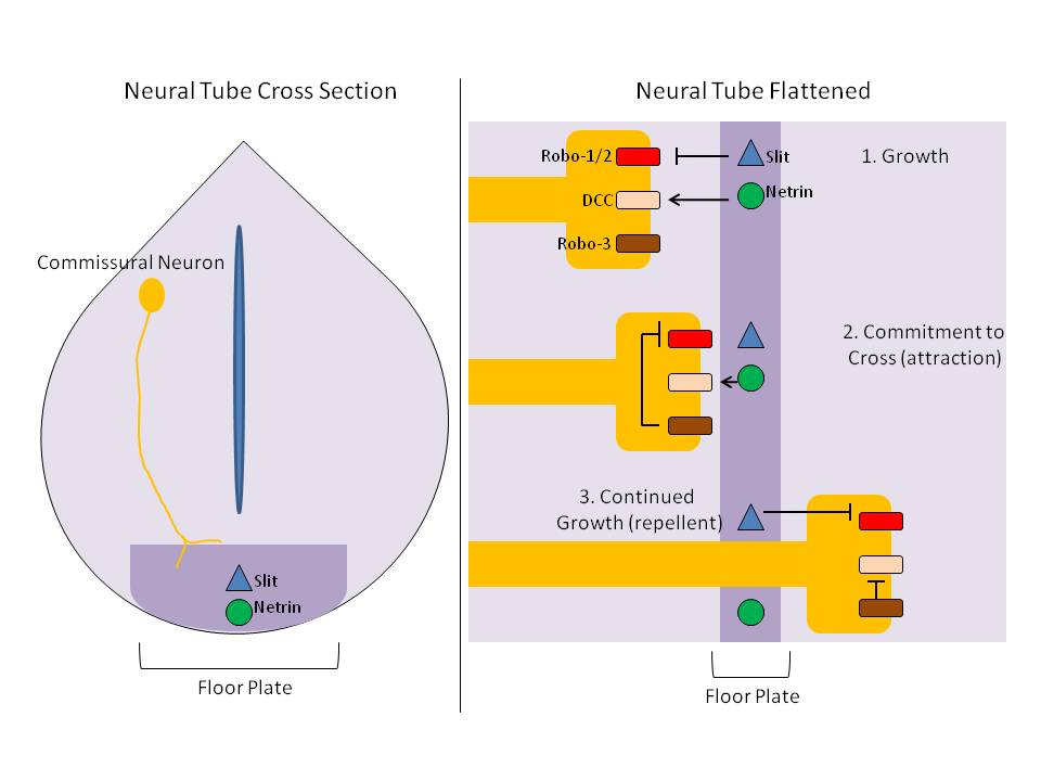
The floor plate is a structure integral to the developing nervous system of vertebrate organisms. Located on the ventral midline of the embryonic neural tube, the floor plate is a specialized glial structure that spans the anteroposterior axis from the midbrain to the tail regions. It has been shown that the floor plate is conserved among vertebrates, such as zebrafish and mice, with homologous structures in invertebrates such as the fruit fly ''Drosophila'' and the nematode ''C. elegans''. Functionally, the structure serves as an organizer to ventralize tissues in the embryo as well as to guide neuronal positioning and differentiation along the dorsoventral axis of the neural tube. Induction Induction of the floor plate during embryogenesis of vertebrate embryos has been studied extensively in chick and zebrafish and occurs as a result of a complex signaling network among tissues, the details of which have yet to be fully refined. Currently there are several competing lines o ... [...More Info...] [...Related Items...] OR: [Wikipedia] [Google] [Baidu] |
Neurulation
Neurulation refers to the folding process in vertebrate embryos, which includes the transformation of the neural plate into the neural tube. The embryo at this stage is termed the neurula. The process begins when the notochord induces the formation of the central nervous system (CNS) by signaling the ectoderm germ layer above it to form the thick and flat neural plate. The neural plate folds in upon itself to form the neural tube, which will later differentiate into the spinal cord and the brain, eventually forming the central nervous system. Computer simulations found that cell wedging and differential proliferation are sufficient for mammalian neurulation. Different portions of the neural tube form by two different processes, called primary and secondary neurulation, in different species. * In primary neurulation, the neural plate creases inward until the edges come in contact and fuse. * In secondary neurulation, the tube forms by hollowing out of the interior of a solid precur ... [...More Info...] [...Related Items...] OR: [Wikipedia] [Google] [Baidu] |
Lower Motor Neuron
Lower motor neurons (LMNs) are motor neurons located in either the anterior grey column, anterior nerve roots (spinal lower motor neurons) or the cranial nerve nuclei of the brainstem and cranial nerves with motor function (cranial nerve lower motor neurons). Many voluntary movements rely on spinal lower motor neurons, which innervate skeletal muscle fibers and act as a link between upper motor neurons and muscles. Cranial nerve lower motor neurons also control some voluntary movements of the eyes, face and tongue, and contribute to chewing, swallowing and vocalization. Damage to the lower motor neurons can lead to flaccid paralysis, absent deep tendon reflexes and muscle atrophy. Classification Lower motor neurons are classified based on the type of muscle fiber they innervate: * Alpha motor neurons (α-MNs) innervate extrafusal muscle fibers, the most numerous type of muscle fiber and the one involved in muscle contraction. * Beta motor neurons (β-MNs) innervate intrafusal fi ... [...More Info...] [...Related Items...] OR: [Wikipedia] [Google] [Baidu] |
Alar Plate
Daminozide—also known as aminozide, Alar, Kylar, SADH, B-995, B-nine, and DMASA,—is a plant growth regulator, a chemical sprayed on fruit to regulate growth, make harvest easier, and keep apples from falling off the trees before they ripen so they are red and firm for storage. It was produced in the U.S. by the Uniroyal Chemical Company, Inc, (now integrated into the Chemtura Corporation), which registered daminozide for use on fruits intended for human consumption in 1963. In addition to apples and ornamental plants, they also registered it for use on cherries, peaches, pears, Concord grapes, tomato transplants, and peanut vines. Alar was first approved for use in the U.S. in 1963. It was primarily used on apples until 1989, when the manufacturer voluntarily withdrew it after the U.S. Environmental Protection Agency proposed banning it based on concerns about cancer risks to consumers. On fruit trees, daminozide affects flow-bud initiation, fruit-set maturity, fruit firmness ... [...More Info...] [...Related Items...] OR: [Wikipedia] [Google] [Baidu] |
Spinal Cord
The spinal cord is a long, thin, tubular structure made up of nervous tissue, which extends from the medulla oblongata in the brainstem to the lumbar region of the vertebral column (backbone). The backbone encloses the central canal of the spinal cord, which contains cerebrospinal fluid. The brain and spinal cord together make up the central nervous system (CNS). In humans, the spinal cord begins at the occipital bone, passing through the foramen magnum and then enters the spinal canal at the beginning of the cervical vertebrae. The spinal cord extends down to between the first and second lumbar vertebrae, where it ends. The enclosing bony vertebral column protects the relatively shorter spinal cord. It is around long in adult men and around long in adult women. The diameter of the spinal cord ranges from in the cervical and lumbar regions to in the thoracic area. The spinal cord functions primarily in the transmission of nerve signals from the motor cortex to the body, ... [...More Info...] [...Related Items...] OR: [Wikipedia] [Google] [Baidu] |
Nervous System
In biology, the nervous system is the highly complex part of an animal that coordinates its actions and sensory information by transmitting signals to and from different parts of its body. The nervous system detects environmental changes that impact the body, then works in tandem with the endocrine system to respond to such events. Nervous tissue first arose in wormlike organisms about 550 to 600 million years ago. In vertebrates it consists of two main parts, the central nervous system (CNS) and the peripheral nervous system (PNS). The CNS consists of the brain and spinal cord. The PNS consists mainly of nerves, which are enclosed bundles of the long fibers or axons, that connect the CNS to every other part of the body. Nerves that transmit signals from the brain are called motor nerves or '' efferent'' nerves, while those nerves that transmit information from the body to the CNS are called sensory nerves or '' afferent''. Spinal nerves are mixed nerves that serve both fu ... [...More Info...] [...Related Items...] OR: [Wikipedia] [Google] [Baidu] |
Mesencephalon
The midbrain or mesencephalon is the forward-most portion of the brainstem and is associated with vision, hearing, motor control, sleep and wakefulness, arousal (alertness), and temperature regulation. The name comes from the Greek ''mesos'', "middle", and ''enkephalos'', "brain". Structure The principal regions of the midbrain are the tectum, the cerebral aqueduct, tegmentum, and the cerebral peduncles. Rostrally the midbrain adjoins the diencephalon (thalamus, hypothalamus, etc.), while caudally it adjoins the hindbrain (pons, medulla and cerebellum). In the rostral direction, the midbrain noticeably splays laterally. Sectioning of the midbrain is usually performed axially, at one of two levels – that of the superior colliculi, or that of the inferior colliculi. One common technique for remembering the structures of the midbrain involves visualizing these cross-sections (especially at the level of the superior colliculi) as the upside-down face of a bea ... [...More Info...] [...Related Items...] OR: [Wikipedia] [Google] [Baidu] |
Sulcus Limitans (neural Tube)
A shallow, longitudinal groove separating the developing gray matter into a basal and alar plates along the length of the neural tube. The sulcus limitans extends the length of the spinal cord and through the mesencephalon The midbrain or mesencephalon is the forward-most portion of the brainstem and is associated with vision, hearing, motor control, sleep and wakefulness, arousal (alertness), and temperature regulation. The name comes from the Greek ''mesos'', "m .... References Embryology of nervous system {{Developmental-biology-stub ... [...More Info...] [...Related Items...] OR: [Wikipedia] [Google] [Baidu] |


