|
Babinski Sign
The plantar reflex is a reflex elicited when the sole of the foot is stimulated with a blunt instrument. The reflex can take one of two forms. In healthy adults, the plantar reflex causes a downward response of the hallux (flexion). An upward response (extension) of the hallux is known as the Babinski response or Babinski sign, named after the neurologist Joseph Babinski. The presence of the Babinski sign can identify disease of the spinal cord and brain in adults, and also exists as a primitive reflex in infants. While first described in the medical literature by Babinski in 1896, the reflex has been identified in art at least as early as Botticelli's '' Virgin and Child with an Angel'', painted in the mid-15th century. Methods The lateral side of the sole of the foot is rubbed with a blunt instrument or device so as not to cause pain, discomfort, or injury to the skin; the instrument is run from the heel along a curve to the toes (metatarsal pads). Many reflex hammers tap ... [...More Info...] [...Related Items...] OR: [Wikipedia] [Google] [Baidu] |
Reflex
In biology, a reflex, or reflex action, is an involuntary, unplanned sequence or action and nearly instantaneous response to a stimulus. Reflexes are found with varying levels of complexity in organisms with a nervous system. A reflex occurs via neural pathways in the nervous system called reflex arcs. A stimulus initiates a neural signal, which is carried to a synapse. The signal is then transferred across the synapse to a motor neuron which evokes a target response. These neural signals do not always travel to the brain, so many reflexes are an automatic response to a stimulus that does not receive or need conscious thought. Many reflexes are fine-tuned to increase organism survival and self-defense. This is observed in reflexes such as the startle reflex, which provides an automatic response to an unexpected stimuli, and the feline righting reflex, which reorients a cat's body when falling to ensure safe landing. The simplest type of reflex, a short-latency reflex, has a ... [...More Info...] [...Related Items...] OR: [Wikipedia] [Google] [Baidu] |
Reflex Hammer
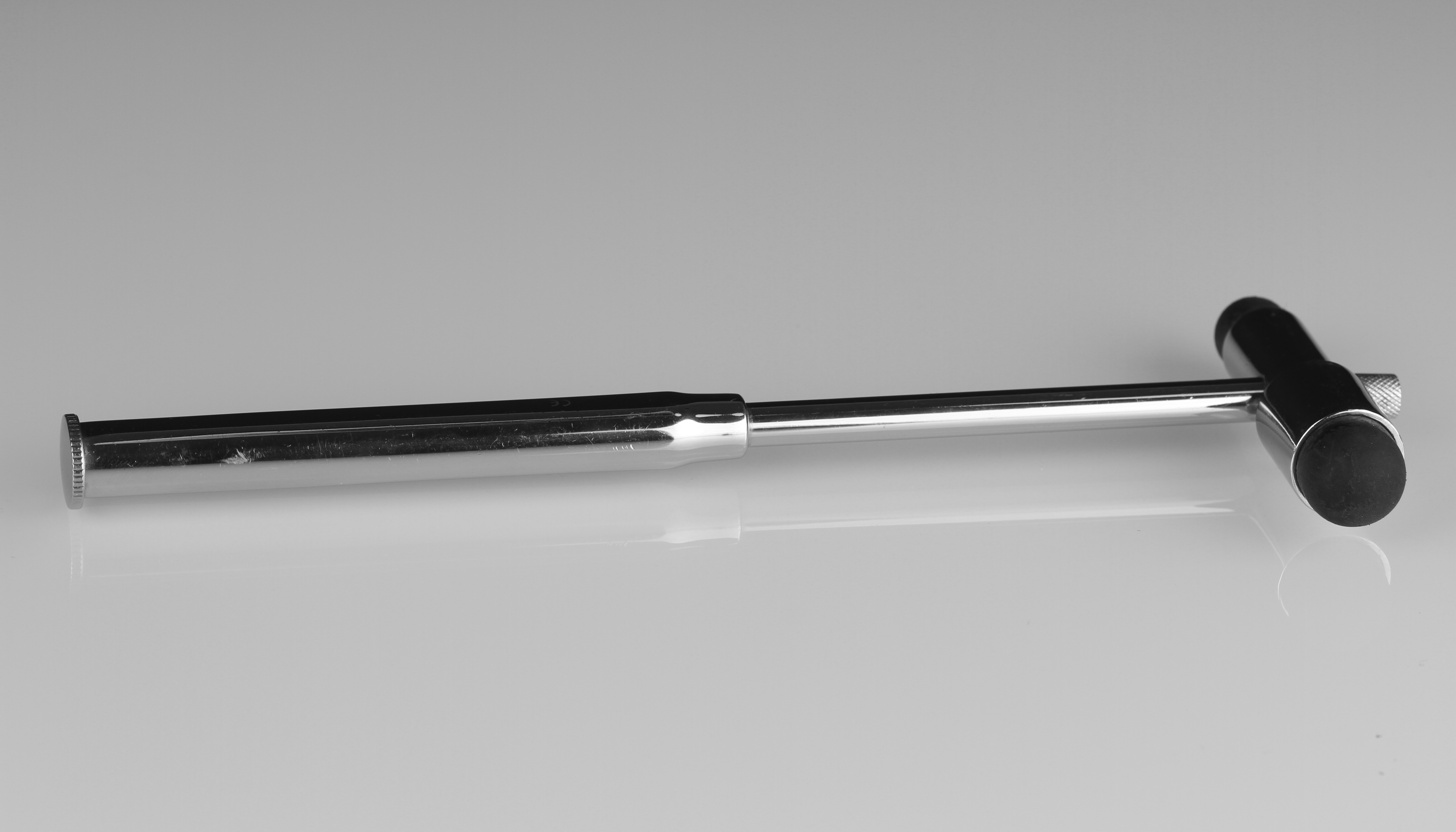
A reflex hammer is a medical instrument used by practitioners to test deep tendon reflexes. Testing for reflexes is an important part of the neurological physical examination in order to detect abnormalities in the central or peripheral nervous system. Reflex hammers can also be used for chest percussion.Swartz MH. ''Textbook of Physical Diagnosis: History and Examination.'' Third edition. Philadelphia: WB Saunders; 1998 Models of reflex hammer Prior to the development of specialized reflex hammers, hammers specific for percussion of the chest were used to elicit reflexes.Lanska DJ. The history of reflex hammers. ''Neurology''. 1989 Nov;39(11):1542-9. However, this proved to be cumbersome, as the weight of the chest percussion hammer was insufficient to generate an adequate stimulus for a reflex. Starting in the late 19th century, several models of specific reflex hammers were created: *The Taylor or tomahawk reflex hammer was designed by John Madison Taylor in 1888 and ... [...More Info...] [...Related Items...] OR: [Wikipedia] [Google] [Baidu] |
Lumbar Puncture
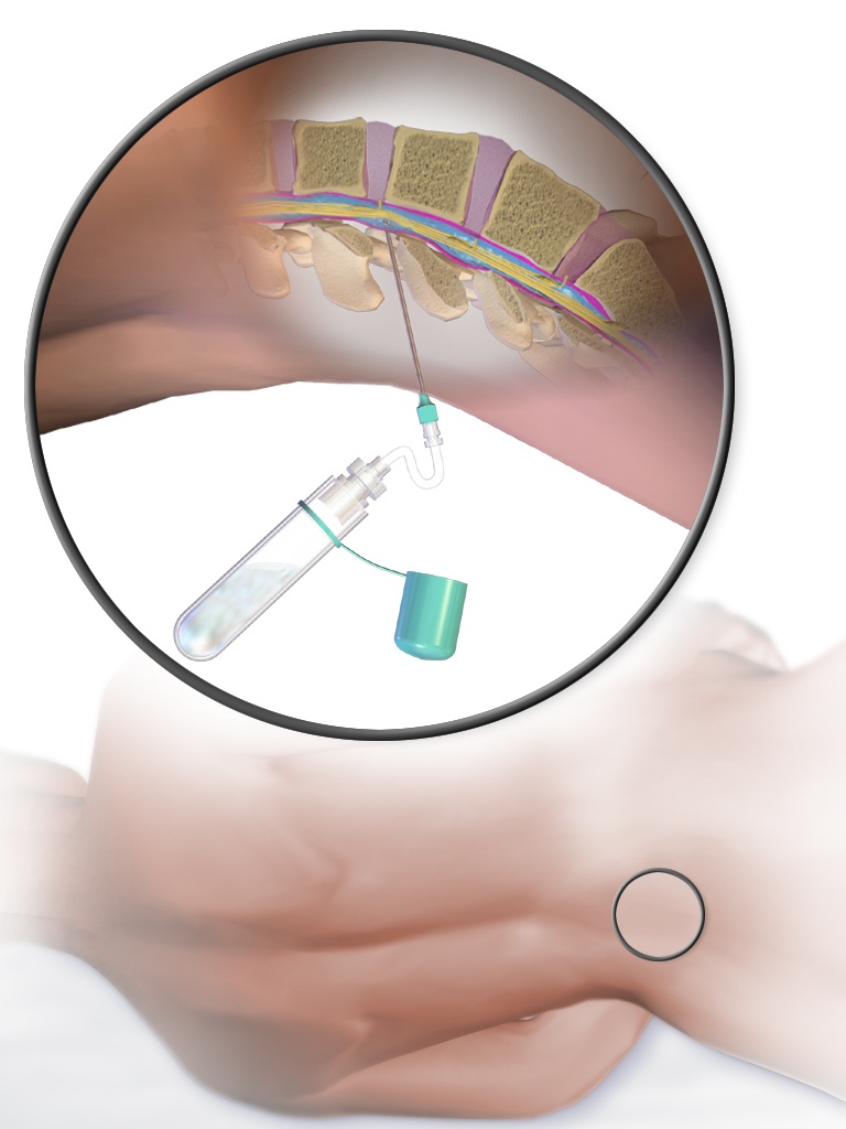
Lumbar puncture (LP), also known as a spinal tap, is a medical procedure in which a needle is inserted into the spinal canal, most commonly to collect cerebrospinal fluid (CSF) for diagnostic testing. The main reason for a lumbar puncture is to help diagnose diseases of the central nervous system, including the brain and spine. Examples of these conditions include meningitis and subarachnoid hemorrhage. It may also be used therapeutically in some conditions. Increased intracranial pressure (pressure in the skull) is a contraindication, due to risk of brain matter being compressed and pushed toward the spine. Sometimes, lumbar puncture cannot be performed safely (for example due to a severe bleeding tendency). It is regarded as a safe procedure, but post-dural-puncture headache is a common side effect if a small atraumatic needle is not used. The procedure is typically performed under local anesthesia using a sterile technique. A hypodermic needle is used to access the suba ... [...More Info...] [...Related Items...] OR: [Wikipedia] [Google] [Baidu] |
Magnetic Resonance Imaging
Magnetic resonance imaging (MRI) is a medical imaging technique used in radiology to form pictures of the anatomy and the physiological processes of the body. MRI scanners use strong magnetic fields, magnetic field gradients, and radio waves to generate images of the organs in the body. MRI does not involve X-rays or the use of ionizing radiation, which distinguishes it from CT and PET scans. MRI is a medical application of nuclear magnetic resonance (NMR) which can also be used for imaging in other NMR applications, such as NMR spectroscopy. MRI is widely used in hospitals and clinics for medical diagnosis, staging and follow-up of disease. Compared to CT, MRI provides better contrast in images of soft-tissues, e.g. in the brain or abdomen. However, it may be perceived as less comfortable by patients, due to the usually longer and louder measurements with the subject in a long, confining tube, though "Open" MRI designs mostly relieve this. Additionally, implants and ... [...More Info...] [...Related Items...] OR: [Wikipedia] [Google] [Baidu] |
Neurology
Neurology (from el, νεῦρον (neûron), "string, nerve" and the suffix -logia, "study of") is the branch of medicine dealing with the diagnosis and treatment of all categories of conditions and disease involving the brain, the spinal cord and the peripheral nerves. Neurological practice relies heavily on the field of neuroscience, the scientific study of the nervous system. A neurologist is a physician specializing in neurology and trained to investigate, diagnose and treat neurological disorders. Neurologists treat a myriad of neurologic conditions, including stroke, seizures, movement disorders such as Parkinson's disease, autoimmune neurologic disorders such as multiple sclerosis, headache disorders like migraine and dementias such as Alzheimer's disease. Neurologists may also be involved in clinical research, clinical trials, and basic or translational research. While neurology is a nonsurgical specialty, its corresponding surgical specialty is neurosurger ... [...More Info...] [...Related Items...] OR: [Wikipedia] [Google] [Baidu] |
Corticospinal Tract
The corticospinal tract is a white matter motor pathway starting at the cerebral cortex that terminates on lower motor neurons and interneurons in the spinal cord, controlling movements of the limbs and trunk. There are more than one million neurons in the corticospinal tract, and they become myelinated usually in the first two years of life. The corticospinal tract is one of the pyramidal tracts, the other being the corticobulbar tract. Anatomy The corticospinal tract originates in several parts of the brain, including not just the motor areas, but also the primary somatosensory cortex and premotor areas. Most of the neurons originate in the primary motor cortex (precentral gyrus, Brodmann area 4) or the premotor frontal areas.Purves, D. et al. (2012). Neuroscience: Fifth edition. Sunderland, MA: Sinauer Associates, Inc.Kolb, B. & Whishaw, I. Q. (2014). An introduction to brain and behavior: Fourth edition. New York, NY: Worth Publishers. About 30% of corticospinal neurons origi ... [...More Info...] [...Related Items...] OR: [Wikipedia] [Google] [Baidu] |
Upper Motor Neuron
Upper motor neurons (UMNs) is a term introduced by William Gowers in 1886. They are found in the cerebral cortex and brainstem and carry information down to activate interneurons and lower motor neurons, which in turn directly signal muscles to contract or relax. UMNs in the cerebral cortex are the main source of voluntary movement. They are the larger pyramidal cells in the cerebral cortex. There is a type of giant pyramidal cell called '' Betz cells'' and are found just below the surface of the cerebral cortex within layer V of the primary motor cortex. The cell bodies of Betz cell neurons are the largest in the brain, approaching nearly 0.1 mm in diameter. The primary motor cortex, or precentral gyrus, is the most posterior gyrus of the frontal lobe and lies anterior to the central sulcus. The pyramidal cells of the precentral gyrus are also called upper motor neurons. The fibers of the upper motor neurons project out of the precentral gyrus ending in the brainstem, ... [...More Info...] [...Related Items...] OR: [Wikipedia] [Google] [Baidu] |
Afferent Nerve
A sensory nerve, or afferent nerve, is a general anatomic term for a nerve which contains predominantly somatic afferent nerve fibers. Afferent nerve fibers in a sensory nerve carry sensory information toward the central nervous system (CNS) from different sensory receptors of sensory neurons in the peripheral nervous system. A motor nerve carries information from the CNS to the PNS, and both types of nerve are called peripheral nerves. Afferent nerve fibers link the sensory neurons throughout the body, in pathways to the relevant processing circuits in the central nervous system. Afferent nerve fibers are often paired with efferent nerve fibers from the motor neurons (that travel from the CNS to the PNS), in mixed nerves. Stimuli cause nerve impulses in the receptors and alter the potentials, which is known as sensory transduction. Spinal cord entry Afferent nerve fibers leave the sensory neuron from the dorsal root ganglia of the spinal cord, and motor commands carried ... [...More Info...] [...Related Items...] OR: [Wikipedia] [Google] [Baidu] |
Lesion
A lesion is any damage or abnormal change in the tissue of an organism, usually caused by disease or trauma. ''Lesion'' is derived from the Latin "injury". Lesions may occur in plants as well as animals. Types There is no designated classification or naming convention for lesions. Since lesions can occur anywhere in the body and the definition of a lesion is so broad, the varieties of lesions are virtually endless. Generally, lesions may be classified by their patterns, their sizes, their locations, or their causes. They can also be named after the person who discovered them. For example, Ghon lesions, which are found in the lungs of those with tuberculosis, are named after the lesion's discoverer, Anton Ghon. The characteristic skin lesions of a varicella zoster virus infection are called '' chickenpox''. Lesions of the teeth are usually called dental caries. Location Lesions are often classified by their tissue types or locations. For example, a "skin lesion" or a " b ... [...More Info...] [...Related Items...] OR: [Wikipedia] [Google] [Baidu] |
Central Nervous System
The central nervous system (CNS) is the part of the nervous system consisting primarily of the brain and spinal cord. The CNS is so named because the brain integrates the received information and coordinates and influences the activity of all parts of the bodies of bilaterally symmetric and triploblastic animals—that is, all multicellular animals except sponges and diploblasts. It is a structure composed of nervous tissue positioned along the rostral (nose end) to caudal (tail end) axis of the body and may have an enlarged section at the rostral end which is a brain. Only arthropods, cephalopods and vertebrates have a true brain (precursor structures exist in onychophorans, gastropods and lancelets). The rest of this article exclusively discusses the vertebrate central nervous system, which is radically distinct from all other animals. Overview In vertebrates, the brain and spinal cord are both enclosed in the meninges. The meninges provide a barrier to chemicals ... [...More Info...] [...Related Items...] OR: [Wikipedia] [Google] [Baidu] |


