|
Axo-axonic Synapse
An axo-axonic synapse is a type of synapse, formed by one neuron projecting its axon terminals onto another neuron's axon. Axo-axonic synapses have been found and described more recently than the other more familiar types of synapses, such as axo-dendritic synapses and axo-somatic synapses. The spatio-temporal properties of neurons get altered by the type of synapse formed between neurons. Unlike the other types, the axo-axonic synapse does not contribute towards triggering an action potential in the postsynaptic neuron. Instead, it affects the probability of neurotransmitter release in the response to any action potential passing through the axon of the postsynaptic neuron. Thus, axo-axonic synapses appear to be very important for the brain in achieving a specialized neural computation. Axo-axonic synapses are found throughout the central nervous system, including in the hippocampus, cerebral cortex and striatum in mammals; in the Neuromuscular junction, neuro-muscular junctions i ... [...More Info...] [...Related Items...] OR: [Wikipedia] [Google] [Baidu] [Amazon] |
Synapse
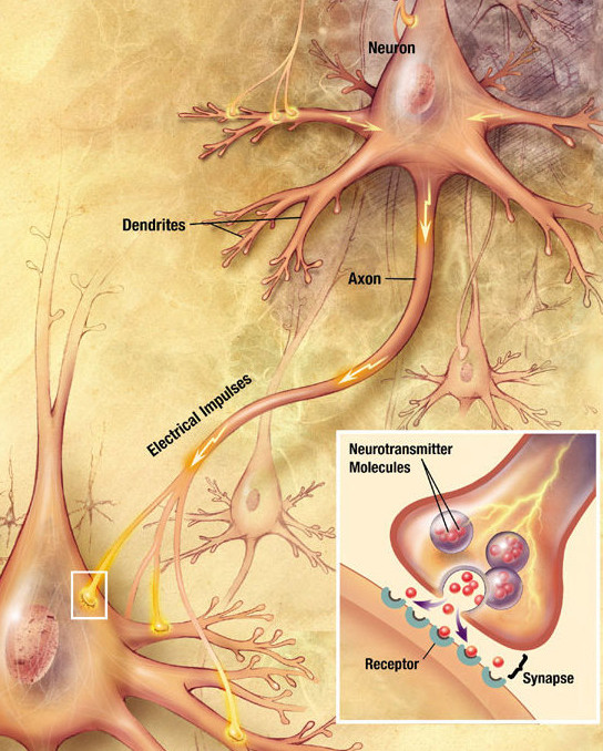
In the nervous system, a synapse is a structure that allows a neuron (or nerve cell) to pass an electrical or chemical signal to another neuron or a target effector cell. Synapses can be classified as either chemical or electrical, depending on the mechanism of signal transmission between neurons. In the case of electrical synapses, neurons are coupled bidirectionally with each other through gap junctions and have a connected cytoplasmic milieu. These types of synapses are known to produce synchronous network activity in the brain, but can also result in complicated, chaotic network level dynamics. Therefore, signal directionality cannot always be defined across electrical synapses. Chemical synapses, on the other hand, communicate through neurotransmitters released from the presynaptic neuron into the synaptic cleft. Upon release, these neurotransmitters bind to specific receptors on the postsynaptic membrane, inducing an electrical or chemical response in the target neuron ... [...More Info...] [...Related Items...] OR: [Wikipedia] [Google] [Baidu] [Amazon] |
Axo-axonic Synapse
An axo-axonic synapse is a type of synapse, formed by one neuron projecting its axon terminals onto another neuron's axon. Axo-axonic synapses have been found and described more recently than the other more familiar types of synapses, such as axo-dendritic synapses and axo-somatic synapses. The spatio-temporal properties of neurons get altered by the type of synapse formed between neurons. Unlike the other types, the axo-axonic synapse does not contribute towards triggering an action potential in the postsynaptic neuron. Instead, it affects the probability of neurotransmitter release in the response to any action potential passing through the axon of the postsynaptic neuron. Thus, axo-axonic synapses appear to be very important for the brain in achieving a specialized neural computation. Axo-axonic synapses are found throughout the central nervous system, including in the hippocampus, cerebral cortex and striatum in mammals; in the Neuromuscular junction, neuro-muscular junctions i ... [...More Info...] [...Related Items...] OR: [Wikipedia] [Google] [Baidu] [Amazon] |
Slice Preparation
The slice preparation or brain slice is a laboratory technique in electrophysiology that allows the study of neurons from various brain regions in isolation from the rest of the brain, in an ex-vivo condition. Brain tissue is initially sliced via a tissue slicer then immersed in artificial cerebrospinal fluid (aCSF) for stimulation and/or recording. The technique allows for greater scientific control, experimental control, through elimination of the effects of the rest of the brain on the neural circuit, circuit of interest, careful control of the physiological conditions through perfusion of substrates through the incubation fluid, to precise manipulation of neurotransmitter activity through perfusion of agonists and antagonists. However, the increase in control comes with a decrease in the ease with which the results can be applied to the whole neural system. Slice preparation techniques Free hand sectioning is a type of preparation techniques where a skilled operator uses razo ... [...More Info...] [...Related Items...] OR: [Wikipedia] [Google] [Baidu] [Amazon] |
Reflex Arc
A reflex arc is a neural pathway that controls a reflex. In vertebrates, most sensory neurons synapse in the spinal cord and the signal then travels through it into the brain. This allows for faster reflex actions to occur by activating spinal motor neurons without the delay of routing signals through the brain. The brain will receive the input while the reflex is being carried out and the analysis of the signal takes place after the reflex action. There are two types: autonomic nervous system, autonomic reflex arc (affecting inner organs) and somatic nervous system, somatic reflex arc (affecting muscles). Autonomic reflexes sometimes involve the spinal cord and some somatic reflexes are mediated more by the brain than the spinal cord. During a somatic reflex, nerve signals travel along the following pathway: # ''Somatic receptors'' in the skin, muscles and tendons # ''Afferent nerve fibers'' carry signals from the somatic receptors to the Posterior horn of spinal cord, posterior ... [...More Info...] [...Related Items...] OR: [Wikipedia] [Google] [Baidu] [Amazon] |
Afferent Nerve Fiber
Afferent nerve fibers are axons (nerve fibers) of sensory neurons that carry sensory information from sensory receptors to the central nervous system. Many afferent projections ''arrive'' at a particular brain region. In the peripheral nervous system, afferent nerve fibers are part of the sensory nervous system and arise from outside of the central nervous system. Sensory and mixed nerves contain afferent fibers. Structure Afferent neurons are pseudounipolar neurons that have a single process leaving the cell body dividing into two branches: the long one towards the sensory organ, and the short one toward the central nervous system (e.g. spinal cord). These cells do have sensory afferent dendrites, similar to those typically inherent in neurons. They have a smooth and rounded cell body located in the ganglia of the peripheral nervous system. Just outside the spinal cord, thousands of afferent neuronal cell bodies are aggregated in a swelling in the dorsal root known a ... [...More Info...] [...Related Items...] OR: [Wikipedia] [Google] [Baidu] [Amazon] |
Micrograph
A micrograph is an image, captured photographically or digitally, taken through a microscope or similar device to show a magnify, magnified image of an object. This is opposed to a macrograph or photomacrograph, an image which is also taken on a microscope but is only slightly magnified, usually less than 10 times. Micrography is the practice or art of using microscopes to make photographs. A photographic micrograph is a photomicrograph, and one taken with an electron microscope is an electron micrograph. A micrograph contains extensive details of microstructure. A wealth of information can be obtained from a simple micrograph like behavior of the material under different conditions, the phases found in the system, failure analysis, grain size estimation, elemental analysis and so on. Micrographs are widely used in all fields of microscopy. Types Photomicrograph A light micrograph or photomicrograph is a micrograph prepared using an optical microscope, a process referred to ... [...More Info...] [...Related Items...] OR: [Wikipedia] [Google] [Baidu] [Amazon] |
Synaptic Vesicle
In a neuron, synaptic vesicles (or neurotransmitter vesicles) store various neurotransmitters that are exocytosis, released at the chemical synapse, synapse. The release is regulated by a voltage-dependent calcium channel. Vesicle (biology), Vesicles are essential for propagating nerve impulses between neurons and are constantly recreated by the cell (biology), cell. The area in the axon that holds groups of vesicles is an axon terminal or "terminal bouton". Up to 130 vesicles can be released per bouton over a ten-minute period of stimulation at 0.2 Hz. In the visual cortex of the human brain, synaptic vesicles have an average diameter of 39.5 nanometers (nm) with a standard deviation of 5.1 nm. Structure Synaptic vesicles are relatively simple because only a limited number of proteins fit into a sphere of 40 nm diameter. Purified vesicles have a protein:phospholipid ratio of 1:3 with a lipid composition of 40% phosphatidylcholine, 32% phosphatidylethanola ... [...More Info...] [...Related Items...] OR: [Wikipedia] [Google] [Baidu] [Amazon] |
Hyperpolarization (biology)
Hyperpolarization is a change in a cell's membrane potential that makes it more negative. Cells typically have a negative resting potential, with neuronal action potentials depolarizing the membrane. When the resting membrane potential is made more negative, it increases the minimum stimulus needed to surpass the needed threshold. Neurons naturally become hyperpolarized at the end of an action potential, which is often referred to as the relative refractory period. Relative refractory periods typically last 2 milliseconds, during which a stronger stimulus is needed to trigger another action potential. Cells can also become hyperpolarized depending on channels and receptors present on the membrane, which can have an inhibitory effect. Hyperpolarization is often caused by efflux of K+ (a cation) through K+ channels, or influx of Cl– (an anion) through Cl– channels. On the other hand, influx of cations, e.g. Na+ through Na+ channels or Ca2+ through Ca2+ cha ... [...More Info...] [...Related Items...] OR: [Wikipedia] [Google] [Baidu] [Amazon] |
Depolarization
In biology, depolarization or hypopolarization is a change within a cell (biology), cell, during which the cell undergoes a shift in electric charge distribution, resulting in less negative charge inside the cell compared to the outside. Depolarization is essential to the function of many cells, communication between cells, and the overall physiology of an organism. Most cells in higher organisms maintain an internal environment that is negatively charged relative to the cell's exterior. This difference in charge is called the cell's membrane potential. In the process of depolarization, the negative internal charge of the cell temporarily becomes more positive (less negative). This shift from a negative to a more positive membrane potential occurs during several processes, including an action potential. During an action potential, the depolarization is so large that the potential difference across the cell membrane briefly reverses polarity, with the inside of the cell becoming p ... [...More Info...] [...Related Items...] OR: [Wikipedia] [Google] [Baidu] [Amazon] |
Membrane Potential
Membrane potential (also transmembrane potential or membrane voltage) is the difference in electric potential between the interior and the exterior of a biological cell. It equals the interior potential minus the exterior potential. This is the energy (i.e. work) per charge which is required to move a (very small) positive charge at constant velocity across the cell membrane from the exterior to the interior. (If the charge is allowed to change velocity, the change of kinetic energy and production of radiation must be taken into account.) Typical values of membrane potential, normally given in units of milli volts and denoted as mV, range from −80 mV to −40 mV. For such typical negative membrane potentials, positive work is required to move a positive charge from the interior to the exterior. However, thermal kinetic energy allows ions to overcome the potential difference. For a selectively permeable membrane, this permits a net flow against the gradient. This is a kind ... [...More Info...] [...Related Items...] OR: [Wikipedia] [Google] [Baidu] [Amazon] |
Soma (biology)
In cellular neuroscience, the soma (: somata or somas; ), neurocyton, or cell body is the bulbous, non-process portion of a neuron or other brain cell type, containing the cell nucleus. Although it is often used to refer to neurons, it can also refer to other cell types as well, including astrocytes, oligodendrocytes, and microglia. The part of the soma without the nucleus is called perikaryon (: perikarya). There are many different specialized types of neurons, and their sizes vary from as small as about 5 micrometres to over 10 millimetres for some of the smallest and largest neurons of invertebrates, respectively. The soma of a neuron (i.e., the main part of the neuron in which the dendrites branch off of) contains many organelles, including granules called Nissl granules, which are composed largely of rough endoplasmic reticulum and free polyribosomes. The cell nucleus is a key feature of the soma. The nucleus is the source of most of the RNA that is produced in ne ... [...More Info...] [...Related Items...] OR: [Wikipedia] [Google] [Baidu] [Amazon] |
Chemical Synapse
Chemical synapses are biological junctions through which neurons' signals can be sent to each other and to non-neuronal cells such as those in muscles or glands. Chemical synapses allow neurons to form circuits within the central nervous system. They are crucial to the biological computations that underlie perception and thought. They allow the nervous system to connect to and control other systems of the body. At a chemical synapse, one neuron releases neurotransmitter molecules into a small space (the synaptic cleft) that is adjacent to another neuron. The neurotransmitters are contained within small sacs called synaptic vesicles, and are released into the synaptic cleft by exocytosis. These molecules then bind to neurotransmitter receptors on the postsynaptic cell. Finally, the neurotransmitters are cleared from the synapse through one of several potential mechanisms including enzymatic degradation or re-uptake by specific transporters either on the presynaptic cell or ... [...More Info...] [...Related Items...] OR: [Wikipedia] [Google] [Baidu] [Amazon] |


